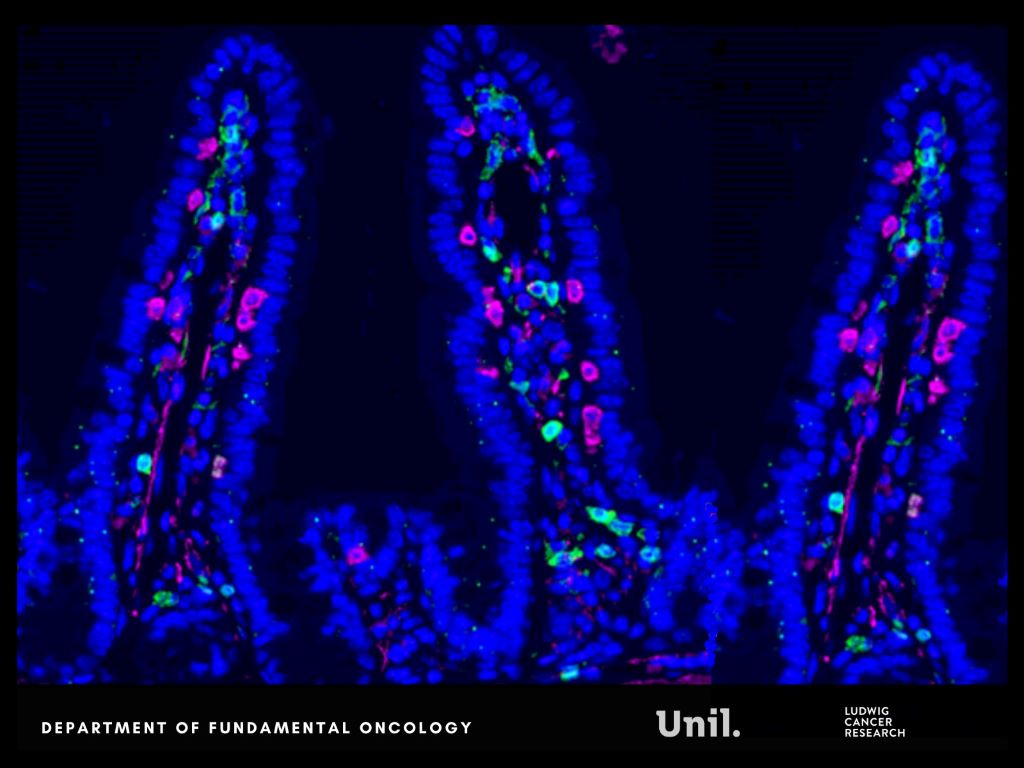
Microscopy image of intestinal lining in mice, shows CD4 (green), CD8 (magenta) and DAPI (blue). Ludivine Bersier 2025
In Nature Communications, researchers from Lusanne University reveal that chemotherapy alters gut microbes and bone marrow immune cell development, unexpectedly reprogramming systemic immunity in ways that help restrict metastatic progression.
Chemotherapy commonly damages the intestinal lining, a well-known side effect. But this injury does not remain confined to the gut. It reshapes nutrient availability for intestinal bacteria, forcing the microbiota to adapt.
The researchers report that chemotherapy-induced damage to the intestinal lining alters nutrient availability for gut bacteria, reshaping the microbiota and increasing the production of indole-3-propionic acid (IPA), a tryptophan-derived microbial metabolite.
Rather than acting locally, IPA functions as a systemic messenger. It travels from the gut to the bone marrow, where it rewires immune cell production. Elevated IPA levels reprogram myelopoiesis, reducing the generation of immunosuppressive monocytes that facilitate immune evasion and metastatic growth.
“We were surprised by how a side effect often seen as collateral damage of chemotherapy can trigger such a structured systemic response. By reshaping the gut microbiota, chemotherapy sets off a cascade of events that rewires immunity and makes the body less permissive to metastasis.” says Ludivine Bersier, first author of the study.
This immune reconfiguration enhances T-cell activity and remodels immune interactions within metastatic niches, particularly in the liver, resulting in a metastasis-refractory state in preclinical models.
Experimental findings are mirrored in patients. Clinical relevance is supported by patient data obtained in collaboration with Dr Thibaud Koessler (Geneva University Hospitals, HUG). In patients with colorectal cancer, higher circulating IPA levels following chemotherapy are associated with reduced monocyte levels, a feature of improved survival outcomes.
“This work shows that the effects of chemotherapy extend far beyond the tumor itself. By uncovering a functional axis linking the gut, the bone marrow and metastatic sites, we highlight systemic mechanisms that could be harnessed to durably limit metastatic progression.” says Tatiana Petrova, corresponding author of the study.
This research was supported by multiple funders, including the Swiss National Science Foundation and Swiss Cancer League. An ISREC Foundation Tandem Grant supported close collaboration between clinical and fundamental research, led at Unil by Professor Tatiana Petrova and Dr Thibaud Koessler at HUG. The project posits that chemotherapy can induce a form of biological “memory”, mediated by gut microbiome–derived metabolites that durably inhibit metastatic growth.
Together, these findings reveal a previously underappreciated gut–bone marrow–liver metastasis axis through which chemotherapy can exert durable systemic effects, opening new avenues to harness microbiota-derived metabolites as adjuvant strategies to limit metastasis.
Cedars-Sinai Led Clinical trial showed combo treatment reduced deaths in patients with an aggressive form of the disease
Credit: Darryl Leja National Human Genome Research Institute National Institutes Of Health
Men whose prostate cancer returns after surgery or radiation therapy may now benefit from a new drug combination shown in clinical trials to cut the risk of death by more than 40%.
The combination therapy, which adds the drug enzalutamide to commonly prescribed hormone therapy, reduced deaths in patients with recurrent prostate cancer after surgery or radiation for whom other treatments are no longer an option. The trial results were published in The New England Journal of Medicine (NEJM) with simultaneous presentation during the European Society for Medical Oncology Congress (ESMO) Oct. 19 in Berlin.
“After initial treatment, some patients see their prostate cancer come back in an aggressive way and are at risk for their disease to spread quickly,” said Stephen Freedland, MD, director of the Center for Integrated Research in Cancer and Lifestyle at Cedars-Sinai Cancer and co-principal investigator of the study. “Hormone therapy, which is what we’ve been offering patients for 30 years, has not improved survival and neither has anything else. That makes these findings a real game changer.”
The trial included more than 1000 patients from 244 sites in 17 countries. All the patients were diagnosed with what is known as high-risk biochemically recurrent prostate cancer. Following the patients’ surgery or radiation therapy, the levels of prostate specific antigen, or PSA, in their blood had risen rapidly. PSA is a protein used to detect prostate cancer, and a rapid rise in PSA levels after treatment indicates a patient’s cancer is likely to return and metastasise, often to the bones or spine.
“We know these patients are at high risk of developing metastatic disease and dying of their cancer unless we offer a meaningful treatment option,” said Freedland, professor of Urology and the Warschaw, Robertson, Law Families Chair in Prostate Cancer.
Patients were randomly selected to receive standard hormone therapy alone, enzalutamide alone, or a combination of the two. After eight years, the risk of death was 40.3% lower in the combination group than in the other two groups, Freedland said.
“This clinical trial, one of many that Cedars-Sinai Cancer has offered to its patients, is an example of the translational work being done by our physician-scientists,” said Robert Figlin, MD, interim director of Cedars-Sinai Cancer. “The result will be improved treatment and better outcomes for patients everywhere.”
Freedland noted that, based on previous results published by the team, enzalutamide is approved by the Food and Drug Administration and listed in National Comprehensive Cancer Network treatment guidelines. These latest results, he said, are likely to strengthen the network’s recommendation and solidify this drug combination as the standard of care for patients with high-risk biochemically recurrent prostate cancer.
“These important findings identify a treatment that prolongs survival in men with aggressive prostate cancer,” said Hyung Kim, MD, a urologic oncologist and chair of the Department of Urology at Cedars-Sinai. “The latest analysis complements previous studies that found enzalutamide significantly improved survival in other prostate cancer settings, and will change how we take care of our patients.”
A new study from researchers at The University of Texas MD Anderson Cancer Center shows that the glucose-fructose mix found in sugary drinks directly fuels metastasis in preclinical models of advanced colorectal cancer. The study was published in Nature Metabolism.
A research team led by Jihye Yun, PhD, assistant professor of Genetics, studied how sugary drinks may affect late-stage colorectal cancer. Using laboratory cancer models, they compared the effects of the glucose-fructose mix found in most sugary drinks with those of glucose or fructose alone. Only the sugar mix made cancer cells more mobile, leading to faster spread to the liver – the most common site of colorectal cancer metastasis.
The sugar mix activated an enzyme called sorbitol dehydrogenase (SORD), which boosts glucose metabolism and triggers the cholesterol pathway, ultimately driving metastasis. This is the same pathway targeted by statins, common heart drugs that inhibit cholesterol production. Blocking SORD slowed metastasis, even with the sugar mix present. These findings suggest that targeting SORD could also offer an opportunity to block metastasis.
“Our findings highlight that daily diet matters not only for cancer risk but also for how the disease progresses once it has developed,” Yun said. “While these findings need further investigation, they suggest that reducing sugary drinks, targeting SORD or repurposing statins may benefit patients with colorectal cancer.”
The Yun Laboratory is interested in studying how diet affects the intestine and cancer development, and they have made important discoveries on the impacts of sugary drinks on colorectal cancer.
Sugar has long been indirectly linked to an increase in cancer risk through obesity. However, a previous study by Yun’s lab challenged that view, showing that even moderate intake of sugary drinks directly fuelled tumour growth in early-stage colorectal cancer, independent of obesity. The current study was done to determine how sugary drinks may impact later-stage disease.
While this study needs further clinical investigation, the results suggest that reducing sugary drinks and targeting the SORD enzyme may offer opportunities to reduce colorectal cancer metastasis. Additional studies are warranted to confirm these results outside of preclinical models.
Further, Yun explained it may be worthwhile to consider revisions to current dietary recommendations to reduce sugary drink consumption in this patient population. To meet nutritional needs, many patients with cancer are encouraged to have nutritional supplement drinks and concentrated juices that contain high glucose and fructose content.
A Swedish-led research team at Karolinska Institutet and Karolinska University Hospital has shown in a new randomised clinical trial that a low dose of the well-known medicine aspirin halves the risk of recurrence after surgery in patients with colon and rectal cancer with a certain type of genetic alteration in the tumour.
Every year, nearly two million people worldwide are diagnosed with colorectal cancer, and 20–40% develop metastases.
Previous observational studies have suggested that aspirin may reduce the risk of certain cancers and possibly also the risk of recurrence after surgery in patients with colorectal cancer harbouring mutations in genes within the PIK3 signaling pathway. These genes regulate key cellular processes such as growth and division. When mutated, these processes can become dysregulated, leading to uncontrolled cell proliferation and cancer development.
Randomised clinical trials were lacking
Prior findings have been inconsistent and no randomised clinical trials had previously confirmed the association. To address this gap, the ALASCCA trial was initiated, with the results now been published in The New England Journal of Medicine.
The current study included more than 3500 patients with colon and rectal cancer from 33 hospitals in Sweden, Norway, Denmark, and Finland. Patients whose tumours showed a specific genetic mutation in the PIK3 signalling pathway – a mutation found in approximately 40% of patients – were randomised to receive either 160mg of aspirin daily or a placebo for three years after surgery.
For patients with the genetic mutation in PIK3, the risk of recurrence was reduced by 55% in those who received aspirin compared with the placebo group.
“Aspirin is being tested here in a completely new context as a precision medicine treatment. This is a clear example of how we can use genetic information to personalise treatment and at the same time save both resources and suffering,” says first author Anna Martling, professor at the Department of Molecular Medicine and Surgery, Karolinska Institutet, and senior consultant surgeon at Karolinska University Hospital.
Less favourable environment for cancer
So how does aspirin reduce the risk of recurrence of colon and rectal cancer? The researchers believe that the effect is likely due to aspirin acting through several parallel mechanisms – it reduces inflammation, inhibits platelet function and tumour growth. This combination makes the environment less favourable for cancer.
“Although we do not yet fully understand all the molecular links, the findings strongly support the biological rationale and suggest that the treatment may be particularly effective in genetically defined subgroups of patients,” says Anna Martling.
The researchers believe that the results could have global significance and influence treatment guidelines for colon and rectal cancer worldwide. Anna Martling sees the fact that the drug is well established as a major advantage.
“Aspirin is a drug that is readily available globally and extremely inexpensive compared to many modern cancer drugs, which is very positive,” says Anna Martling.
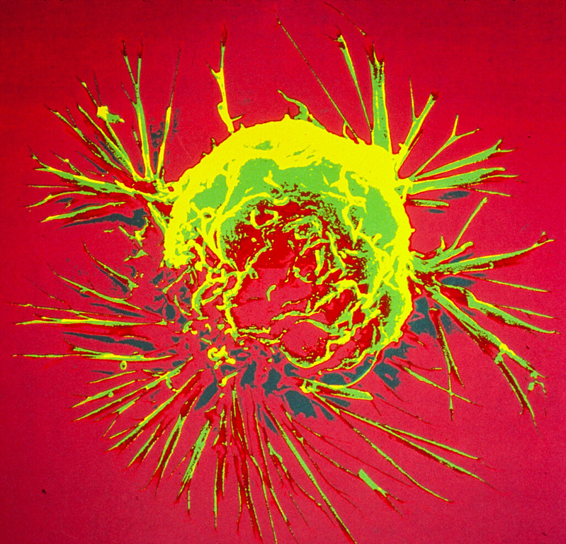
Colourised scanning electron micrograph of a breast cancer cell. Credit: NIH
Researchers from Tampere University, Finland, and Izmir Institute of Technology, Turkey, have developed an in vitro cancer model to investigate why breast cancer spreads to bone. Their findings, published in PLOS One, hold promise for advancing the development of preclinical tools to predict breast cancer bone metastasis.
Breast canceris a significant global public health challenge, with 2.3 million new cases and 700 000 deaths every year. Approximately 80% of patients with primary breast cancer can be cured, if they are diagnosed and treated promptly. However, in many cases, the cancer has already metastasised at the time of diagnosis.
Metastatic cancer is incurable and accounts for more than 90% of cancer-related deaths. Currently, there are no reliable in vitro models to study how breast cancer spreads to secondary organs such as bone, lung, liver or brain. Now, researchers from the Precision Nanomaterials Group at Tampere University in Finland, and the Cancer Molecular Biology Lab at Izmir Institute of Technology in Turkey, have used lab-on-a-chip platforms to create a physiologically relevant metastasis model to study the factors controlling breast cancer bone metastasis.
“Breast cancer most frequently spreads to bone, with an estimated rate of 53%, resulting in severe symptoms such as pain, pathological bone fractures, and spinal cord compressions. Our research provides a laboratory model that estimates the likelihood and mechanism of bone metastasis occurring within a living organism. This advances the understanding of molecular mechanisms in breast cancer bone metastasis and provides the groundwork for developing preclinical tools for predicting bone metastasis risk,” says Burcu Firatligil-Yildirir, postdoctoral researcher at Tampere University and the first author of the paper.
According to Nonappa, Associate Professor and leader of the Precision Nanomaterials Group at Tampere University, developing sustainable in vitro models that mimic the complexity of the native breast and bone microenvironment is a multidisciplinary challenge.
“Our work shows that physiologically relevant in vitro models can be generated by combining cancer biology, microfluidics and soft materials. The results open new possibilities for developing predictive disease, diagnostic and treatment models,” he says.
The largest population-based study to date supports the survival benefits of immunotherapy for people with metastatic non–small cell lung cancer.
Squamous cancer cell being attacked by cytotoxic T cells. Image by National Cancer Institute on Unsplash
Since the first immunotherapy drug to boost the body’s immune response against advanced lung cancer was introduced in the United States in 2015, survival rates of patients with the disease have improved significantly. That’s the conclusion of a recent real-world study published by Wiley online in CANCER, a peer-reviewed journal of the American Cancer Society.
For the research, a team led by Dipesh Uprety, MD, FACP, of the Barbara Ann Karmanos Cancer Institute and the Wayne State University School of Medicine, analysed data from the National Cancer Institute Surveillance, Epidemiology, and End Results database, which compiles cancer-related data covering approximately 48% of the US population. The investigators’ analysis focused on non–small cell lung cancer (NSCLC), which accounts for up to 90% of all cases of lung cancer and is the leading cause of cancer-related death among both men and women in the United States.
In a comparison of 100 995 patients with metastatic NSCLC treated in 2015–2020 (after immunotherapy was deemed the standard of care) and 90 807 patients with metastatic NSCLC in the pre-immunotherapy era of 2010–2014, patients in the immunotherapy era were less likely to die from any cause. The overall survival rates at one, three, and five years were 40.1% versus 33.5%, 17.8% versus 11.7%, and 10.7% versus 6.8%. The median overall survival was eight months in patients in the immunotherapy era and seven months in those in the pre-immunotherapy era.
Similarly, patients treated after immunotherapy was available were less likely to die specifically from cancer than those treated before immunotherapy. The one-, three-, and five-year cancer-specific survival rates were 44.0% versus 36.8%, 21.7% versus 14.4%, and 14.3% versus 9.0%, with a median survival of 10 months versus eight months.
Survival rates remained significantly better in the immunotherapy era even after accounting for factors including age, sex, race, income, and geographical area.
“By utilizing a large national database, our study provided real-world evidence of the positive impact of immunotherapy in patients with lung cancer,” said Dr Uprety. The investigators stressed that additional studies are needed, however. “Immunotherapy provides long-term benefits. Since the durable benefits of immunotherapy are limited to a small subset of patients, future research should aim to optimize immunotherapy with new agents that can benefit a broader population,” said lead author Yating Wang, MD, of Ascension Providence Hospital.
There is a surprising dearth of research about how breast cancer cells can go dormant, spread and then resurface years or even decades later, according to a new review of in vitro breast cancer studies conducted by researchers at the University of Massachusetts Amherst.
“[Our review found that] less than 1% of all these studies that combine cells with designer environments look at dormancy,” says Shelly Peyton, Provost Professor of Chemical Engineering. “It’s not enough. We just don’t understand what’s happening – and it’s killing patients.”
Breast cancer dormancy is a phenomenon in which breast cancer cells metastasise (typically to the liver, lungs, brain or bones) but don’t grow. “They’re not detectable or symptomatic tumours,” Peyton explains. “A patient will have their primary tumour removed and appear to be disease-free for months, years, even decades. And for reasons we don’t understand, something changes about the environment that causes those cells to start regrowing, and then you have a deadly metastasis.”
Patients with metastatic breast cancer have a 30% five-year survival rate, compared to a 99% survival rate for localised breast cancer. “Early detection is key, particularly in the Western world,” says Peyton. “You can have lumpectomies, radiation, small surgeries. And women can survive. It’s when that cancer has spread that it becomes much harder to treat.”
This relapse in distant organs impacts 40% of early-stage breast cancer patients, and breast cancer dormancy is a contributing factor. But while metastasis has known biomarkers, dormant cancer cells are very hard to identify.
“When you have a single dormant breast cancer cell that’s hiding in a distant tissue, it’s really hard to detect that,” says Nate Richbourg, lead author on the paper and postdoctoral researcher in the Peyton Lab. “And you don’t want to do an invasive biopsy or prescribe toxic chemotherapy for something that might not be a problem.”
With these challenges in mind, the review, published in Science Advances, aimed to identify gaps in the research, particularly focusing on in vitro studies, or research using benchtop-model environments instead of animal models or humans. In vitro studies allow for the precise control of the environment, which Peyton’s research group says may play a deciding role in whether a cell remains dormant or reactivates into a deadly metastatic tumor.
“What can we control in these artificial environments that will give us insight into how breast cancer dormancy happens, and what we can do to treat it as well?” Richbourg asks, describing the importance of in vitro modelling. “When we create this artificial dormancy, we can see how many of those cells could turn back into proliferating and potentially deadly cells.”
Their review highlights just how complex the role of the environment is. “If you have a [breast cancer] cell somewhere in the bone marrow, you’re going to have other cells there, the physical factors in your environment, and the biochemical factors,” Richbourg gives as an example. “We try to use reductive models to separate the thing that is influencing this behaviour. But what we’re seeing is that everything works together to create this breast cancer dormancy effect. The better we can create models that capture all that nuance, the better we’re going to be able to understand it.”
For Peyton, their work is also a call to action. “The paper is calling out to the field that we need to do more,” she says. This includes being more creative with the materials that already exist and developing new materials; identifying ways to model the decades-long progression of dormancy that is impossible to recreate in a single study; and expanding the diversity of cell lines used for research (Richbourg points out that many of the studies they reviewed used the same cell line, MDA-MB-231, derived from one 40-to-50-year-old white woman).
Finally, the researchers have an eye to the ultimate goal: better treatments to save patients. “We see that that there are some clinical trials that are happening that are derived from some of those in vitro models,” says Ninette Irakoze, graduate student in the Peyton Lab. “The paper gives hope that, with more development of these in vitro models, eventually we could find treatments to eradicate dormant cancer.”
Deaths from breast cancer dropped 58% between 1975 and 2019 due to a combination of screening mammography and improvements in treatment, according to a new study led by Stanford Medicine clinicians and biomedical data scientists.
Nearly one-third of the decrease (29%) is due to advances in treating metastatic breast cancer, also known as stage 4 breast cancer or recurrent cancer. Although these advanced cancers are not considered curable, women with metastatic disease are living longer than ever.
The analysis helps cancer researchers assess where to focus future efforts and resources.
“We’ve known that deaths from breast cancer have been decreasing over the past several decades, but it’s been difficult or impossible to quantify which of our interventions have been most successful, and to what extent,” said Jennifer Caswell-Jin, MD, assistant professor of medicine. “This type of study allows us to see which of our efforts are having the most impact and where we still need to improve.”
Caswell-Jin and Liyang Sun are co-first authors of the study, which was published in the Journal of the American Medical Association. Sylvia Plevritis, PhD, professor and chair of biomedical data science, and Allison Kurian, MD, MSc, professor of medicine and of epidemiology and population health, are co-senior authors.
The study was a collaborative effort by a national consortium of researchers called CISNET, or the Cancer Intervention and Surveillance Modeling Network. CISNET was established in 2000 by the National Cancer Institute to understand the impact of cancer surveillance, screening and treatment on incidence and mortality. Doing so requires sophisticated computer algorithms capable of modelling the natural course of the disease and the typical treatment paths of individual patients, then translating that information to population-level data collected by the national Surveillance, Epidemiology, and End Results Program, or SEER registry, from 1975 to 2019.
The study is the third in a trio of papers from CISNET published since 2005 that assess the relative contributions of regular screening and treatment advances on breast cancer deaths. The previous two papers informed national guidelines and helped cancer researchers focus their efforts on the most intractable problems.
“Twenty years ago, there was a question whether routine screening mammography actually decreased the number of deaths from breast cancer,” Plevritis said. But in 2005, she and other CISNET researchers published a paper in the New England Journal of Medicine that conclusively demonstrated that screening was responsible for anywhere from 28% to 65% (different models came up with varying degrees of impact) of the reduction in mortality by 2000 between 1975 and 2000.
The second paper, published in 2018 in the Journal of the American Medical Association, highlighted the differences in treatment responsiveness and survival outcomes among women with differing breast cancer subtypes from 2000 to 2012, pinpointing subgroups with poorer survival.
“We found that, while screening still had an important impact, most of the decline in annual deaths was due to improvements in treating early-stage breast cancer based on each cancer’s molecular profile,” Plevritis said.
The current study is the first to explicitly include patients with metastatic breast cancer in its models. The finding that 29% of the decrease in mortality is due to advances in treating metastatic breast cancer both surprised and gratified the researchers.
“Initially, we assumed that treatment of advanced disease was unlikely to make a significant contribution to the declines in mortality we documented in the previous two papers,” Caswell-Jin said. “But our treatments have improved, and it’s clear that they are having a significant impact on annual mortality.”
The CISNET researchers used four computer models to assess the SEER data from 1975 to 2019 — one developed at Stanford Medicine in the Plevritis Lab, one by researchers at the Dana-Farber Cancer Institute, one at MD Anderson Cancer Center, and another jointly developed by researchers at the University of Wisconsin and Harvard Medical School. The four models came up with remarkably similar estimates for the impact of each intervention: screening mammography, treatment of early-stage (stages 1, 2 or 3) breast cancer and treatment of metastatic breast cancer.
The models reproduced the decline in mortality in breast cancer known from SEER data, from 48 per 100 000 women dying of breast cancer each year in 1975 to 27 per 100,000 in 2019, a decrease of about 44%. The models arrived at a larger estimated reduction in mortality of about 58% because the incidence of breast cancer has risen during the same period and more women would have died had screening and treatments not improved.
The models concluded that about 47% of this reduction in mortality is the result of improved treatments for early-stage breast cancer, and about 25% is attributed to screening mammography. The remainder, or about 29%, is due to improvements in treating metastatic disease.
“Designing the new model, which had to account for individuals with non-metastatic cancer who underwent treatment but later progressed to metastatic cancer, and who may have been treated with multiple drugs over the course of their disease, was extremely complex,” Plevritis said. “It took about four years. But it was really satisfying when we were able to validate the model’s behaviour and see that all four models from different institutions, which used the new model inputs in different ways, delivered consistent findings. The models not only make sense, but also produce meaningful insights.”
The impact of treating metastatic disease is exemplified by the increases in median survival time after metastasis: Patients diagnosed in 2000 with metastatic disease lived an average of 1.9 years versus an average of 3.2 years for those diagnosed in 2019. Survival time varies by subgroup status, however. Patients with what are known as oestrogen receptor-positive and HER2 positive cancers saw an average increase in survival time of 2.5 years. Those with oestrogen receptor-positive and HER2-negative cancers lived an average of 1.6 years longer, but those with cancers that are oestrogen receptor-negative and HER2-negative lived about 0.5 years longer in 2019 than in 2000.
“It was meaningful as a breast oncologist to spend time with this history and see real progress over the past decades,” Caswell-Jin said. “There is much more work to be done; metastatic breast cancer isn’t yet curable. But it is rewarding to see that advances have made a difference in these numbers,” she added. “Our scientific and clinical work is helping our patients live longer, and I believe deaths from breast cancer will continue to steadily decline as innovation continues to grow.”
The vertebral bones that constitute the spine are derived from a distinct type of stem cell that secretes a protein favouring tumour metastases, according to a study led by researchers at Weill Cornell Medicine. The discovery, published in Nature, opens up a new line of research on spinal disorders and helps explain why solid tumours so often spread to the spine, and could lead to new orthopaedic and cancer treatments.
Vertebral bone was found to be derived from a stem cell that is different from other bone-making stem cells. Using bone-like “organoids” made from vertebral stem cells, they showed that the known tendency of tumours to spread to the spine rather than long bones is due largely to a protein called MFGE8, secreted by these stem cells.
“We suspect that many bone diseases preferentially involving the spine are attributable to the distinct properties of vertebral bone stem cells,” said study senior author Dr Matthew Greenblatt.
In recent years, Dr Greenblatt and other scientists have found that different types of bone are derived from different types of bone stem cells. Since vertebrae develop along a different pathway early in life, and also appear to have had a distinct evolutionary trajectory, Dr Greenblatt and his team hypothesised that a distinct vertebral stem cell probably exists.
The researchers started out by isolating what are broadly known as skeletal stem cells, which give rise to all bone and cartilage, from different bones in lab mice based on known surface protein markers of such cells. They then analysed gene activity in these cells to see if they could find a distinct pattern for the ones associated with vertebral bone.
This effort yielded two key findings. The first was a new and more accurate surface-marker-based definition of skeletal stem cells as a whole. This new definition excluded a set of cells that are not stem cells that had been included in the old stem cell definition, thus clouding some prior research in this area.
The second finding was that skeletal stem cells from different bones do indeed vary systematically in their gene activity. From this analysis, the team identified a distinct set of markers for vertebral stem cells, and confirmed these cells’ functional roles to form spinal bone in further experiments in mice and in lab-dish cell culture systems.
The researchers next investigated the phenomenon of the spine’s relative attraction for tumour metastases, including breast, prostate and lung tumours, compared to other types of bone. The traditional theory, dating to the 1940s, is that this “spinal tropism” relates to patterns of blood flow that preferentially convey metastases to the spine versus long bones. But when the researchers reproduced the spinal tropism phenomenon in animal models, they found evidence that blood flow isn’t the explanation, finding instead a clue pointing to vertebral stem cells as the possible culprits.
“We observed that the site of initial seeding of metastatic tumour cells was predominantly in an area of marrow where vertebral stem cells and their progeny cells would be located,” said study first author Dr Jun Sun, a postdoctoral researcher in the Greenblatt laboratory.
Subsequently, the team found that removing vertebral stem cells eliminated the difference in metastasis rates between spine bones and long bones. Ultimately, they determined that MFGE8, a protein secreted in higher amounts by vertebral compared to long bone stem cells, is a major contributor to spinal tropism. To confirm the relevance of the findings in humans, the team collaborated with investigators at Hospital for Special Surgery to identify the human counterparts of the mouse vertebral stem cells and characterise their properties.
The researchers are now exploring methods for blocking MFGE8 to reduce the risk of spinal metastasis in cancer patients. More generally, said Dr Greenblatt, they are studying how the distinctive properties of vertebral stem cells contribute to spinal disorders.
“There’s a subdiscipline in orthopaedics called spinal orthopaedics, and we think that most of the conditions in that clinical category have to do with this stem cell we’ve just identified,” Dr Greenblatt said.
In a paper published in the journal Biomolecules, UK and Chinese researchers report their creation of a biomedical compound that has the potential to stop breast cancer metastasis.
The scientists from the Chemistry and Biochemistry Departments at the University of Liverpool and Nanjing Medical School in China have discovered a possible way to block proteins produced by cancer cells that promote metastasis – the chief impediment to successful cancer treatment.
Prof Philip Rudland from the University of Liverpool explained: “As a general rule, cancer that has spread is treated with chemotherapy, but this treatment can rarely be given without severely harming or becoming toxic to the patient. The importance of our work was to identify a specific and important target to attack, without toxic side effects.”
The University’s research team have in the past discovered that specific proteins are involved in the metastatic process; these proteins are different from those involved in the production of the primary tumour. One such example is a protein called ‘S100A4’, and is the protein chosen by the research team to target for the identification of chemical inhibitors of metastasis, using model systems of cells from the highly metastatic and incurable hormone receptor-free breast cancer.
Using these model systems, researchers at the University’s Department of Biochemistry discovered a novel compound that can specifically block the interaction of this metastasis-inducing protein S100A4 with its target inside the cell. Researchers in the Department of Chemistry then synthesised a simpler chemical and connected it to a warhead which stimulates cells’ normal protein-degrading machinery. This compound now works at very low doses to inhibit properties associated with metastasis, an improvement of over 20 000-fold on the original unarmed inhibitor, with virtually no toxic side effects. Moreover, in collaboration with Chinese researchers at Nanjing Medical School, they have shown that this compound inhibits metastasis in similar metastatic tumours in mice, suggesting a potential therapeutic role.
Dr Gemma Nixon, Senior Lecturer in Medicinal Chemistry at the University of Liverpool said: “This is an exciting breakthrough in our research. We now hope to take the next steps, and repeat this study in a large group of animals with similar metastatic cancers so that the efficacy and stability of the compounds can be thoroughly investigated and if necessary improved by further design and syntheses, prior to any clinical trials.”
“Significantly, this particular protein we’re investigating occurs in many different cancers, which could mean this approach may be valid for many other commonly occurring human cancers.”