Opioid use is a significant cause of premature death, caused by supressing respiratory activity. New research, published in The Journal of Physiology, points to a novel treatment for respiratory depression associated with opioid use that administers electrical pulses to the back of the neck, helping patients regain respiratory control following high dosage opioid use.
Breathing problems can occur after opioid use or post-operative complications from anaesthesia because opioids desensitise the brain stem to rises in carbon dioxide. This can cause respiratory failure, which can be fatal. Current treatments, such as manual lung inflation and medication, can work in the short term to combat breathing problems following opioid use, but getting patients to breathe independently remains a challenge. Therefore, this new research, which administers epidural electrical stimulation (EES) offers an alternative, non-pharmacological treatment.
EES administered at the cervical spinal cord, which is located at the back of the neck, activates a network of neurons in the brainstem that stimulates and coordinates respiratory muscles and improves the rate and depth of breathing.
Researchers from the University of California, Los Angeles (UCLA), targeted sensory-motor circuits in the cervical spinal cord of 18 patients with degenerative spine diseases who were anaesthetised for surgical treatment. They delivered 30 Hertz of EES to the cervical spinal cord continuously for no longer than 90 seconds.
They found that short periods of continuous low-intensity EES not only increased the volume of breath but also actively controlled the frequency and rhythm during opioid-induced breathing problems. The rhythmic breathing pattern was sustained briefly after the EES stopped in the presence of high-dose opioids.
Senior author Dr Daniel Lu, UCLA professor and vice chair of neurosurgery, said: “Our results provide proof of principle that cervical EES could improve respiration following opioid use. We can compare the human body to a car, our goal is to jump start the body so it can run by itself without periodic pushes. We hope to use EES to provide novel approaches to restore breathing for healthcare providers as we are now using defibrillation devices for restoring cardiac activities.”
Future human trials with larger cohorts will be conducted to further assess the practical application and impact of EES to determine whether EES can alleviate or reduce the need for ventilator support in acute pathological conditions such as OIRD, stroke, and traumatic brain, brain stem or spinal cord injury. Experimental studies in mice will be carried out to further investigate the role specific neurons play in response to EES.
Schematic diagram of the alveolar chip (upper left), photograph of the chip (upper middle), CAD drawing of the multi-generation alveolar structure (upper right), and two typical flow patterns in the alveolar chip (bottom). CREDIT: Yonggang Zhu
Understanding how air and particulates through the alveoli is important to better treat respiratory disease. In Biomicrofluidics, researchers detail a model alveolar system that they built to mimic the breathing action of the human lung and allows visualisation of flow patterns within the alveoli. They observed that flow changes after the 20th branching of the alveoli.
The scientists, from Harbin Institute of Technology in China, designed a chip that includes tubes arranged like the structure of a bifurcation point in the bronchial network. The upper layer of the chip is made of a flexible polymer moulded into small tubes that mimic the alveolar structure. The lower layer is glass, which allows the authors to visualise fluid flow through the tubes.
To mimic inhalation and exhalation, the scientists devised a system in which gas was pressurised in a sinusoidal fashion and pumped around the flexible tubes. Small red polystyrene spheres were added to the fluid flowing through tubes. These spheres allowed them to photograph movement of the fluid as it was pushed through the tubes by the artificial breathing apparatus.
Subsequent branches in the bronchial network are termed ‘generations’, and the team found different flow patterns for different generations. In the human lung, alveoli appear at the 15th generation and remain present for generations up to 23. The researchers found a change in flow pattern between the 19th–20th and the 21st–22nd generations.
“The alveolar flow pattern of the 19th generation is dominated by vortex flow,” author Yonggang Zhu said. “Alveolar flow patterns in the 20th generation are similar to those in the 19th, but somewhat compressed.”
The investigators observed a change in the next generation.
“The alveolar flow pattern in the 21st generation has both vortex flow and radial flow. The vortex region is much smaller than the radial flow region. By the time the flow reaches the 22nd generation, vortex flow disappears completely, and we observe only radial flow,” Zhu said.
The authors also found evidence of chaotic behaviour near the vortex centre. They said more research is needed to fully understand this, but they felt the current study provides a good baseline for deeper investigations.
With the model, researchers will be able to study changes in flow patterns in the alveoli due to diseases such as emphysema and COPD.
A team of researchers working on developing oral insulin tablets as a replacement for daily insulin injections have made a game-changing discovery, which they published in Scientific Reports. The University of British Columbia team found that it’s not so much the composition of the pill so much as where it’s absorbed.
Researchers have discovered that insulin from the latest version of their oral tablets is absorbed by rats in the same way that injected insulin is.
“These exciting results show that we are on the right track in developing an insulin formulation that will no longer need to be injected before every meal, improving the quality of life, as well as mental health, of more than nine million Type 1 diabetics around the world.” said Professor Anubhav Pratap-Singh, the principal investigator.
He said the inspiration behind the search for a non-injectable insulin comes from his diabetic father, who has had to inject insulin for the past 15 years.
According to Dr Alberto Baldelli, they are now seeing nearly 100% of the insulin from their tablets go straight into the liver. In previous attempts to develop a drinkable insulin, most of the insulin would accumulate in the stomach.
“Even after two hours of delivery, we did not find any insulin in the stomachs of the rats we tested. It was all in the liver and this is the ideal target for insulin – it’s really what we wanted to see,” said PhD candidate Yigong Guo, first author of the study.
Changing the mode of delivery
When it comes to insulin delivery, injections are not the most comfortable or convenient for diabetes patients. But with several other oral insulin alternatives also being tested and developed, the UBC team worked to solve where and how to facilitate a higher absorption rate.
The team instead developed a different kind of tablet that isn’t made for swallowing, but instead dissolves when placed between the gum and cheek.
This method makes use of the buccal mucosa to deliver all the insulin to the liver without wasting or decomposing any insulin along the way.
“For injected insulin we usually need 100iu per shot. Other swallowed tablets being developed that go to the stomach might need 500iu of insulin, which is mostly wasted, and that’s a major problem we have been trying to work around,” explained Yigong.
Most swallowed insulin tablets in development tend to release insulin slowly over two to four hours, while fast-release injected insulin can be fully released in 30–120 minutes.
“Similar to the rapid-acting insulin injection, our oral delivery tablet absorbs after half an hour and can last for about two to four hours long,” said Dr Baldelli.
Potential broad benefits
The study is yet to go into human trials, and for this to happen Prof Pratap-Singh says they will require more time, funding and collaborators. But beyond the clear potential benefits to diabetics, he says the tablet they are developing could also be more sustainable, cost-effective and accessible.
“More than 300 000 Canadians have to inject insulin multiple times per day,” Prof Pratap-Singh said. “That is a lot of environmental waste from the needles and plastic from the syringe that might not be recycled and go to landfill, which wouldn’t be a problem with an oral tablet.”
He explains that their hope is to reduce the cost of insulin per dose since their oral alternative could be cheaper and easier to make. Pills would be easier for diabetics as well, since currently their doses need to be kept cool.
Researchers have discovered that spider silk proteins can be fused to biologically active proteins and then converted into a gel at body temperature. This could allow for injectable protein solutions that form a gel inside the body, which could be used in tissue engineering and for drug release. Their study is published in Nature Communications.
“We have developed a completely new method for creating a three-dimensional gel from spider silk that can be designed to deliver different functional proteins,” says Anna Rising, research group leader at the Department of Biosciences and Nutrition, Karolinska Institutet (KI) and professor at the Department of Anatomy, Physiology and Biochemistry, Swedish University of Agricultural Sciences (SLU). “The proteins in the gel are very close together and the method is so mild that it can be used even for sensitive proteins.”
An injectable protein solution
In the future, the researchers hope to develop an injectable protein solution that forms a gel inside the body. The ability to design hydrogels with specific functions opens up for a range of possible applications, for example, achieving a controlled release of drugs into the body. In the chemical industry, it could be fused to enzymes, a form of proteins used to speed up various chemical processes.
“In the slightly longer term, I think injectable gels can become very useful in regenerative medicine,” says the study’s first author Tina Arndt, PhD student in Prof Rising’s research group at Karolinska Institutet. “We have a long way to go, but the fact that the protein solution quickly forms a gel at body temperature and that the spider silk has been shown to be well tolerated by the body is promising.”
Mimics spider silk spinning
The researchers have been particularly interested in the spiders’ ability to keep proteins soluble so that they do not clump together before the spinning of the spider silk. They have previously developed a method for the production of valuable proteins which mimics the process the spider uses to produce and store its silk proteins.
“We have previously shown that a specific part of the spider silk protein called the N-terminal domain is produced in large quantities and can keep other proteins soluble, and we can exploit this for medical applications,” said Anna Rising. “We have let bacteria produce this part of the protein linked to functional proteins, including various drugs and enzymes.”
Transformed into a gel
The new study shows that the N-terminal domain also has the ability to change shape and transition to small fibrils that cause the protein solution to be converted into a gel if incubated at 37 °C. In addition, it can be fused to functional proteins that preserve their function in the gel.
Genetic mutations behind a genetic kidney disease affecting children and young adults have been fixed in patient-derived kidney cells with a high-capacity DNA ‘repair kit’. The advance, developed by University of Bristol scientists, is published in Nucleic Acids Research.
In this new study, the international team describe how they created a DNA repair vehicle to genetically fix faulty podocin, a common genetic cause of inheritable Steroid Resistant Nephrotic Syndrome (SRNS).
Podocin is a protein normally located on the surface of specialised kidney cells and is essential for kidney function. Faulty podocin, however, remains stuck inside the cell and never makes it to the surface, terminally damaging the podocytes. Since the disease cannot be cured with medications, gene therapy which repairs the genetic mutations causing the faulty podocin offers hope for patients.
Typically, human viruses have been utilised in gene therapy applications to carry out genetic repairs. These are used as a ‘Trojan Horse’ to enter cells carrying the errors. Currently dominating systems include lentivirus (LV), adenovirus (AV) and adeno-associated virus (AAV), which are all relatively harmless viruses that readily infect humans. Their viral shells however restrict the amount of cargo they can carry and deliver, namely the DNA kit necessary for efficient genetic repair. This limits the scope of their application in gene therapy.
By applying synthetic biology techniques, the team led by Dr Francesco Aulicino and Professor Imre Berger, re-engineered baculovirus, a insect virus which has a nearly unlimited cargo capacity.
“What sets apart baculovirus from LV, AV, and AAV is the lack of a rigid shell encapsulating the cargo space.” said Dr Francesco Aulicino, who led the study. The shell of baculovirus resembles a hollow stick, simply lengthening when the cargo increases. This allows a much more sophisticated tool-kit can be delivered by the baculovirus.
First, baculovirus had to be equipped to penetrate human cells which it normally would not do. “We decorated the baculovirus with proteins that enabled it to enter human cells very efficiently.” explained Dr Aulicino. The scientists then used their engineered baculovirus to deliver much larger DNA pieces than was previously possible, and build these into the genomes of a whole range of human cells.
The DNA in the human genome comprises 3 billion base-pairs making up ~25,000 genes, which encode for the proteins that are essential for cellular functions. If faulty base-pairs occur in our genes, faulty proteins are made which can make us ill, resulting in hereditary disease. Gene therapy promises repair of hereditary disease at its very root, by rectifying such errors in our genomes. Gene editing approaches, in particular CRISPR/Cas-based methods, have greatly advanced the field by enabling genetic repair with base-pair precision.
The team used patient-derived podocytes carrying the disease-causing error in the genome to demonstrate the aptitude of their technology. By creating a DNA repair kit, comprising protein-based scissors and the nucleic acid molecules that guide them – and the DNA sequences to replace the faulty gene, the team delivered with a single engineered baculovirus a healthy copy of the podocin gene concomitant with the CRISPR/Cas machinery to insert it with base-pair precision into the genome. This was able to reverse the disease-causing phenotype and restore podocin to the cell surface.
Professor Imre Berger explained: “We had previously used baculovirus to infect cultured insect cells to produce recombinant proteins for studying their structure and function.” This method, called MultiBac, has been highly successful to make very large multiprotein complexes with many subunits, in laboratories world-wide. “MultiBac already exploited the flexibility of the baculovirus shell to deliver large pieces of DNA into the cultured insect cells, instructing them to assemble the proteins we were interested in.” When the scientists realised that the same property could potentially transform gene therapy in human cells, they created this new DNA repair kit.
Dr Aulicino added: “There are many avenues to utilise our system. In addition to podocin repair, we could show that we can simultaneously correct many errors in very different places in the genome efficiently, by using our single baculovirus delivery system and the most recent editing techniques available.”
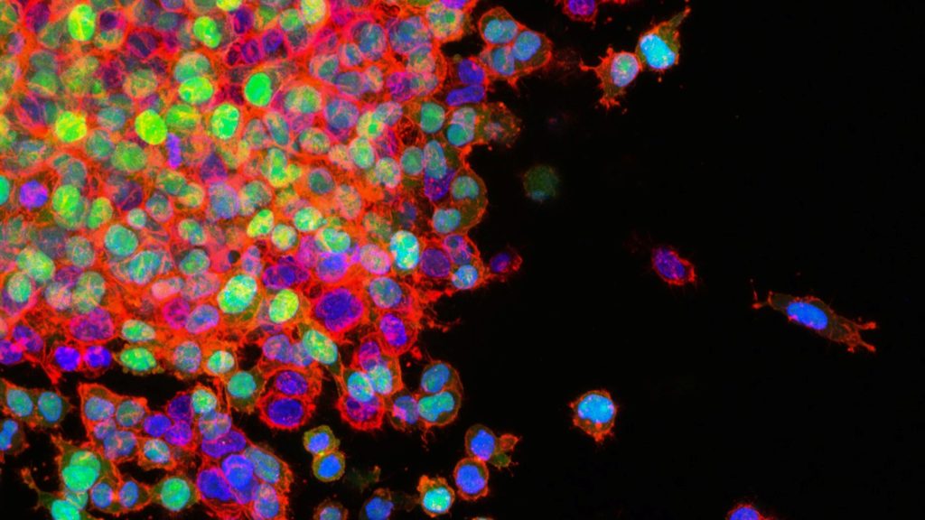
Researchers have discovered that, counterintuitively, certain cells move faster in thicker fluid – such as mucus as opposed to blood – because their ruffled edges sense the viscosity of their environment and adapt to increase their speed.
The researchers’ combined results in cancer and fibroblast cells suggest that the viscosity of a cell’s surrounding environment is an important contributor to disease. The findings, published in Nature Physics, may help explain tumour progression, scarring in mucus-filled lungs affected by cystic fibrosis, and the wound-healing process.
“This link between cell viscosity and attachment has never been demonstrated before,” noted Sergey Plotnikov, assistant professor at the University of Toronto and a co-corresponding author of the study. “We found that the thicker the surrounding environment, the stronger the cells adhere to the substrate and the faster they move – much like walking on an icy surface with shoes that have spikes, versus shoes with no grip at all.”
Understanding why cells behave in this surprising way is important because cancer tumours create a viscous environment, which means spreading cells can move into tumours faster than non-cancerous tissues. Since the researchers observed that cancer cells speed up in a thickened environment, they concluded that the development of ruffled edges in cancer cells may contribute to cancer spreading to other areas of the body.
Targeting the spreading response in fibroblasts, on the other hand, may reduce tissue damage in the mucus-filled lungs affected by cystic fibrosis. Because ruffled fibroblasts move quickly, they are the first type of cells to move through the mucus to the wound, contributing to scarring rather than healing. These results also imply that cell movement might be controlled by changing the viscosity of the lung’s mucus.
“By showing how cells respond to what’s around them, and by describing the physical properties of this area, we can learn what affects their behaviour and eventually how to influence it,” says Ernest Iu, PhD student at the University of Toronto and study co-author.
Plotnikov added, “For example, perhaps if you put a liquid as thick as honey into a wound, the cells will move deeper and faster into it, thereby healing it more effectively.”
Asst Prof Plotnikov and Iu used advanced microscopy techniques to measure the traction that cells exert to move, and changes in structural molecules inside the cells. They compared cancer and fibroblast cells, which have ruffled edges, to cells with smooth edges. They determined that ruffled cell edges sense the thickened environment, triggering a response that allows the cell to pull through the resistance – the ruffles flatten down, spread out and latch on to the surrounding surface.
The experiment originated at Johns Hopkins, where assistant professor Yun Chen, lead author of the study, and Matthew Pittman, PhD student and first author, were first examining the movement of cancer cells. Pittman created a viscous, mucus-like polymer solution, deposited it on different cell types, and saw that cancer cells moved faster than non-cancerous cells when migrating through the thick liquid. To further probe this behaviour, Asst Prof Chen collaborated with U of T’s Plotnikov, who specialises in the push and pull of cell movement.
Plotnikov was amazed at the change in speed going into thick, mucus-like liquid. “Normally, we’re looking at slow, subtle changes under the microscope, but we could see the cells moving twice as fast in real time, and spreading to double their original size,” he explained.
Typically, cell movement depends on myosin proteins, which help muscles contract. Asst Prof Plotnikov and Iu reasoned that stopping myosin would prevent cells from spreading, however were surprised when evidence showed the cells still sped up despite this action. They instead found that columns of the actin protein inside the cell, which contributes to muscle contraction, became more stable in response to the thick liquid, further pushing out the edge of the cell.
The teams are now investigating how to slow the movement of ruffled cells through thickened environments, which may open the door to new treatments for people affected by cancer and cystic fibrosis.
Leading vaccinologist Professor Shabir Madhi received the Lifetime Award from South Africa’s prestigious ‘Science Oscars’ held by the National Science and Technology Foundation. He received the honour for his leadership in research on vaccines against life-threatening diseases in Africa and globally, and he has been at the cutting edge of research in this area since 1997.
As well as being the Dean of the Faculty of Health Sciences and Professor of Vaccinology at Wits, Prof Madhi is also the director of the South African Medical Research Council (SAMRC) Vaccine and Infectious Diseases Analytics Research Unit (Wits-VIDA); and is co-director of African Leadership in Vaccinology Expertise, Wits. During the COVID pandemic he became one of the most-cited expert by the media as the public looked to the healthcare sector for advice and guidance during this crisis.
A number of awards also went to those in the field of healthcare or who contributed to healthcare, an area especially marked by SA’s response to the COVID pandemic.
CEO of SA Health Products Regulatory Authority (SAHPRA), Dr Boitumelo Semete-Makokotlela, received the Management Award for successfully leading the authorisation of a number of COVID diagnostic tests, vaccines and therapies during the COVID.
The Network for Genomics Surveillance (NGS-SA) in SA received the Data for Research award for NGS-SA, which generated of genomic surveillance data of SARS-CoV-2 aimed at informing SA’s public health response to this virus. It was represnted by its co-founders, Dr Jinal Bhiman, Scientific Lead for Global Immunology and Immune Sequencing for Epidemic Response South Africa (GIISER-SA); and Professor Tulio de Oliveira, SU.
Other recipients in the field of healthcare included Dr Wynand Goosen, who received an Emerging Researcher aware for leadership of research in SA on the surveillance of zoonotic TB in domestic cattle and wild animals as potential infection sources in susceptible people in rural areas.
Decades of research has provided no clear evidence that serotonin levels or serotonin activity are responsible for depression, according to a major review of existing literature.
Published in Molecular Psychiatry, this new umbrella review is an overview of existing meta-analyses and systematic reviews. It suggests that depression is not likely to be caused by a chemical imbalance. It also calls into question what antidepressants do: most antidepressants are selective serotonin reuptake inhibitors (SSRIs), whose mechanism of action was supposedly to correct abnormally low serotonin levels. But there is no other accepted pharmacological mechanism by which antidepressants affect the symptoms of depression.
Lead author Professor Joanna Moncrieff, at University College London said: “It is always difficult to prove a negative, but I think we can safely say that after a vast amount of research conducted over several decades, there is no convincing evidence that depression is caused by serotonin abnormalities, particularly by lower levels or reduced activity of serotonin.
“The popularity of the ‘chemical imbalance’ theory of depression has coincided with a huge increase in the use of antidepressants. Prescriptions for antidepressants have risen dramatically since the 1990s, with one in six adults in England and 2% of teenagers now being prescribed an antidepressant in a given year.
“Many people take antidepressants because they have been led to believe their depression has a biochemical cause, but this new research suggests this belief is not grounded in evidence.”
The umbrella review aimed to capture all relevant studies, encompassing tens of thousands of participants, that have been published in the most important fields of research on serotonin and depression.
Research that compared levels of serotonin and its breakdown products in the blood or brain fluids found no difference between participants diagnosed with depression and healthy controls.
Research on serotonin receptors and the serotonin transporter, the protein targeted by most antidepressants, found weak and inconsistent evidence suggestive of higher levels of serotonin activity in people with depression. However, the researchers say the findings are likely explained by the use of antidepressants among people diagnosed with depression, since such effects were not reliably ruled out.
Some studies artificially lowered serotonin levels were by depriving participant’s diets of the necessary amino acid. These studies have been cited as demonstrating that a serotonin deficiency is linked to depression. A meta-analysis conducted in 2007 and a sample of recent studies found that lowering serotonin in this way did not produce depression in hundreds of healthy volunteers, however. There was very weak evidence in a small subgroup of people with a family history of depression, but this only involved 75 participants, and more recent evidence was inconclusive.
Very large studies involving tens of thousands of patients looked at gene variation, including the gene for the serotonin transporter, and found no difference between people with depression and healthy controls. These studies also examined stressful life events, and found these to strongly increase people’s risk of becoming depressed. A famous early study found a relationship between stressful events, the type of serotonin transporter gene a person had and the chance of depression. But larger, more comprehensive studies suggest this was a false finding.
These findings together led the authors to conclude that there is “no support for the hypothesis that depression is caused by lowered serotonin activity or concentrations.”
The researchers say their findings are important as studies show that as many as 85–90% of the public believes that depression is caused by low serotonin or a chemical imbalance. A growing number of scientists and professional bodies are recognising the chemical imbalance framing as an over-simplification. Evidence also suggests that believing that low mood is caused by a chemical imbalance leads to pessimism about recovery, and the possibility of managing moods without medical help. This is important because most people will at some point in their lives meet criteria for anxiety or depression.
A large meta-analysis provided evidence that people who used antidepressants actually had lower levels of serotonin in their blood. The researchers concluded that some evidence was consistent with the possibility that long-term antidepressant use reduces serotonin concentrations. The researchers say this may imply that the increase in serotonin that some antidepressants produce in the short term could lead to compensatory changes in the brain that produce the opposite effect in the long term.
Though antidepressants’ efficacies was not examined, the authors encourage looking into treatments such psychotherapy, alongside other practices such as exercise or mindfulness, or addressing underlying contributors such as poverty, stress and loneliness.
Professor Moncrieff said: “Our view is that patients should not be told that depression is caused by low serotonin or by a chemical imbalance, and they should not be led to believe that antidepressants work by targeting these unproven abnormalities. We do not understand what antidepressants are doing to the brain exactly, and giving people this sort of misinformation prevents them from making an informed decision about whether to take antidepressants or not.”
Co-author Dr Mark Horowitz said: “I had been taught that depression was caused by low serotonin in my psychiatry training and had even taught this to students in my own lectures. Being involved in this research was eye-opening and feels like everything I thought I knew has been flipped upside down.
“One interesting aspect in the studies we examined was how strong an effect adverse life events played in depression, suggesting low mood is a response to people’s lives and cannot be boiled down to a simple chemical equation.”
Professor Moncrieff added: “Thousands of people suffer from side effects of antidepressants, including the severe withdrawal effects that can occur when people try to stop them, yet prescription rates continue to rise. We believe this situation has been driven partly by the false belief that depression is due to a chemical imbalance. It is high time to inform the public that this belief is not grounded in science.”
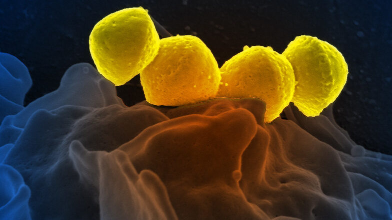
Streptococcus pyogenese bound to a human neutrophil. Credit: National Institute of Allergy and Infectious Diseases, National Institutes of Health
Bordering on science fiction, medicinal microrobots could help physicians better treat and prevent diseases. But a serious problem is the synthetic materials they are made of trigger immune responses. Now, for the first time, researchers report in ACS Central Science that they achieved precise control neutrophils as a natural, biocompatible microrobot by using lasers. By getting the ‘neutrobots’ to perform multiple tasks, the researchers demonstrated they could one day deliver drugs to precise locations in the body.
Microrobots being developed for medical applications would need to be administered in injections or oral capsules to get them inside the body. But these microscopic objects are often found to trigger immune reactions in small animals, resulting in the the microrobots being ejected from the body before they can carry out their tasks. By using the body’s own cells, such as neutrophils, drugs could be delivered less invasively without provoking an immune response.
Neutrophils already naturally pick up nanoparticles and dead red blood cells and can migrate through blood vessels into adjacent tissues, so they are good candidates for becoming microrobots. Previously, researchers have guided neutrophils with lasers in lab dishes, moving them around as ‘neutrobots’. However, this had not been tried in living animals. So, researchers set out to demonstrate the feasibility of light-driven neutrobots in animals using live zebrafish.
The researchers manipulated and moved neutrophils in zebrafish tails, using focused laser beams as optical tweezers. The ‘neutrobots’ could be moved up to a velocity of 1.3 µm/s, three times faster than a neutrophil’s natural speed. The optical tweezers were able to precisely and actively control the functions that neutrophils conduct as part of the immune system. For example, moving through a blood vessel wall into the surrounding tissue; carrying a plastic nanoparticle, showing potential for delivering medicine; and pushed towards red blood cell debris, a neutrophil engulfed the pieces. Surprisingly, at the same time, a different neutrophil, which wasn’t controlled by a laser, tried to naturally remove the cellular debris. Because they successfully controlled neutrobots in vivo, the researchers say this study advances the possibilities for targeted drug delivery and precise treatment of diseases.
From left to right: Junior doctor Aniket Bharadwaj with trainers Dr Ruby Woodard and Dr Jonny Martin, diagnosing a hologram patient. Credit: University of Cambridge
A new effort from Cambridge University brings medical training in ‘mixed reality’ one step closer with modules that allow student doctors to interact with a ‘holographic’ patient.
Traditional simulation has numerous costs including maintaining simulation centres, their equipment and the faculty and staff hours to operate the labs and hire and train patient actors. This new technology could provide more flexible, cost-effective training that can be accessed all over the world.
HoloScenarios is a new training application based on life-like holographic patient scenarios, is being developed by Cambridge University Hospitals NHS Foundation Trust (CUH), in partnership with the University of Cambridge and US tech company GigXR. The first module focuses on common respiratory conditions and emergencies.
“Mixed reality is increasingly recognised as a useful method of simulator training,” said project leader Dr Arun Gupta, consultant anaesthetist at CUH and director of postgraduate education at CUH.
“As institutions scale procurement, the demand for platforms that offer utility and ease of mixed reality learning management is rapidly expanding,” he said.
Learners wearing mixed-reality headsets can interact with each other and a multi-layered, medically accurate ‘holographic’ patient. This creates a unique environment to learn and practice vital, real-time decision making and treatment choices.
Medical instructors with their own headsets can make changes on the fly, by changing patient responses or introducing complications – whether in person in a teaching group or over the internet.
Learners can also watch, contribute to and assess the holographic patient scenarios from Android, iOS smartphone or tablet. This means true-to-life, safe-to-fail immersive learning can be accessed, delivered and shared across the world, with the technology now available for license to learning institutions everywhere.
Professor Riikka Hofmann at Cambridge’s Faculty of Education is leading an analysis of the technology as a teaching method.
“Our research is aimed at uncovering how such simulations can best support learning and accelerate the adoption of effective mixed reality training while informing ongoing development,” said Prof Hofmann.
“We hope that it will help guide institutions in implementing mixed reality into their curricula, in the same way institutions evaluate conventional resources, such as textbooks, manikins, models or computer software, and, ultimately, improve patient outcomes.”
Junior doctor Aniket Bharadwaj is one of the first to try out the new technology. “Throughout medical school we would have situations where actors would come in an act as patients. With the pandemic a lot of that changed to tablet based interactions because of the risk to people of the virus,” he said.
“Having a hologram patient you can see, hear and interact with is really exciting and will really make a difference to student learning.”
The first module features a hologram patient with asthma, followed by anaphylaxis, pulmonary embolism and pneumonia. Further modules in cardiology and neurology are in development.
Delivered by the Gig Immersive Learning Platform, HoloScenarios aims to centralise and streamline access and management of mixed reality learning, and encapsulate the medical experience of world-leading doctors at CUH and across the University of Cambridge.