Category: Medical Research & Technology

Smartwatch Equals Treadmill Test in Detecting HF

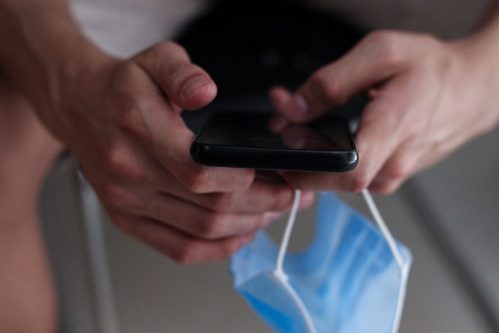
A smartwatch ECG can accurately detect heart failure (HF) in nonclinical environments, according to a study published in Nature Medicine. Researchers analysed Apple Watch ECG recordings with AI to identify patients with ventricular dysfunction. Study participants were able to remotely record their smartwatch ECGs at any time, with the data automatically and securely uploaded to their electronic health records via a smartphone app.

“Currently, we diagnose ventricular dysfunction – a weak heart pump – through an echocardiogram, CT scan or an MRI, but these are expensive, time consuming and at times inaccessible. The ability to diagnose a weak heart pump remotely, from an ECG that a person records using a consumer device, such as a smartwatch, allows a timely identification of this potentially life-threatening disease at massive scale,” says senior study author Paul Friedman, MD, chair of the Department of Cardiovascular Medicine at Mayo Clinic.

Ventricular dysfunction might not cause symptoms, but affects about 2% of the population and 9% of people over 60. Symptoms may develop with a low ejection fraction, including shortness of breath, a rapid heart rate and swelling in the legs. Early diagnosis is important because once identified, there are numerous treatments to improve quality of life and decrease the risks of heart failure and death.

Mayo researchers interpreted Apple Watch single-lead ECGs by modifying an earlier algorithm developed for 12-lead ECGs that is proven to detect a low ejection fraction.

While the data are early, the modified AI algorithm using single-lead ECG data had an area under the curve of 0.88 to detect low ejection fraction. By comparison, this measure of accuracy is as good as or slightly better than a medical treadmill diagnostic test.

“These data are encouraging because they show that digital tools allow convenient, inexpensive, scalable screening for important conditions. Through technology, we can remotely gather useful information about a patient’s heart in an accessible way that can meet the needs of people where they are,” says first author Zachi Attia, PhD, the lead AI scientist in the Department of Cardiovascular Medicine at Mayo Clinic.

“Building the capability to ingest data from wearable consumer electronics and provide analytic capabilities to prevent disease or improve health remotely in the manner demonstrated by this study can revolutionize health care. Solutions like this not only enable prediction and prevention of problems, but also will eventually help diminish health disparities and the burden on health systems and clinicians,” says co-author Bradley Leibovich, MD, the medical director for the Mayo Clinic Center for Digital Health.

Approximately 420 of the 2454 participants had an echocardiogram within 30 days of logging an Apple Watch ECG in the app. Of those, 16 patients had low ejection fraction confirmed by the echocardiogram, which provided a comparison for accuracy.

Source: Mayo Clinic

Delivering Cancer and Diabetes Drugs in Pills Instead of Injections

Source: Danilo Alvesd on Unsplash

In a new Journal of the American Chemical Society paper, researchers describe how they are developing a new way for diabetes and cancer patients to manage their conditions by enabling drugs to be delivered in pill form instead of through injections.

Some drugs for these diseases are water soluble, so transporting them through the intestines, is not feasible and makes them impossible to administer orally. However, UCR scientists have created a chemical “tag” that can be added to these drugs, allowing them to enter blood circulation via the intestines.

This tag is composed of a small peptide. “Because they are relatively small molecules, you can chemically attach them to drugs, or other molecules of interest, and use them to deliver those drugs orally,” said research leader Min Xue, UC Riverside chemistry professor.

Xue’s laboratory was testing something unrelated when the researchers observed these peptides making their way into cells.

“We did not expect to find this peptide making its way into cells. It took us by surprise,” Xue said. “We always wanted to find this kind of chemical tag, and it finally happened serendipitously.”

This observation was unexpected, Xue said, because previously, the researchers believed that this type of delivery tag needed to carry positive charges to be accepted into the negatively charged cells. Their work with this neutral peptide tag, called EPP6, shows that belief was not accurate.

Testing the peptide’s ability to move through a body, the Xue group teamed up with Kai Chen’s group in the Keck School of Medicine at the University of Southern California and fed the peptide to mice. With PET scans, the team observed the peptide accumulating in the intestines, and documented its ultimate transfer into the animals’ organs via the blood.

Having proven the tag successfully navigated the circulatory systems through oral administration, the team now plans to demonstrate that the tag can do the same thing when attached to a selection of drugs. “Quite compelling preliminary results make us think we can push this further,” Xue said.

Many drugs, including insulin, must be injected. The researchers are hopeful their next set of experiments will change that, allowing them to add this tag to a wide variety of drugs and chemicals, changing the way those molecules move through the body.

“This discovery could lift a burden on people who are already burdened with illness,” Xue said.

Source: University of California – Riverside

World First Trial of Lab-grown Red Blood Cells for Transfusion

https://www.pexels.com/photo/a-close-up-shot-of-bags-of-blood-4531306/
Photo by Charlie-Helen Robinson on Pexels

In a world first, researchers have launched a clinical trial of lab-grown red blood cells for transfusion into another person. These manufactured blood cells were grown from stem cells from donors, for transfusion into volunteers in the RESTORE randomised controlled clinical trial.

If our trial is successful, it will mean that patients who currently require regular long-term blood transfusions will need fewer transfusions in future, helping transform their care

Professor Cedric Ghevaert, chief investigator

If the technique is proven safe and effective, manufactured blood cells could in time revolutionise treatments for people with blood disorders such as sickle cell and rare blood types. It can be difficult to find enough well-matched donated blood for some people with these disorders.

To produce the lab-grown blood cells, stem cells are first magnetically extracted from a normal 470ml blood donation. These stem cells are then coaxed into becoming red blood cells. Over the three week process, an initial pool of about half a million stem cells generates 50 billion red blood cells.

Chief Investigator Professor Cedric Ghevaert, Professor in Transfusion Medicine and Consultant Haematologist at the University of Cambridge and NHS Blood and Transplant, said: “We hope our lab grown red blood cells will last longer than those that come from blood donors. If our trial, the first such in the world, is successful, it will mean that patients who currently require regular long-term blood transfusions will need fewer transfusions in future, helping transform their care.”

Professor Ashley Toye, Professor of Cell Biology at the University of Bristol and Director of the NIHR Blood and Transplant Unit in red cell products, said: “This challenging and exciting trial is a huge stepping stone for manufacturing blood from stem cells. This is the first-time lab grown blood from an allogeneic donor has been transfused and we are excited to see how well the cells perform at the end of the clinical trial.”

The trial is studying the lifespan of the lab grown cells compared with infusions of standard red blood cells from the same donor. The lab-grown blood cells are all fresh, so the trial team expect them to perform better than a similar transfusion of standard donated red cells, which contains cells of varying ages.

Additionally, if manufactured cells last longer in the body, patients who regularly need blood may not need transfusions as often. That would reduce iron overload from frequent blood transfusions, which can lead to serious complications.

The trial is the first step towards making lab grown red blood cells available as a future clinical product. For the foreseeable future, manufactured cells could only be used for a very small number of patients with very complex transfusions needs. NHSBT continues to rely on the generosity of donors.

Co-Chief Investigator Dr Rebecca Cardigan, Head of Component Development NHS Blood and Transplant and Affiliated Lecturer at the University of Cambridge, said: “It’s really fantastic that we are now able to grow enough red cells to medical grade to allow this trial to commence. We are really looking forward to seeing the results and whether they perform better than standard red cells.”

Thus far, two people have been transfused with the lab grown red cells. They are well and healthy, and were closely monitored with no untoward side effects were reported. The amount of lab grown cells being infused varies but is around 5-10mls.

Donors were recruited from NHSBT’s blood donor base. They donated blood to the trial and stem cells were separated out from their blood. These stem cells were then grown to produce red blood cells in a laboratory at NHS Blood and Transplant’s Advanced Therapies Unit in Bristol. The recipients of the blood were recruited from healthy members of the NIHR BioResource.

A minimum of 10 participants will receive two mini transfusions at least four months apart, one of standard donated red cells and one of lab grown red cells, to see if the young lab-made red blood cells last longer than cells made in the body.

Further trials are needed before clinical use, but this research marks a significant step in using lab grown red blood cells to improve treatment for patients with rare blood types or people with complex transfusion needs.

Source: University of Cambridge

Study Links Vitamin D Deficiency to Premature Death

Photo by Julian Jagtenberg on Pexels

New research in the  Annals of Internal Medicine provides strong evidence that vitamin D deficiency is associated with premature death, prompting calls for people to follow healthy vitamin D level guidelines.

The study by the University of South Australia found that premature mortality increased in line with the severity of Vitamin D deficiency.

First author and UniSA PhD candidate, Josh Sutherland, says that while vitamin D has been connected with mortality, it has been challenging to establish causal effects.

“While severe vitamin D deficiency is rarer in Australia than elsewhere in the world, it can still affect those who have health vulnerabilities, the elderly, and those who do not acquire enough vitamin D from healthy sun exposure and dietary sources,” Sutherland says.

“Our study provides strong evidence for the connection between low levels of vitamin D and mortality, and this is the first study of its kind to also include respiratory disease related mortality as an outcome.

“We used a new genetic method to explore and affirm the non-linear relationships that we’ve seen in observational settings, and through this we’ve been able give strong evidence for the connection between low vitamin D status and premature death.

“Vitamin D deficiency has been connected with mortality, but as clinical trials have often failed to recruit people with low vitamin D levels – or have been prohibited from including vitamin deficient participants – it’s been challenging to establish causal relationships.”

The Mendelian randomisation study (an alternative to the gold standard of a randomised controlled trial) evaluated 307 601 records from the UK Biobank. Low levels of vitamin D were noted as less than <25 nmol/L with the average concentration found to be 45.2 nmol/L. Over a 14-year follow up period, researchers found that the risk for death significantly decreased with increased vitamin D concentrations, with the strongest effects seen among those with severe deficiencies.

Senior investigator Professor Elina Hyppönen says more research is now needed to establish effective public health strategies that can help achieve national guidelines and reduce the risk of premature death associated with low vitamin D levels.

“The take-home message here is simple – the key is in the prevention. It is not good enough to think about vitamin D deficiency when already facing life-challenging situations, when early action could make all the difference,” Prof Hyppönen says.

“It is very important to continue public health efforts to ensure the vulnerable and elderly maintain sufficient vitamin D levels throughout the year.”

Source: University of South Australia

Discovery Could Lead to Blood Pressure Drugs with Fewer Side Effects

Credit: Thirdman on Pexels

Researchers at the University of Virginia have identified a key contributor to hypertension that could lead to new treatments with fewer side effects. Their findings were published in the journal Circulation.

The discovery, from scientist Swapnil Sonkusare, PhD, and colleagues, sheds new light on blood pressure regulation and how problems with this critical biological process drive hypertension.

Blood pressure is partly controlled by calcium levels in smooth muscle cells that line blood vessel walls. Smooth muscle cells take in calcium and use it to regulate the contraction of blood vessels as needed. Hypertension is commonly treated with calcium blockers that reduce the movement of calcium, but since multiple organs also use this calcium mechanism, these drugs have many side effects. So a treatment option that targets the harmful effects of calcium but not its beneficial effects could be very helpful for patients with hypertension.

Dr Sonkusare and his team discovered two critical signalling centres in smooth muscle cells that bring in calcium and regulate blood pressure. These ‘nanodomains’, the researchers found, act like symphony conductors for blood vessels, directing them to contract or relax as needed. These signalling centres, the researchers determined, are a key regulator of healthy blood pressure.

Further, the UVA scientists found that disruptions in this process contribute to high blood pressure. In both mouse models of the disease and hypertensive patients, the fine balance between constrictor and dilator signalling centres is lost. This caused the blood vessels to become too constricted, driving up blood pressure.

“Our work identifies a new mechanism that helps maintain healthy blood pressure and shows how abnormalities in this mechanism can lead to hypertension,” said Dr Sonkusare. “The discovery of a new mechanism for elevation of blood pressure could provide therapeutic targets for treating hypertension.”

The research identifies a “new paradigm in hypertension,” according to an accompanying editorial. The editorial says UVA’s “innovative” discoveries fill “major gaps” in our understanding of the fundamental molecular causes of high blood pressure.

The new findings help us better understand how our bodies maintain proper blood pressure and provide enticing targets for scientists seeking to develop treatments targeting underlying causes of hypertension. Developing treatments that do not affect the beneficial effects of calcium will require additional research and a deeper understanding of the calcium-use process, but Dr Sonkusare’s team is already working toward that goal.

“We’ve shown that smooth muscle cells use ‘spatial separation’ of signalling centres to achieve constriction or dilation of arteries. We are now investigating the individual components of these signalling centres,” Dr Sonkusare said. “Understanding these components will help us target them to lower or raise the blood pressure in disease conditions that show high or low blood pressure, respectively.”

Source: University of Virginia

Sleeping with Weighted Blankets Increases Melatonin

Sleeping woman
Photo by Cottonbro on Pexels

A small study has shown that young adults sleeping using weighted blankets had increased levels of the hormone melatonin, which increases in response to darkness, and which some research suggests promotes sleep. The findings are published in the Journal of Sleep Research.

Weighted blankets have been suggested to ease insomnia in humans in previous research but underlying mechanisms are not fully understood. To address this, Uppsala University researchers recruited 26 young men and women to examine if the bedtime use of a weighted blanket increases the production of sleep-promoting and anti-stress hormones like melatonin and oxytocin. They also investigated whether the bedtime use of a weighted blanket (12% of participants’ body weight) reduced the activity of stress systems in the body. Saliva samples were collected from participants while they were covered with either a weighted or a light blanket to measure melatonin, oxytocin, cortisol, and sympathetic nervous system activity.

“Using a weighted blanket increased melatonin concentrations in saliva by about 30%. However, no differences in oxytocin, cortisol, and the activity of the sympathetic nervous system were observed between the weighted and light blanket conditions,” reported Elisa Meth, first author and PhD student.

“Our study may offer a mechanism explaining why weighted blankets may exert some therapeutic benefits, such as improved sleep. However, our findings rely on a small sample and investigated only the acute effects of a weighted blanket. Thus, larger trials are needed, including an investigation of whether the observed effects of a weighted blanket on melatonin are sustained over longer periods,” said senior author Christian Benedict, Associate Professor at Uppsala University.

Source: Uppsala University

Microscopic Robots Kill Pneumonia Bacteria in Lungs

Pseudomonas
Scanning Electron Micrograph of Pseudomonas aeruginosa. Credit: CDC/Janice Carr

Nanoengineers have developed microscopic robots, called microrobots, that can swim around in the lungs, deliver medication and be used to clear up life-threatening cases of bacterial pneumonia.

In mice, the microrobots safely eliminated Pseudomonas aeruginosa in the lungs of infected mice, resulting in a 100% survival rate. By contrast, untreated mice all died within three days after infection. The scientists describe the technology in Nature Materials.

The microrobots are not actually made of metal and plastic: instead they are algae cells armed with antibiotic-filled nanoparticles on their surfaces. The algae provide movement, which allows the microrobots to swim around and deliver antibiotics directly to more bacteria in the lungs. The nanoparticles are also coated with the neutrophil cell membranes, which absorb and neutralise inflammatory molecules produced by bacteria and the body’s immune system. This gives the microrobots a powerful anti-inflammatory tool, and the algae are biodegradable in the body, leaving no toxic traces.

The work is a joint effort between the labs of nanoengineering professors Joseph Wang and Liangfang Zhang, both at the UC San Diego Jacobs School of Engineering, both world leaders in nanoengineering.

“Our goal is to do targeted drug delivery into more challenging parts of the body, like the lungs. And we want to do it in a way that is safe, easy, biocompatible and long lasting,” said Prof Zhang. “That is what we’ve demonstrated in this work.”

The team used the microrobots to treat mice with an acute and potentially fatal form of pneumonia caused by P. aeruginosa. This is commonly seen in mechanically ventilated ICU patients. The researchers administered the microrobots to the lungs of the mice through a tube inserted in the windpipe. The infections fully cleared up after one week. All mice treated with the microrobots survived past 30 days, while untreated mice died within three days.

The microrobots enabled targeted drug delivery of only 500 nanograms of antibiotics per mouse, while an IV injection provided 1.644 milligrams of antibiotics per mouse.

“These results show how targeted drug delivery combined with active movement from the microalgae improves therapeutic efficacy,” said Wang.

“With an IV injection, sometimes only a very small fraction of antibiotics will get into the lungs. That’s why many current antibiotic treatments for pneumonia don’t work as well as needed, leading to very high mortality rates in the sickest patients,” said Professor Victor Nizet, co-author on the study and a physician-scientist collaborator of Profs Wang and Zhang. “Based on these mouse data, we see that the microrobots could potentially improve antibiotic penetration to kill bacterial pathogens and save more patients’ lives.”

The work is still at the proof-of-concept stage. The team plans to do more basic research to understand exactly how the microrobots interact with the immune system. Next steps also include studies to validate the microrobot treatment and scaling it up before testing it in larger animals and eventually, in humans.

“We’re pushing the boundary further in the field of targeted drug delivery,” said Zhang.

Source: University of California – San Diego

Long-standing Theory of Hearing Turned on its Ear

The sensory cells of hearing, outer and inner hair cells, are located in the cochlea, where the arrival sound waves cause the ‘hairs’ of the inner hair cells to bend, sending a signal through the nerves to the brain, which interprets the sound we hear.

For the past century, scientific belief was that each sensory cell has its own ‘optimal frequency’, to which the hair cell responds most strongly. This idea means that a sensory cell with an optimal frequency of 1000Hz would be much less responsive to sounds of slightly lower or higher frequency. It has also been assumed that all parts of the cochlea work in the same way. Now, however, researchers have discovered that this is not so for sensory cells that process sound with frequencies under 1000Hz, considered to be low-frequency sound, where the vowel sounds in human speech lie.

“Our study shows that many cells in the inner ear react simultaneously to low-frequency sound. We believe that this makes it easier to experience low-frequency sounds than would otherwise be the case, since the brain receives information from many sensory cells at the same time,” said Professor Anders Fridberger at Linköping University, senior author of the study published in Science Advances.

The scientists believe that this construction of our hearing system makes it more robust. If some sensory cells are damaged, many others remain that can send nerve impulses to the brain.

As well as the vowel sounds of human speech, many of the sounds that go to make up music also lie in this low-frequency area. Middle C on a piano, for example, has a frequency of 262Hz.

These results may eventually be significant for people with severe hearing impairments. The most successful treatment currently available in such cases is a cochlear implant, in which electrodes are placed into the cochlea.

“The design of current cochlear implants is based on the assumption that each electrode should only give nerve stimulation at certain frequencies, in a way that tries to copy what was believed about the function of our hearing system. We suggest that changing the stimulation method at low frequencies will be more similar to the natural stimulation, and the hearing experience of the user should in this way be improved,” says Anders Fridberger.

The researchers now plan to examine how their new knowledge can be applied in practice. One of the projects they are investigating concerns new methods to stimulate the low-frequency parts of the cochlea.

These results come from experiments on the cochlea of guinea pigs, whose hearing in the low-frequency region is similar to that of humans.

Source: Linköping University

Simple, Painless Microneedle Tattoos

Photo by Benjamin Lehman on Unsplash

Researchers from the Georgia Institute of Technology have developed a low-cost patch of microneedles that can be applied just by pressing it into the skin, with none of the pain and blood of traditional tattooing. The team development presented their research in the journal iScience.

These tattoos, which can be self-administered, have many potential applications, from medical alerts to tracking neutered animals to cosmetics. 

“We’ve miniaturised the needle so that it’s painless, but still effectively deposits tattoo ink in the skin,” said principal investigator Mark Prausnitz “This could be a way not only to make medical tattoos more accessible, but also to create new opportunities for cosmetic tattoos because of the ease of administration.” 

Medical applications of tattoos include covering up scars, guiding radiotherapy, or restoring nipples after breast surgery. Tattoos also serve instead of bracelets as medical alerts to communicate serious medical conditions such as diabetes, epilepsy, or allergies.  

<p>Medical alert tattoo: microneedle patch (above) and tattoo on skin (below).</p><p>Credit: Song Li, Georgia Tech</p>
Medical alert tattoo: microneedle patch (above) and tattoo on skin (below). Credit: Song Li, Georgia Tech

Various cosmetic products using microneedles are already on the market – mostly for anti-ageing – but developing microneedle technology for tattoos is new. Prausnitz, a veteran in this area, has studied microneedle patches for years to painlessly administer drugs and vaccines to the skin without the need for hypodermic needles. 

“We saw this as an opportunity to leverage our work on microneedle technology to make tattoos more accessible,” Prausnitz said. “While some people are willing to accept the pain and time required for a tattoo, we thought others might prefer a tattoo that is simply pressed onto the skin and does not hurt.”   

Transforming tattooing 

Tattoos typically use large needles to puncture repeatedly into the skin to get a good image, a time-consuming and painful process. The Georgia Tech team has developed microneedles that are smaller than a grain of sand and are made of tattoo ink encased in a dissolvable matrix.  

“Because the microneedles are made of tattoo ink, they deposit the ink in the skin very efficiently,” said former Georgia Tech postdoctoral fellow Song Li, the lead author of the study. 

In this way, the microneedles can be pressed into the skin just once and then dissolve, leaving the ink in the skin after a few minutes without bleeding.   

Creating the tattoo 

Although most microneedle patches for pharmaceuticals or cosmetics have dozens or hundreds of microneedles arranged in a square or circle, microneedle patch tattoos imprint a design that can include letters, numbers, symbols, and images. By arranging the microneedles in a specific pattern, each microneedle acts like a pixel to create a tattoo image in any shape or pattern.  

The researchers start with a mold containing microneedles in a pattern that forms an image. They fill the microneedles in the mold with tattoo ink and add a patch backing for convenient handling. The resulting patch is then applied to the skin for a few minutes, during which time the microneedles dissolve and release the tattoo ink. Tattoo inks of various colors can be incorporated into the microneedles, including black-light ink that can only be seen when illuminated with ultraviolet light.  

Prausnitz’s lab has been researching microneedles for vaccine delivery for years and realised they could be equally applicable to tattoos. Prausnitz’s team started working on tattoos to identify spayed and neutered pets, but then realised the technology could be effective for people, too. 

The tattoos were also designed with privacy in mind. The researchers even created patches sensitive to environmental factors such as light or temperature changes, where the tattoo will only appear with ultraviolet light or higher temperatures. This provides patients with privacy, revealing the tattoo only when desired. 

The study showed that the tattoos could last for at least a year and are likely to be permanent, which also makes them viable cosmetic options for people who want an aesthetic tattoo without risk of infection or the pain associated with traditional tattoos. Microneedle tattoos could alternatively be loaded with temporary tattoo ink to address short-term needs in medicine and cosmetics.  

Microneedle patch tattoos can also be used to encode information in the skin of animals. Rather than clipping the ear or applying an ear tag to animals to indicate sterilisation status, a painless and discreet tattoo can be applied instead.  

However, the technology does not aim to put tattoo artists out of business.

“The goal isn’t to replace all tattoos, which are often works of beauty created by tattoo artists,” Prausnitz said. “Our goal is to create new opportunities for patients, pets, and people who want a painless tattoo that can be easily administered.”  

Source: Georgia Institute of Technology

Smartphones Could Serve as Pulse Oximeters in the Home

Photo by Asterfolio on Unsplash

Researchers have demonstrated that smartphones are capable of detecting blood oxygen saturation levels down to 70% – the lowest value that pulse oximeters should be able to measure, as recommended by the US Food and Drug Administration. The team published these results in npj Digital Medicine.

The technique involves participants placing their finger over the camera and flash of a smartphone, which uses a deep-learning algorithm to decipher the blood oxygen levels. When the team delivered a controlled mixture of nitrogen and oxygen to six subjects to artificially bring their blood oxygen levels down, the smartphone correctly predicted whether the subject had low blood oxygen levels 80% of the time.

“Other smartphone apps that do this were developed by asking people to hold their breath. But people get very uncomfortable and have to breathe after a minute or so, and that’s before their blood-oxygen levels have gone down far enough to represent the full range of clinically relevant data,” said co-lead author Jason Hoffman, a UW doctoral student in the Paul G. Allen School of Computer Science & Engineering. “With our test, we’re able to gather 15 minutes of data from each subject. Our data shows that smartphones could work well right in the critical threshold range.”

Another benefit of measuring blood oxygen levels on a smartphone is that almost everyone has one.

“This way you could have multiple measurements with your own device at either no cost or low cost,” said co-author Dr. Matthew Thompson, professor of family medicine in the UW School of Medicine. “In an ideal world, this information could be seamlessly transmitted to a doctor’s office. This would be really beneficial for telemedicine appointments or for triage nurses to be able to quickly determine whether patients need to go to the emergency department or if they can continue to rest at home and make an appointment with their primary care provider later.”

The team recruited six participants ranging in age from 20 to 34. Three identified as female, three identified as male. One participant identified as being African American, while the rest identified as being Caucasian.

To gather data to train and test the algorithm, the researchers had each participant wear a standard pulse oximeter on one finger and then place another finger on the same hand over a smartphone’s camera and flash. Each participant had this same set up on both hands simultaneously.

“The camera is recording a video: Every time your heart beats, fresh blood flows through the part illuminated by the flash,” said Assistant Professor Edward Wang, who started this project as a doctoral student.

“The camera records how much that blood absorbs the light from the flash in each of the three color channels it measures: red, green and blue,” said Wang, who also directs the UC San Diego DigiHealth Lab. “Then we can feed those intensity measurements into our deep-learning model.”

Each participant breathed in a controlled mixture of oxygen and nitrogen to slowly reduce oxygen levels. For all six participants, the team acquired more than 10 000 blood oxygen level readings between 61% and 100%.

The researchers used data from four of the participants to train a deep learning algorithm to extract the blood oxygen levels, and the rest of the data was used to validate the method and then test it to see how well it performed on new subjects.

“Smartphone light can get scattered by all these other components in your finger, which means there’s a lot of noise in the data that we’re looking at,” said co-lead author Varun Viswanath. “Deep learning is a really helpful technique here because it can see these really complex and nuanced features and helps you find patterns that you wouldn’t otherwise be able to see.”

The team hopes to continue this research by testing the algorithm on more people.

“One of our subjects had thick calluses on their fingers, which made it harder for our algorithm to accurately determine their blood oxygen levels,” Hoffman said. “If we were to expand this study to more subjects, we would likely see more people with calluses and more people with different skin tones. Then we could potentially have an algorithm with enough complexity to be able to better model all these differences.”

But, the researchers said, this is a good first step toward developing biomedical devices that are aided by machine learning.

“It’s so important to do a study like this,” Wang said. “Traditional medical devices go through rigorous testing. But computer science research is still just starting to dig its teeth into using machine learning for biomedical device development and we’re all still learning. By forcing ourselves to be rigorous, we’re forcing ourselves to learn how to do things right.”

Source: University of Washington