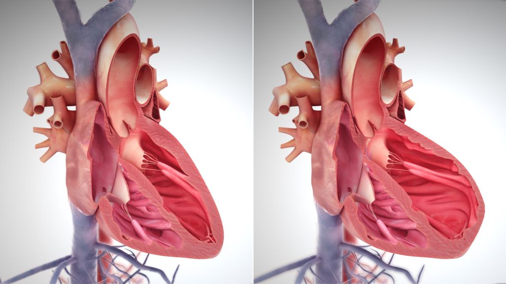
Category: Cardiovascular Disease

Nurses Play a Key Role in Addressing Post-stroke Mental Well-being
Credit: American Heart Association

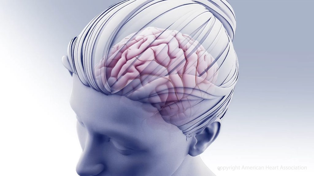
Nurses play a key role in helping patients manage emotional and social health challenges, or psychosocial health, after a stroke, and improved screening and assessment for psychosocial needs are essential to provide optimal patient care. These findings are highlighted in a new statement from the American Stroke Association, a division of the American Heart Association, published in the Association’s peer-reviewed scientific journal Stroke.

While there have been significant advances in stroke prevention and treatment, stroke remains the second leading cause of death globally and a major cause of disability. The latest research indicates that 16% to 85% of stroke survivors experience psychosocial symptoms, such as depression, anxiety, stress, fatigue and/or a decreased quality of life during their recovery.

“Stigma often surrounds discussions about psychosocial health. Therefore, it is crucial for nurses and all health care professionals to create a safe and therapeutic environment for patients and offer hope and comprehensive education on the topic,” said Chair of the scientific statement’s writing group Patricia A. Zrelak, PhD, RN, FAHA, a regional stroke program quality nurse consultant for Kaiser Permanente Northern California and a member of the American Heart Association’s Council on Cardiovascular and Stroke Nursing.  

The scientific statement details a comprehensive review of the latest evidence published from 2018-2023 about psychological health in patients who experienced a stroke. The statement addresses the effects, underlying causes, screening, diagnosis and treatment for five key emotional and social health factors, including depression, stress, anxiety, fatigue and quality of life. The scientific statement aims to establish a guide for nursing care throughout a patient’s recovery after a stroke, from prevention of adverse psychosocial health conditions to identifying and managing symptoms.

“Emotional, cognitive, behavioural and/or personality changes may occur after a stroke,” Zrelak said. “These conditions can emerge immediately after a stroke or have a delayed onset, sometimes occurring more than a year later, and they may also fluctuate in intensity over time. In addition, psychosocial symptoms are interrelated, and patients who experience one are at higher risk of developing other mental health conditions. Effective and regular screening are vital for early detection and treatment.”

Depression

Depression affects about 30% of stroke survivors and is particularly common within the first three months after a stroke. Symptoms of depression may include persistent sadness, anxious or “empty” mood; restlessness and irritability; loss of interest or pleasure in hobbies and activities; difficulty in concentrating and thinking; increased or decreased sleep; changes in appetite; and weight gain or loss. Post-stroke depression worsens cognitive and functional recovery and increases the risks of death and/or another stroke.

The AHA/ASA Guidelines for the Early Management of Patients With Acute Ischemic Stroke recommend routine depression screening for all patients after a stroke. Nurses can help educate stroke survivors and their families on symptom recognition, prevention and treatment options, such as medication management and/or cognitive behavioural therapy.

Stress

A 2022 study found that post-stroke stress and post-traumatic stress disorder (PTSD) affects about one in six (about 16.5%) stroke survivors. These conditions may increase the risk of additional health issues, including anxiety and poor medication adherence. Screening stroke patients for stress and PTSD should occur when they are hospitalised and continue during rehabilitation and outpatient visits after hospital discharge.

Nursing interventions that may help lower patients’ distress include stroke education and self-management strategies, such as mindfulness and meditation. Nurses may also consider stroke survivors’ coping styles. People with high-anxious coping styles face a significantly higher risk of experiencing PTSD after a stroke in comparison to people with low-anxious coping styles.

Anxiety

The frequency of anxiety ranges from 20%-25% in the first months after stroke, increasing to 32% as the year progresses, with a five-year prevalence of 34%. Factors such as younger age at the time of the stroke, lower income, inability to work, social isolation, previous mental health conditions and/or severity of the stroke are factors that increase the risk of developing anxiety. Anxiety is also linked to a higher risk and severity of depression.

Standard screening for anxiety and prompt detection may lead to early treatment, greater patient engagement and improved recovery for stroke survivors. Although established clinical guidelines for treating general anxiety exist, more research is needed on anxiety interventions after different types of strokes.

Fatigue

Post-stroke fatigue may develop anytime, however, it is most common within the first six months after a stroke. Symptoms of fatigue may include reduced physical and mental energy levels that interfere with daily activities and difficulty with self-control, emotions and memory. Women and people with depression, sleep problems, anxiety and/or multiple health conditions are at higher risk for developing post-stroke fatigue.

More research is needed for effective management strategies for post-stroke fatigue, as there are currently no proven treatments. However, interventions focused on improving general physical fitness may help prevent, reduce or treat post-stroke fatigue and other components of psychosocial health.

Quality of life

Returning to the same quality of life after a stroke is challenging and even more so after a severe stroke. Physical strength, speech, depression, anxiety and the ability to return to work and social activities are factors that contribute to a stroke survivor’s quality of life. However, conditions such as chronic pain can negatively impact recovery and return to independent living.

Physical activities that also include interpersonal engagement, such as yoga and tai chi, have shown positive effects on patients’ quality of life. Nurses can help stroke survivors improve their post-stroke quality of life by linking patients to social services in their local area, such as post-stroke support groups and community-based organisations.

 “Mental and emotional well-being are crucial for recovery, and nurses play an important role in supporting patients after a stroke,” Zrelak said. “It’s important to engage stroke survivors and their caregivers so they are aware of these psychosocial conditions and ways they can help. Early detection of symptoms and treatment have the potential to improve post-stroke recovery.”

The statement also highlights existing research that shows stroke outcomes vary significantly among people in different racial and ethnic groups. Social determinants of health, such as structural racism, socioeconomic status, inadequate housing and/or limited access to health care including mental health services, may all influence a stroke survivor’s recovery.

Zrelak added, “The stroke care team is crucial in addressing these health inequities, using targeted interventions and customised treatments to improve mental health support and overall care coordination for those most at risk. More research is needed to help us understand how best to support psychosocial well-being for people after a stroke, so they are better able to return to their routine daily activities and have a better quality of life.”

Source: American Heart Association

Benefits of UV Exposure may Outweigh Risks in Low-sunlight Countries

Photo by Julian Jagtenberg on Pexels

The health benefits of spending time in the sun could outweigh the risks for those living in areas with limited sunshine, a UK study suggests. In low-sunlight locations such as parts of the UK, exposure to higher levels of ultraviolet (UV) radiation was linked to a drop in deaths due to cardiovascular disease and cancer.

Adapting public health advice to reflect both the risks and benefits of UV exposure may help to reduce disease burden and improve life expectancy in low-sunlight countries, the research team says.

Experts caution that measures should still be taken to protect the skin when UV levels are high, to prevent sunburn and the development of skin cancer.

Volunteer data

University of Edinburgh scientists used genetic and health information from the UK BioBank – an anonymised database of health details from volunteers – to examine the UV exposure of 395 000 people across the UK. Participants were restricted to those of white European descent, due to the role skin pigmentation plays in the body’s response to UV exposure.

The team applied two measures to identify those exposed to higher levels of UV. They used the geographical location of participants to calculate their average annual exposure to solar energy and, separately, whether they used sunbeds.

The findings were adjusted for other factors that might influence health – including smoking, exercise, social deprivation and gender – to reduce the chance that these factors were responsible for any of the changes observed.

Health impact

Living in locations with higher UV levels, for example Cornwall, was associated with a lower risk of death from cardiovascular disease and cancer – 19% and 12%, respectively – than living in areas with lower UV levels, such as Edinburgh or Glasgow.

Sunbed use was linked to a 23% lower risk of death from cardiovascular disease and a 14% lower risk of death from cancer, compared to non-users. It is possible that people who use sunbeds may also seek out greater sun exposure and so this result may reflect broader sun seeking behaviour, the team says.

Those with a higher estimated UV exposure had a slightly increased risk of being diagnosed with melanoma, but their risk of dying from the condition was not raised.

As the study is based on UK data from a white European population, the findings are of most relevance to similar groups in low-sunlight countries. Further research into locations with higher UV exposure is needed to build a clearer picture of the potential benefits to health, experts say.

The study, funded by Health Data Research UK, is published in the journal Health and Place.

Our paper adds to a growing body of evidence suggesting that in lower light environments, relatively higher exposure to UV is good for your health. Though there may be an increased risk of skin cancer incidence with higher UV exposure, this risk appears to be outweighed by a larger reduction in the risk of death from cancer and cardiovascular related disease.

Professor Chris Dibben, University of Edinburgh’s School of GeoSciences

Dermatologists have traditionally only considered possible harm to the skin caused by sunlight, much of which dates from the experience of white-skinned individuals in sunny countries such as Australia. When the UV index is very high, protecting skin is important.

However, this research shows that in the UK, the balance of benefit and risk from sunlight exposure is probably very different from that in sunnier countries.

Professor Richard Weller, University of Edinburgh’s Centre for Inflammation Research

Source: The University of Edinburgh

Most Anticoagulant Dosing Problems Emerge after Initial Prescription

Photo by Towfiqu Barbhuiya on Unsplash

Direct oral anticoagulant (DOACs), such as rivaroxaban and apixaban, are under- or over-prescribed in up to one in eight patents, finds a new study. These prescribing issues can have life threatening consequences, and they most often occur after a provider writes the initial prescription, according to a Michigan Medicine-led study in Thrombosis and Haemostasis. 

“Direct oral anticoagulants may be viewed as simpler to manage than traditional blood thinners, like warfarin, but our results highlight why providers need to be consistently monitoring anticoagulant medications before a patient experiences thrombotic or bleeding harms,” said Geoffrey Barnes, MD, MSc, senior author and associate professor of cardiology-internal medicine at U-M Medical School.

At hospitals across Michigan, off-label dosing of DOACs was relatively common among patients being treated for atrial fibrillation and venous thromboembolism, when blood clots form in the veins. 

Researchers evaluated five years of prescribing data from 2018–2022 through the Michigan Anticoagulant Improvement Initiative, a statewide quality improvement collaborative funded by Blue Cross Blue Shield and Blue Care Network of Michigan. 

Nearly 70% of the alerts to off-label dosing occurred during a follow up visit compared to the time of the initial prescription, according to the study results  

When prescribers were contacted about the dosing issue, they made changes three-quarters of the time. 

However, only 18% of dosing alerts resulted in contact to a prescriber. 

“While many clinical decision support tools are designed to ensure accurate medication dosing at the time of an initial prescription, few address the need for ongoing monitoring,” said first author Grace C. Herron, a fourth-year student at U-M Medical School. 

“Any health system that aims to improve safe and effective DOAC prescribing must address the ongoing prescribing period which can last months to years.”

Direct oral anticoagulants became available in 2010 and quickly gained popularity because, unlike conventional blood thinners, they do not require routine monitoring to test their effectiveness. 

However, these medications have their own complicated dosing schemes that can vary based on factors such as kidney function and select interactions between drugs. 

“The hospital systems in the Michigan Anticoagulation Quality Improvement Initiative are leading national efforts to develop, implement and test anticoagulation stewardship teams that ensure patients are always receiving the safest and most appropriate blood thinner possible,” Barnes said. 

“The nurses and pharmacists on these teams play a critical role in helping to monitor for any prescription issue that might develop, even months or years after a patient starts on a blood thinner medication.”

Source: University of Michigan

Study Uncovers Connections Between Obesity and Heart Failure at the Cellular Level

Right side heart failure. Credit: Scientific Animations CC4.0

A new small study led by Johns Hopkins Medicine researchers recently published in the journal Nature Cardiovascular Research has revealed the impact of obesity on muscle structure in patients having a form of heart failure called heart failure with a preserved ejection fraction (HFpEF). They observed swollen mitochondria, lipid droplets and tattered muscle fibre bundles, all independent of diabetes status.

According to the Journal of Cardiac Failure, HFpEF represents more than half of all heart failure world-wide. Originally, this form of heart disease was associated with hypertension and along with this, excess muscle growth (hypertrophy) to help counter the pressures. Over the past two decades, HFpEF is occurring more often in patients with severe obesity and diabetes according to the Journal of the American College of Cardiology. However, there are still very few effective HFpEF therapies, and a challenge in developing therapies has been the lack of studies in human heart tissue to determine exactly what is abnormal. As hospitalisation and death rates in HFpEF patients are quite high, (30–40% over 5 years), understanding its underlying causes is critical.  

“HFpEF is a complex syndrome, involving abnormalities in many different organs”, says lead investigator David Kass, MD, Professor of Medicine at the Johns Hopkins University School of Medicine. “We call it heart failure (HF) because its symptoms are similar to those found in patients with hearts that are weak. However, with HFpEF, heart contraction seems fine, yet heart failure symptoms still exist. While many prior efforts to treat HFpEF using standard HF drugs have not worked, success has since come from drugs used to treat diabetes and obesity.”

More specifically, the drug used to treat diabetes, known as an SGLT2 inhibitor (sodium glucose transporter 2 inhibitor) is currently the only evidence-based drug for HFpEF that has improved not only its symptoms but also reduced long-term rehospitalisation rates and endpoints of mortality. The weight loss drug GLP1-receptor agonist has been tested and found to improve symptoms in patients with HFpEF, and ongoing studies are determining if a similar hard end-point (mortality reduction, hospitalisation for HF reduction) are also possible outcomes. As such, these drugs have already been shown to be effective not only in diabetes where they started, but also in HFpEF.

To perform the study, the research team obtained a small piece of muscle tissue from 25 patients who had been diagnosed with varying degrees of HFpEF caused by diabetes and obesity and compared them to heart tissue from 14 organ donors whose hearts were considered to be normal. They examined the muscle using an electron microscope that shows muscle structure at a very high magnification.  

Mariam Meddeb, MD, MS, cardiovascular disease specialist at the Johns Hopkins University School of Medicine, who conducted the study says that a scanning electron micrograph “provides a very clear picture inside the muscle cell, what we call ultrastructure, such as mitochondria that are the energy power plants, and sarcomeres (unit of muscle fibre) that generate force”.

The researchers found notable ultrastructural abnormalities were particularly present in tissue of the most obese patients who had HEpEF, which had mitochondria that were swollen, pale, and disrupted, had many fat droplets, and their sarcomeres appeared tattered. These abnormalities were not related to whether the patient had diabetes, and were less prominent in patients who were less obese.

“These results will help those trying to develop animal models of HFpEF, since they show what one wants to generate at this microscopic level,” notes Dr Kass. “It also raises the key question of whether reducing obesity, as is now being done with several drug therapies, will reverse these ultrastructural abnormalities, and in turn improve HFpEF outcome.”  

Source: John Hopkins Medicine

Daily Physical Activity not Sufficient to Protect Against Stroke

Photo by Emmanuel Ikwuegbe on Unsplash

Research conducted at the University of Gothenburg shows that daily physical activities, at work or in the home, are not sufficient to protect against stroke. Fortunately, the findings, published in JAMA Network Open, suggest that exercising in free time and using active modes of transport are associated with a decreased risk of stroke.

“Physical activity during leisure time and as transportation is becoming increasingly important now that many jobs and domestic activities are becoming more sedentary,” says lead author of the study Adam Viktorisson, researcher at Sahlgrenska Academy at the University of Gothenburg, Sweden.

Twenty year follow-up

The research study covers 3614 people from the region of Västra Götaland, 269 of whom suffered a stroke in the twenty years spanned by the study. Three months after the stroke, 120 of these had died or were dependent on help to carry out activities of daily living.

Physical activity data was gathered from surveys. Some participants were also given a pedometer to wear. Physical activity during leisure time or for transportation showed a link to the objective measurements from the pedometers, while physical activity at work did not.

Occupational physical activity not protective

The health benefits of physical activity are well known, but earlier studies tend to mainly focus on physical activity during leisure time. Research in recent years has shown that physical activity at work can instead have negative health impacts, increasing the risk of cardiovascular disease.

“How and when we carry out physical activity seems to play a crucial role in determining its health benefits. In our study leisure time and transport related physical activities were associated with a lower risk of stroke, whereas activities during work time or in the household were not” Adam Viktorisson points out.

“Physically demanding jobs are often linked to stress, little opportunity for recovery, air pollution and generally poorer socioeconomic conditions, which can counteract the positive effects of physical activity.”

Promote public health

The study used data from the INTERGENE cohort at the University of Gothenburg. Study participants were surveyed and data was collected from 2001 to 2004, consisting of both clinical and questionnaire data. The researchers hope that these results will bring greater awareness and lead to changes in public health policy to encourage physical activity in society.

“Encouraging people to be physically active in their daily lives, for example by walking, cycling and doing other types of exercise, can be an important strategy in reducing the number of strokes and improving the prognosis of people who suffer a stroke,” says Adam Viktorisson.

Source: University of Gothenburg

Do Non-statin Cholesterol-lowering Drugs Affect Liver Cancer Risk?

Like statins, cholesterol absorption inhibitors are linked with a lower risk of developing liver cancer.

Photo by Towfiqu Barbhuiya on Unsplash

Past studies have suggested that taking cholesterol-lowering statin drugs may lower the risk of developing liver cancer. In a new study of non-statin cholesterol-lowering medications, one type was linked to lower risks of liver cancer. The findings are published by Wiley online in CANCER, a peer-reviewed journal of the American Cancer Society.

Cholesterol absorption inhibitors, bile acid sequestrants, fibrates, niacin, and omega-3 fatty acids are types of non-statin cholesterol-lowering medications prescribed to manage cholesterol and lipid levels. The different classes of drugs work in different ways. A team led by Katherine A. McGlynn, PhD, MPH, of the National Cancer Institute, looked for associations between these five types of non-statin cholesterol-lowering medications and risk of liver cancer, the sixth most commonly occurring cancer globally and the third leading cause of cancer mortality.

The investigators used information from the Clinical Practice Research Datalink (CPRD), a primary care database that covers approximately 7% of the United Kingdom population. Their analysis included 3719 liver cancer cases and 14 876 matched controls without cancer. Additional matches were also made based on individuals’ type 2 diabetes and chronic liver disease status.

Use of cholesterol absorption inhibitors was associated with 31% lower odds of liver cancer risk in the overall analysis. These medications were also linked with a lower risk of liver cancer in analyses based on diabetes and liver disease status. The study also confirmed that statins were associated with 35% lower odds of liver cancer.

No associations with liver cancer risk were observed for fibrates, omega-3 fatty acids, or niacin. While bile acid sequestrant use was associated with higher odds of liver cancer risk in the overall analysis, the results of analyses based on diabetes and liver disease status were inconsistent, suggesting that replication of these observations is important.

“As few studies have examined the effects of non-statin cholesterol-lowering drugs on liver cancer risk, the results of our study require replication in other populations. If our findings are confirmed in other studies, however, our results may inform liver cancer prevention research,” said Dr. McGlynn.

Source: Wiley

Study Discovers Immune System Changes from Stroke Impact the Heart

Human heart. Credit: Scientific Animations CC4.0

Why do new comorbidities arise because of ischaemic stroke? A study from Germany recently published in the journal Cell has discovered why this can happen – and ways in which it might be countered. The findings from the study show that the immune system is involved in damage to other organs, including the heart.

Besides the early mortality and morbidity resulting from the ischaemic brain injury itself, long-term morbidity after stroke is also due to the high prevalence of secondary comorbidities and complications, such as cognitive impairment and dementia, post-stroke depression, cardiac events, persistent vascular inflammation, and stroke-induced metabolic disturbances.

“However, there has been little research to date on the effects of brain injuries on systemic immunity,” says Professor Arthur Liesz from the Institute for Stroke and Dementia Research (ISD) at LMU University Hospital and principal investigator in the cluster of excellence SyNergy.

Liesz is the principal investigator of this new study. The researchers worked on the hypothesis that the high rate of comorbidities that develop after a stroke could have a common immunological cause. And they actually managed to find it: the origin of the dysfunctions in other parts of the body lies in the immunological memory of the blood-forming cells in bone marrow.

Using single-cell sequencing techniques, Liesz and his team demonstrated the presence of permanent proinflammatory changes in the transcriptome of certain immune cells (monocytes/macrophages) in several organs. In other words, certain gene segments are transcribed differently there after the stroke, which unbalances the proteome. These epigenetic modifications occur most frequently in the heart, where they can cause scarring and impair pumping function. “We managed to identify the protein IL-1b as the main culprit for the epigenetic modifications that affect immunological memory after a stroke,” says Liesz.

Promising therapeutic approaches on the horizon

The researchers demonstrated in a mouse model the connection between modified blood formation in bone marrow through overexpressed IL-1b and cardiac dysfunctions. Moreover, they showed that blocking IL-1b and inhibiting migration of the proinflammatory cells to the heart both successfully prevented cardiac problems after a stroke.

“These findings are hugely significant, as they open up the promise of effective therapeutic approaches for the prevention of secondary cardiac conditions after a stroke,” reckons Liesz.

The authors of the study believe that the epigenetic mechanisms they described for the reprogramming of the immune system in the brain-heart axis will create a new framework for explaining the development of various IL-1b-mediated comorbidities.

Source: Ludwig-Maximilians-Universität München

Brainstem Warning Signals in Sleep Apnoea Drive Blood Pressure up

Photo by Andrea Piacquadio

In the US, nearly 40 million adults have sleep apnoea, and more than 30 million of them use a continuous positive airway pressure (CPAP) machine while sleeping. But the machines tend to be expensive, clunky and uncomfortable – resulting in many users giving up on using them.

Hypertension is often linked with sleep apnoea because the brain works harder to regulate blood flow and breathing during sleep. A recent study at the University of Missouri (Mizzou) offers new insight into the underlying mechanisms within the brain contributing to hypertension for those with sleep apnoea.

The findings, which are published in the Journal of Physiology, can help pave the way for new drugs that target the brainstem to bring blood pressure back down to normal levels for those with sleep apnoea.

The study took place in the lab of David Kline, a professor in Mizzou’s College of Veterinary Medicine and researcher at the Dalton Cardiovascular Research Center.

“When oxygen levels in the blood drop during sleep apnoea, the forebrain sends warning signals to the brainstem area that controls heart and lung functions,” Kline said. “By studying these signals, we found that two neurochemicals, oxytocin and corticotropin-releasing hormone (CRH), cause the brainstem to become overactive. Over time, this leads to hypertension.”

Hypertension leads to an increased risk of stroke, complications in the metabolism and a variety of other health issues.

“Not only do those with sleep apnoea often have high blood pressure, but they also lose a lot of sleep, they have more cognitive and memory issues, and they are more prone to injury at work due to sleepiness,” Kline said.

By being the first to identify the role that oxytocin and CRH play in strengthening and overexciting the pathways and mechanisms involved in sleep apnoea, Kline and his fellow researchers hope to pave the way for the design of better therapeutic approaches for humans and animals.

“Our ultimate goal is to eventually help clinicians develop specific drugs to target either these neurochemicals or the proteins they bind to in a way that reduces high blood pressure,” Kline said. “This discovery opens the door for future research to block the pathways these neurochemicals use, ultimately helping to bring blood pressure back to normal levels.”

Source: University of Missouri

Women Lose More Years of Life After a Heart Attack Than Men

Photo by Joice Kelly on Unsplash

A new study shows that women lose more years of life after a heart attack than men. A 50-year-old woman with a large heart attack loses an average of 11 years, while an 80-year-old man with a small heart attack loses an average of 5 months of life. The results of the study, led by researchers at Karolinska Institutet and Danderyd Hospital, are published in the journal Circulation.

The new study examined 335 000 individuals with first-time myocardial infarction registered in the SWEDEHEART quality registry during the period 1991-2022. The individuals with myocardial infarction were compared with 1.6 million individuals without myocardial infarction using data from Statistics Sweden and the National Board of Health and Welfare. Using the comparator population and new statistical methods, the difference in life expectancy between heart attack individuals and comparison individuals could be calculated, providing a measure of how much life expectancy was shortened due to the disease. 

“We found that there were large differences between groups. Women and young individuals lost the most life expectancy when they had a heart attack. If the cardiac function was impaired after the infarction, the effects were even greater. For example, a 50-year-old woman with impaired cardiac function loses an average of 11 years in 2022 compared to an 80-year-old man with normal cardiac function who loses an average of 5 months in life expectancy,” says first author Christian Reitan, researcher at the Department of Clinical Sciences, Danderyd Hospital, Karolinska Institut. 

Parameters affecting heart attack risk

The researchers were also able to take into account differences in income, education, other illnesses and medication at the time of the illness – which helped to measure the effect of the heart attack itself when everything else was taken into account.

“The results showed that a fairly large part of the reduction in life expectancy disappeared, that is, much of the reduction in life expectancy is explained by factors other than the heart attack itself, but which may still be associated with heart attack, such as socioeconomics or other diseases such as hypertension and diabetes. Provided that the patient had preserved cardiac function, we saw that the gender difference had disappeared. We interpret this to mean that the effect of the heart attack, and thus also the care for heart attacks, is similar between the sexes and that the large reduction in life expectancy we see in women is due to differences in risk factors, other diseases and socioeconomics,” says Christian Reitan. 

According to the researchers, there is a lack of individualized heart attack care in Sweden for women. The study shows that women who have a heart attack lose more years of life than men of the same age.

“If a woman had impaired cardiac function, the gender difference was large. We don’t have the data to answer why, but it raises questions about whether women get as good follow-up and treatment for heart failure as men, or whether it is simply a more serious condition for a woman. Our findings are important because they challenge existing guidelines for heart attack treatment today. By identifying high-risk groups, we can hopefully better tailor treatment to the individual. We believe that ‘years of life lost’ is a good and easy-to-understand measure of risk for both doctors and patients. It makes it easier for us to assess and communicate the seriousness of the disease,” concludes Christian Reitan. 

Source: Karolinska Institutet

Is it Time to Stop Recommending Strict Salt Restriction in Heart Failure?

Credit: Pixabay CC0

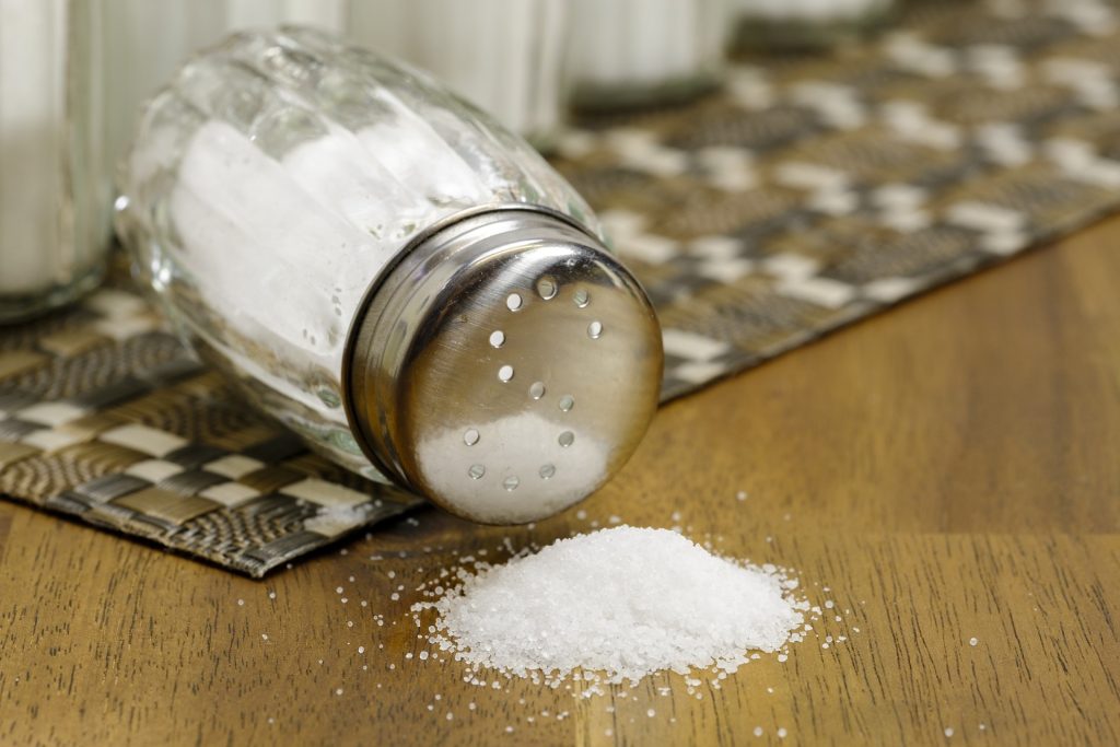
For decades, it’s been thought that people with heart failure should drastically reduce their dietary salt intake, but some studies have suggested that salt restriction could be harmful for these patients. A recent review in the European Journal of Clinical Investigation that assessed all relevant studies published between 2000 and 2023 has concluded that there is no proven clinical benefit to this strategy for patients with heart failure.

Most relevant randomised trials were small, and a single large, randomised clinical trial was stopped early due to futility. Although moderate to strict salt restriction was linked with better quality of life and functional status, it did not affect mortality and hospitalisation rates among patients with heart failure.

“Doctors often resist making changes to age-old tenets that have no true scientific basis; however, when new good evidence surfaces, we should make an effort to embrace it,” said author Paolo Raggi MD, PhD, of the University of Alberta.

Source: Wiley