Category: Cardiovascular Disease

Study Uncovers Connections Between Obesity and Heart Failure at the Cellular Level

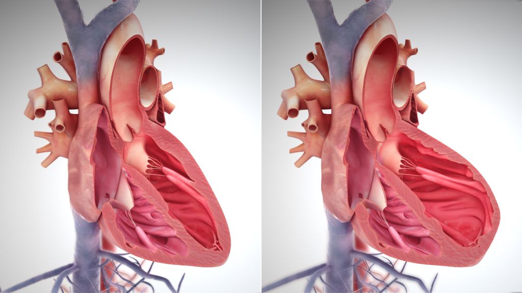
Right side heart failure. Credit: Scientific Animations CC4.0

A new small study led by Johns Hopkins Medicine researchers recently published in the journal Nature Cardiovascular Research has revealed the impact of obesity on muscle structure in patients having a form of heart failure called heart failure with a preserved ejection fraction (HFpEF). They observed swollen mitochondria, lipid droplets and tattered muscle fibre bundles, all independent of diabetes status.

According to the Journal of Cardiac Failure, HFpEF represents more than half of all heart failure world-wide. Originally, this form of heart disease was associated with hypertension and along with this, excess muscle growth (hypertrophy) to help counter the pressures. Over the past two decades, HFpEF is occurring more often in patients with severe obesity and diabetes according to the Journal of the American College of Cardiology. However, there are still very few effective HFpEF therapies, and a challenge in developing therapies has been the lack of studies in human heart tissue to determine exactly what is abnormal. As hospitalisation and death rates in HFpEF patients are quite high, (30–40% over 5 years), understanding its underlying causes is critical.  

“HFpEF is a complex syndrome, involving abnormalities in many different organs”, says lead investigator David Kass, MD, Professor of Medicine at the Johns Hopkins University School of Medicine. “We call it heart failure (HF) because its symptoms are similar to those found in patients with hearts that are weak. However, with HFpEF, heart contraction seems fine, yet heart failure symptoms still exist. While many prior efforts to treat HFpEF using standard HF drugs have not worked, success has since come from drugs used to treat diabetes and obesity.”

More specifically, the drug used to treat diabetes, known as an SGLT2 inhibitor (sodium glucose transporter 2 inhibitor) is currently the only evidence-based drug for HFpEF that has improved not only its symptoms but also reduced long-term rehospitalisation rates and endpoints of mortality. The weight loss drug GLP1-receptor agonist has been tested and found to improve symptoms in patients with HFpEF, and ongoing studies are determining if a similar hard end-point (mortality reduction, hospitalisation for HF reduction) are also possible outcomes. As such, these drugs have already been shown to be effective not only in diabetes where they started, but also in HFpEF.

To perform the study, the research team obtained a small piece of muscle tissue from 25 patients who had been diagnosed with varying degrees of HFpEF caused by diabetes and obesity and compared them to heart tissue from 14 organ donors whose hearts were considered to be normal. They examined the muscle using an electron microscope that shows muscle structure at a very high magnification.  

Mariam Meddeb, MD, MS, cardiovascular disease specialist at the Johns Hopkins University School of Medicine, who conducted the study says that a scanning electron micrograph “provides a very clear picture inside the muscle cell, what we call ultrastructure, such as mitochondria that are the energy power plants, and sarcomeres (unit of muscle fibre) that generate force”.

The researchers found notable ultrastructural abnormalities were particularly present in tissue of the most obese patients who had HEpEF, which had mitochondria that were swollen, pale, and disrupted, had many fat droplets, and their sarcomeres appeared tattered. These abnormalities were not related to whether the patient had diabetes, and were less prominent in patients who were less obese.

“These results will help those trying to develop animal models of HFpEF, since they show what one wants to generate at this microscopic level,” notes Dr Kass. “It also raises the key question of whether reducing obesity, as is now being done with several drug therapies, will reverse these ultrastructural abnormalities, and in turn improve HFpEF outcome.”  

Source: John Hopkins Medicine

Daily Physical Activity not Sufficient to Protect Against Stroke

Photo by Emmanuel Ikwuegbe on Unsplash

Research conducted at the University of Gothenburg shows that daily physical activities, at work or in the home, are not sufficient to protect against stroke. Fortunately, the findings, published in JAMA Network Open, suggest that exercising in free time and using active modes of transport are associated with a decreased risk of stroke.

“Physical activity during leisure time and as transportation is becoming increasingly important now that many jobs and domestic activities are becoming more sedentary,” says lead author of the study Adam Viktorisson, researcher at Sahlgrenska Academy at the University of Gothenburg, Sweden.

Twenty year follow-up

The research study covers 3614 people from the region of Västra Götaland, 269 of whom suffered a stroke in the twenty years spanned by the study. Three months after the stroke, 120 of these had died or were dependent on help to carry out activities of daily living.

Physical activity data was gathered from surveys. Some participants were also given a pedometer to wear. Physical activity during leisure time or for transportation showed a link to the objective measurements from the pedometers, while physical activity at work did not.

Occupational physical activity not protective

The health benefits of physical activity are well known, but earlier studies tend to mainly focus on physical activity during leisure time. Research in recent years has shown that physical activity at work can instead have negative health impacts, increasing the risk of cardiovascular disease.

“How and when we carry out physical activity seems to play a crucial role in determining its health benefits. In our study leisure time and transport related physical activities were associated with a lower risk of stroke, whereas activities during work time or in the household were not” Adam Viktorisson points out.

“Physically demanding jobs are often linked to stress, little opportunity for recovery, air pollution and generally poorer socioeconomic conditions, which can counteract the positive effects of physical activity.”

Promote public health

The study used data from the INTERGENE cohort at the University of Gothenburg. Study participants were surveyed and data was collected from 2001 to 2004, consisting of both clinical and questionnaire data. The researchers hope that these results will bring greater awareness and lead to changes in public health policy to encourage physical activity in society.

“Encouraging people to be physically active in their daily lives, for example by walking, cycling and doing other types of exercise, can be an important strategy in reducing the number of strokes and improving the prognosis of people who suffer a stroke,” says Adam Viktorisson.

Source: University of Gothenburg

Do Non-statin Cholesterol-lowering Drugs Affect Liver Cancer Risk?

Like statins, cholesterol absorption inhibitors are linked with a lower risk of developing liver cancer.

Photo by Towfiqu Barbhuiya on Unsplash

Past studies have suggested that taking cholesterol-lowering statin drugs may lower the risk of developing liver cancer. In a new study of non-statin cholesterol-lowering medications, one type was linked to lower risks of liver cancer. The findings are published by Wiley online in CANCER, a peer-reviewed journal of the American Cancer Society.

Cholesterol absorption inhibitors, bile acid sequestrants, fibrates, niacin, and omega-3 fatty acids are types of non-statin cholesterol-lowering medications prescribed to manage cholesterol and lipid levels. The different classes of drugs work in different ways. A team led by Katherine A. McGlynn, PhD, MPH, of the National Cancer Institute, looked for associations between these five types of non-statin cholesterol-lowering medications and risk of liver cancer, the sixth most commonly occurring cancer globally and the third leading cause of cancer mortality.

The investigators used information from the Clinical Practice Research Datalink (CPRD), a primary care database that covers approximately 7% of the United Kingdom population. Their analysis included 3719 liver cancer cases and 14 876 matched controls without cancer. Additional matches were also made based on individuals’ type 2 diabetes and chronic liver disease status.

Use of cholesterol absorption inhibitors was associated with 31% lower odds of liver cancer risk in the overall analysis. These medications were also linked with a lower risk of liver cancer in analyses based on diabetes and liver disease status. The study also confirmed that statins were associated with 35% lower odds of liver cancer.

No associations with liver cancer risk were observed for fibrates, omega-3 fatty acids, or niacin. While bile acid sequestrant use was associated with higher odds of liver cancer risk in the overall analysis, the results of analyses based on diabetes and liver disease status were inconsistent, suggesting that replication of these observations is important.

“As few studies have examined the effects of non-statin cholesterol-lowering drugs on liver cancer risk, the results of our study require replication in other populations. If our findings are confirmed in other studies, however, our results may inform liver cancer prevention research,” said Dr. McGlynn.

Source: Wiley

Study Discovers Immune System Changes from Stroke Impact the Heart

Human heart. Credit: Scientific Animations CC4.0

Why do new comorbidities arise because of ischaemic stroke? A study from Germany recently published in the journal Cell has discovered why this can happen – and ways in which it might be countered. The findings from the study show that the immune system is involved in damage to other organs, including the heart.

Besides the early mortality and morbidity resulting from the ischaemic brain injury itself, long-term morbidity after stroke is also due to the high prevalence of secondary comorbidities and complications, such as cognitive impairment and dementia, post-stroke depression, cardiac events, persistent vascular inflammation, and stroke-induced metabolic disturbances.

“However, there has been little research to date on the effects of brain injuries on systemic immunity,” says Professor Arthur Liesz from the Institute for Stroke and Dementia Research (ISD) at LMU University Hospital and principal investigator in the cluster of excellence SyNergy.

Liesz is the principal investigator of this new study. The researchers worked on the hypothesis that the high rate of comorbidities that develop after a stroke could have a common immunological cause. And they actually managed to find it: the origin of the dysfunctions in other parts of the body lies in the immunological memory of the blood-forming cells in bone marrow.

Using single-cell sequencing techniques, Liesz and his team demonstrated the presence of permanent proinflammatory changes in the transcriptome of certain immune cells (monocytes/macrophages) in several organs. In other words, certain gene segments are transcribed differently there after the stroke, which unbalances the proteome. These epigenetic modifications occur most frequently in the heart, where they can cause scarring and impair pumping function. “We managed to identify the protein IL-1b as the main culprit for the epigenetic modifications that affect immunological memory after a stroke,” says Liesz.

Promising therapeutic approaches on the horizon

The researchers demonstrated in a mouse model the connection between modified blood formation in bone marrow through overexpressed IL-1b and cardiac dysfunctions. Moreover, they showed that blocking IL-1b and inhibiting migration of the proinflammatory cells to the heart both successfully prevented cardiac problems after a stroke.

“These findings are hugely significant, as they open up the promise of effective therapeutic approaches for the prevention of secondary cardiac conditions after a stroke,” reckons Liesz.

The authors of the study believe that the epigenetic mechanisms they described for the reprogramming of the immune system in the brain-heart axis will create a new framework for explaining the development of various IL-1b-mediated comorbidities.

Source: Ludwig-Maximilians-Universität München

Brainstem Warning Signals in Sleep Apnoea Drive Blood Pressure up

Photo by Andrea Piacquadio

In the US, nearly 40 million adults have sleep apnoea, and more than 30 million of them use a continuous positive airway pressure (CPAP) machine while sleeping. But the machines tend to be expensive, clunky and uncomfortable – resulting in many users giving up on using them.

Hypertension is often linked with sleep apnoea because the brain works harder to regulate blood flow and breathing during sleep. A recent study at the University of Missouri (Mizzou) offers new insight into the underlying mechanisms within the brain contributing to hypertension for those with sleep apnoea.

The findings, which are published in the Journal of Physiology, can help pave the way for new drugs that target the brainstem to bring blood pressure back down to normal levels for those with sleep apnoea.

The study took place in the lab of David Kline, a professor in Mizzou’s College of Veterinary Medicine and researcher at the Dalton Cardiovascular Research Center.

“When oxygen levels in the blood drop during sleep apnoea, the forebrain sends warning signals to the brainstem area that controls heart and lung functions,” Kline said. “By studying these signals, we found that two neurochemicals, oxytocin and corticotropin-releasing hormone (CRH), cause the brainstem to become overactive. Over time, this leads to hypertension.”

Hypertension leads to an increased risk of stroke, complications in the metabolism and a variety of other health issues.

“Not only do those with sleep apnoea often have high blood pressure, but they also lose a lot of sleep, they have more cognitive and memory issues, and they are more prone to injury at work due to sleepiness,” Kline said.

By being the first to identify the role that oxytocin and CRH play in strengthening and overexciting the pathways and mechanisms involved in sleep apnoea, Kline and his fellow researchers hope to pave the way for the design of better therapeutic approaches for humans and animals.

“Our ultimate goal is to eventually help clinicians develop specific drugs to target either these neurochemicals or the proteins they bind to in a way that reduces high blood pressure,” Kline said. “This discovery opens the door for future research to block the pathways these neurochemicals use, ultimately helping to bring blood pressure back to normal levels.”

Source: University of Missouri

Women Lose More Years of Life After a Heart Attack Than Men

Photo by Joice Kelly on Unsplash

A new study shows that women lose more years of life after a heart attack than men. A 50-year-old woman with a large heart attack loses an average of 11 years, while an 80-year-old man with a small heart attack loses an average of 5 months of life. The results of the study, led by researchers at Karolinska Institutet and Danderyd Hospital, are published in the journal Circulation.

The new study examined 335 000 individuals with first-time myocardial infarction registered in the SWEDEHEART quality registry during the period 1991-2022. The individuals with myocardial infarction were compared with 1.6 million individuals without myocardial infarction using data from Statistics Sweden and the National Board of Health and Welfare. Using the comparator population and new statistical methods, the difference in life expectancy between heart attack individuals and comparison individuals could be calculated, providing a measure of how much life expectancy was shortened due to the disease. 

“We found that there were large differences between groups. Women and young individuals lost the most life expectancy when they had a heart attack. If the cardiac function was impaired after the infarction, the effects were even greater. For example, a 50-year-old woman with impaired cardiac function loses an average of 11 years in 2022 compared to an 80-year-old man with normal cardiac function who loses an average of 5 months in life expectancy,” says first author Christian Reitan, researcher at the Department of Clinical Sciences, Danderyd Hospital, Karolinska Institut. 

Parameters affecting heart attack risk

The researchers were also able to take into account differences in income, education, other illnesses and medication at the time of the illness – which helped to measure the effect of the heart attack itself when everything else was taken into account.

“The results showed that a fairly large part of the reduction in life expectancy disappeared, that is, much of the reduction in life expectancy is explained by factors other than the heart attack itself, but which may still be associated with heart attack, such as socioeconomics or other diseases such as hypertension and diabetes. Provided that the patient had preserved cardiac function, we saw that the gender difference had disappeared. We interpret this to mean that the effect of the heart attack, and thus also the care for heart attacks, is similar between the sexes and that the large reduction in life expectancy we see in women is due to differences in risk factors, other diseases and socioeconomics,” says Christian Reitan. 

According to the researchers, there is a lack of individualized heart attack care in Sweden for women. The study shows that women who have a heart attack lose more years of life than men of the same age.

“If a woman had impaired cardiac function, the gender difference was large. We don’t have the data to answer why, but it raises questions about whether women get as good follow-up and treatment for heart failure as men, or whether it is simply a more serious condition for a woman. Our findings are important because they challenge existing guidelines for heart attack treatment today. By identifying high-risk groups, we can hopefully better tailor treatment to the individual. We believe that ‘years of life lost’ is a good and easy-to-understand measure of risk for both doctors and patients. It makes it easier for us to assess and communicate the seriousness of the disease,” concludes Christian Reitan. 

Source: Karolinska Institutet

Is it Time to Stop Recommending Strict Salt Restriction in Heart Failure?

Credit: Pixabay CC0

For decades, it’s been thought that people with heart failure should drastically reduce their dietary salt intake, but some studies have suggested that salt restriction could be harmful for these patients. A recent review in the European Journal of Clinical Investigation that assessed all relevant studies published between 2000 and 2023 has concluded that there is no proven clinical benefit to this strategy for patients with heart failure.

Most relevant randomised trials were small, and a single large, randomised clinical trial was stopped early due to futility. Although moderate to strict salt restriction was linked with better quality of life and functional status, it did not affect mortality and hospitalisation rates among patients with heart failure.

“Doctors often resist making changes to age-old tenets that have no true scientific basis; however, when new good evidence surfaces, we should make an effort to embrace it,” said author Paolo Raggi MD, PhD, of the University of Alberta.

Source: Wiley

Innovative Cuffless Blood Pressure Device Improves Hypertension Management

A new study led by an investigator from Brigham and Women’s Hospital, evaluated a cuffless monitor that uses optical sensors to record blood pressure continually and efficiently, without disruption to the patient. The study, published in Frontiers in Medicine, highlights promising advancements in hypertension diagnosis, risk assessment and management that may be enabled by use of cuffless devices. 

“The successful management of hypertension depends on patients being able to take blood pressure measurements easily and reliably outside of the traditional doctor’s office setting,” said corresponding author Naomi Fisher, MD, of the Division of Endocrinology, Diabetes and Hypertension at Brigham and Women’s Hospital.  “Cuffless devices have the potential to revolutionise hypertension management.  They provide many more readings than traditional devices, during both the day and night, which can help confirm the diagnosis of hypertension and guide medication titration.” 

Medical guidelines increasingly recommend the incorporation of at-home blood pressure monitoring into hypertension diagnosis and management. This is because isolated blood pressure readings taken at a clinician’s office may be inaccurate: for some, blood pressure tends to rise in medical settings (“white coat hypertension”) while others have normal blood pressure during examination despite hypertensive readings at home (“masked hypertension”).  

Time-in-target-range (TTR) describes how often a patient’s blood pressure is in the normal range, and it is emerging as a promising metric of cardiovascular risk. But TTR requires more frequent blood pressure readings that can feasibly be obtained by patients with traditional blood pressure cuffs, which can be inconvenient, burdensome and sometimes uncomfortable for patients.  

Fisher, who designed and led the study, collaborated with co-authors from Aktiia SA, a Swiss biotechnology company, to analyse over 2.2 million blood pressure readings from 5189 subjects in Europe and the U.K. who wore a cuffless wrist monitor manufactured by Aktiia. On average, the Aktiia device collected 29 readings per day, a substantial increase from the number of blood pressure readings patients typically take with home devices (guidelines recommend four per day, which is more than most patients measure). Over a 15-day period, the researchers obtained an average of 434 readings from each patient.  

By calculating TTR over a 15-day period, the researchers were able to risk stratify participants by percentage of readings in target range and compare these classifications to those generated via traditional measurement patterns, using either 24-hour or week-long daytime monitoring schedules. They found that the traditional methods misclassified 26 and 45 percent of subjects, respectively, compared to the reference TTR. They determined that continual monitoring for seven days is required to obtain 90 percent or greater accuracy in hypertension risk classification, a frequency of measurement that may only be possible with cuffless monitors.  

Though the cuffless device studied here has not been approved by the US Food and Drug Administration, it has been validated in multiple studies and is available for over-the-counter purchase in Europe and the UK. Work to evaluate and set standards for such devices in the U.S. is ongoing. 

“For the first time, by using a cuffless device, we can collect continual out-of-office blood pressure readings and use these data to calculate a new metric, time-in-target-range, which shows great promise as a predictor of risk,” Fisher said. “The use of cuffless devices could create a shift in the paradigm of blood pressure monitoring and hypertension management.” 

Source: Brigham and Women’s Hospital

How does Oxygen Depletion Disrupt Memory Formation in the Brain?

Scientists identify a positive molecular feedback loop which could explain stroke-induced memory loss.

Ischaemic and haemorrhagic stroke. Credit: Scientific Animations CC4.0

In learning, neurons communicate with each other, and the connections between them getting stronger with repetition. This is known as long-term potentiation or LTP.  

Another type of LTP occurs when the brain is deprived of oxygen temporarily – anoxia-induced long-term potentiation or aLTP. aLTP blocks the former process, thereby impairing learning and memory. Therefore, some scientists think that aLTP might be involved in memory problems seen in conditions like stroke. 

Researchers at the Okinawa Institute of Science and Technology (OIST) and their collaborators have studied the aLTP process in detail. They found that maintaining aLTP requires the amino acid glutamate, which triggers nitric oxide (NO) production in both neurons and brain blood vessels. This process forms a positive glutamate-NO-glutamate feedback loop. Their study, published in iScience, indicates that the continuous presence of aLTP could potentially hinder the brain’s memory strengthening processes and explain the memory loss observed in certain patients after experiencing a stroke.  

The brain’s response to low oxygen 

When there is a lack of oxygen in the brain, the neurotransmitter glutamate is released from neurons in large amounts. This increased glutamate causes the production of NO. NO produced in neurons and brain blood vessels boosts glutamate release from neurons during aLTP. This glutamate-NO-glutamate loop continues even after the brain gets enough oxygen. 

“We wanted to know how oxygen depletion affects the brain and how these changes occur,” stated Dr Han-Ying Wang, a researcher in the former Cellular and Molecular Synaptic Function Unit at OIST and lead author of the study,. “It’s been known that nitric oxide is involved in releasing glutamate in the brain when there is a shortage of oxygen, but the mechanism was unclear.”  

During a stroke, when the brain is deprived of oxygen, amnesia – the loss of recent memories – can be one of the symptoms. Investigating the effects of oxygen deficiency on the brain is important because of the potential medicinal benefits. “If we can work out what’s going wrong in those neurons when they have no oxygen, it may point in the direction of how to treat stroke patients,” Dr Patrick Stoney, a scientist in OIST’s Sensory and Behavioral Neuroscience Unit, explained. 

Brain tissues from mice were placed in a saline solution, mimicking the natural environment in the living brain. Normally, this solution is oxygenated to meet the high oxygen demands of brain tissue. However, replacing the oxygen with nitrogen allowed the researchers to deprive the cells of oxygen for precise lengths of time.  

The tissues were then examined under a microscope and electrodes were placed on them to record electrical activity of the individual cells. The cells were stimulated in a way that mimics how they would be stimulated in living mice. 

Stopping memory and learning activity 

The aLTP process is activated when the brain is deprived of oxygen
The aLTP process is activated when the brain is temporarily deprived of oxygen and glutamate levels increase. If aLTP is maintained for an extended period, this hijacks the normal functioning of the memory strengthening process (LTP), resulting in memory loss. Blocking nitric oxide (NO) synthesis or the molecular pathways that boost glutamate release eventually stops aLTP. Credit: Wang et al., 2024 

The scientists found that maintaining aLTP requires NO production in both neurons and in blood vessels in the brain. Collaborating scientists from OIST’s Optical Neuroimaging Unit showed that in addition to neurons and blood vessels, aLTP requires the activity of astrocytes, another type of brain cell. Astrocytes connect and support communication between neurons and blood vessels. 

“Long-term maintenance of aLTP requires continuous synthesis of nitric oxide. NO synthesis is self-sustaining, supported by the NO-glutamate loop, but blocking molecular steps for NO-synthesis or those that trigger glutamate release eventually disrupt the loop and stop aLTP,” Prof. Tomoyuki Takahashi, leader of the former Cellular and Molecular Synaptic Function Unit at OIST, explained.  

Notably, the cellular processes that support aLTP are shared by those involved in memory strengthening and learning (LTP). When aLTP is present, it hijacks molecular activities required for LTP and removing aLTP can rescue these memory enhancing mechanisms. This suggests that long-lasting aLTP may obstruct memory formation, possibly explaining why some patients have memory loss after a short stroke. 

Prof Takahashi emphasised that the formation of a positive feedback loop formed between glutamate and NO when the brain is temporarily deprived of oxygen is an important finding. It explains long-lasting aLTP and may offer a solution for memory loss caused by a lack of oxygen.  

Source: Okinawa Institute of Science and Technology

Standardised Protocols Help Speed up Stroke Treatment

Photo by Mat Napo o Unsplash

Research by West Virginia University has demonstrated that American Heart Association and American Stroke Association guidelines are effective at speeding up hospitals’ response times for stroke treatment and can be mastered even by members of ‘ad hoc‘ medical teams that assemble rapidly on the fly.

When a stroke patient arrives at an emergency room, specialists from across hospital departments – emergency medical services, neurologists, pharmacists, physicians, nurses, radiologists and technicians – rush to coordinate a team response. AHA and ASA guidelines put specific limits on how much time can optimally elapse between the onset of ischaemic stroke, in which blood flow to the brain is blocked, and subsequent events like arrival at the hospital and delivery of an infusion.

But experts have questioned whether the communication of those best practices helps medical teams that assemble temporarily and whose members don’t typically collaborate. In a Journal of Operations Management article, WVU associate professor Bernardo Quiroga and coauthors answer that question using data about more than 8000 patients who received stroke care at a large hospital between 2009 and 2017.

“‘Time is brain’ for stroke victims,” Quiroga explained. “Blocked blood flow to the brain kills almost two million neurons a minute, so your life or ability to walk or talk hinges on how quickly multiple professionals coordinate to restore blood flow. If you’re lucky, you’re treated within the first hour of symptom onset. Better yet, you receive a shot of Tissue Plasminogen Activator, which dissolves clots. TPA works better the earlier it’s given and usually isn’t effective after 4.5 hours.”

In 2010, the AHA and ASA launched Target: Stroke, a program that identifies stroke care best practices and standardises each step in the process. Participating hospitals reduced median treatment times from 79 minutes in 2009 to 51 minutes in 2017, but it wasn’t clear if that improvement was driven by adherence to best practices or by clinicians learning through repetition as they handled more stroke cases.

To figure that out, the researchers investigated whether repeated ‘learning by doing’ decreased the hospital’s stroke care time. Then, they evaluated whether deliberate, ‘induced’ learning and implementation of AHA/ASA best practices decreased the time further.

Learning through repetition worked. The more strokes the hospital treated, the faster it responded. For each doubling of cumulative stroke alerts, ‘door-to-needle time’ – the time to get patients from the hospital door to a TPA infusion – decreased by 10.2%.

Best practices also worked. Specifically, the researchers examined two best practices: the Helsinki Model protocol, which directs that EMS staff keep stroke patients on the stretcher for transport to the CT room rather than transferring them to ER beds; and the Rapid Administration of TPA protocol, which requires the pharmacist to be in the CT room with TPA before completion of the CT scan. Those protocols significantly reduced the hospital’s door-to-needle time beyond improvements from repetition-based learning.

According to Quiroga’s coauthor and former PhD student Brandon Lee, that matters because it demonstrates the efficacy of best practices and shows ad hoc teams learning guidelines and implementing them long-term.

However, Lee emphasised the importance of the presence of the hospital’s stroke advisory committee, which set targets, evaluated stroke teams’ performances and gave feedback.

Without similar “countermeasures to organisational forgetting,” Quiroga acknowledged that best practices aren’t always sustainable, especially on ad hoc teams.

“In the case of the best practice indicated by the Helsinki Model, compliance is difficult because the hospital needs to coordinate with multiple independent EMS systems. Some EMS providers may be reluctant to commit resources to extended time in the CT room, and EMS staff turnover may lead to forgetting,” Quiroga said.

Lee added, “Overall, because ad hoc teams are fluid, information sharing is harder. And when a group of people don’t know each other well, group learning slows. But although ad hoc teams learn more slowly, we determined they still learn.”

The research also assessed whether neurologists’ abilities to meet time goals were affected by their recent experiences treating prior stroke patients.

“As team leaders, neurologists can have an outsized influence on performance,” Quiroga said. “Because other members of the ad hoc team aren’t familiar with each other, they lean on their leader.”

But data showed stroke teams improving response times regardless of how many stroke cases the neurologist had treated individually or what the neurologist’s recent success rate was. Quiroga said that’s good news.

“The implication is that learning and sustaining best practices ensures an even quality of care for patients, regardless of individual neurologists’ experience levels.”

Source: West Virginia University