Category: Cancer

CNN Anchor Christiane Amanpour Reveals Her Ovarian Cancer Diagnosis

Photo by Obi Onyeador on Unsplash

CNN anchor Christiane Amanpour told viewers on Monday that she has been diagnosed with ovarian cancer.

The 63 year-old international news veteran told viewers she had had “major successful surgery to remove it” and will now undergo several months of chemotherapy, adding that she was “very confident”.

Amanpour, who works in CNN’s London studio, said she feels “fortunate to have health insurance through work and incredible doctors who are treating me in a country underpinned by, of course, the brilliant NHS,” referencing the UK’s National Health Service.

After four weeks off, she said in Monday’s announcement, “I’m telling you this in the interest of transparency but in truth really mostly as a shoutout to early diagnosis.” Pointing out “millions of women around the world”, she added that she wanted to “urge women to educate themselves on this disease; to get all the regular screenings and scans that you can; to always listen to your bodies; and of course to ensure that your legitimate medical concerns are not dismissed or diminished.”

Amanpour has decades of experience reporting around the world, covering a wide range of conflicts and crises.

Ovarian cancer is the leading cause of death in women diagnosed with gynaecological cancers. It is also the fifth most frequent cause of death in women, in general. The symptoms, such as bloated, swollen or painful stomach, are easily mistaken for less serious health problems, making diagnosis difficult. Most cases are only diagnosed at an advanced stage, leading to poor outcomes. Existing screening tests unfortunately have a low predictive value.

Standard care treatment includes surgery and platinum-based chemotherapy; however, anti-angiogenic bevacizumab and Poly(ADP-ribose) polymerase (PARP) inhibitors are gaining ground in the treatment of this disease.

Source: BBC News

An Easy to Swallow Detection Method for Oesophageal Cancer

Image by Natural Herbs Clinic from Pixabay

In the UK, a “game-changer” method to sample cells for the detection of oesophageal cancer is being trialled in a mobile unit.

The cytosponge, a pill containing a sampling sponge, was developed at the and collects cells which are tested at a laboratory. Details on its development were published in The Lancet. In a previous trial with more than 13 000 participants receiving either the cytosponge or usual care from a GP, the odds of detecting oesophageal cancer were ten times higher than with usual care.

It is hoped the test will be much more efficient and quicker than the current detection method, requiring an endoscopy in hospital.

Prof Rebecca Fitzgerald from the University of Cambridge, which developed the test, said it was “really simple and straightforward”.

Early signs of cancer of the oesophagus are often mistaken for heartburn. It is the sixth most common cause of death from cancer worldwide.

A mobile unit will perform the test at GP surgeries at different locations around the UK.

Prof Fitzgerald, who specialises in cancer prevention, said the cytosponge “can diagnose cancer of the oesophagus really early”.

“Usually you would have to go to the hospital and get an endoscopy, with all that entails, and our idea was could you make something that was so simple you could go to a mobile unit or GP surgery,” Prof Fitzgerald said.

“The simplicity is the absolute key of this – we know the power of diagnosis is in the cells you collect.”

She added that due to COVID, “some endoscopy has been completely on hold so you might have to wait months” for the procedure, where a long, thin tube with a camera is sent down the patient’s mouth and throat.

Prof Fitzgerald explained: “You swallow the capsule on a string with water and it will go down to the top of the stomach.

“The capsule will dissolve in five to seven minutes, and as it dissolves out pops a sponge which has been compressed in that capsule. The nurse simply pulls the sponge out with the string and it will collect about a million cells on its way out.

“We put that sponge into a preservative, send it to the laboratory where it is tested to see whether there are Barrett cells or not and whether the cells look like they are turning to pre-cancer. Then we can let the patient know and if there is anything to worry about they can have an endoscopy and treatment.”

The procedure takes about 10 minutes to perform in total.

Source: BBC News

New Insights into How Kidney Cancer Cells Respond to Treatment

Photo by National Cancer Institute on Unsplash

Researchers from the University of Michigan Rogel Cancer Center have uncovered clues as to why kidney cancers respond so differently to treatment, opening up new tailored treatment options.

Not all kidney cancers behave the same, and some have wildly differing responses to immunotherapy or other treatments – resulting in wildly different outcomes for patients.

By sequencing the RNA of individual cells within multiple benign and cancerous kidney tumors, the researchers have identified the cells from which different subtypes of kidney cancer originate, the pathways involved and how the tumor microenvironment impacts cancer development and response to treatment.

The findings, published in PNAS, could help researchers better understand renal cell carcinoma development and guide oncologists in optimising therapies for each patient.

“Single cell RNA sequencing was key to allowing us to monitor gene expression patterns in each individual cell, revealing the mechanisms at play within the tumour microenvironment that can predict overall survival,” says study author Arul Chinnaiyan, MD, PhD, director of the Michigan Center for Translational Pathology and SP Hicks Professor of Pathology at Michigan Medicine.

Researchers produced gene expression atlases for normal kidney and renal cell carcinoma samples. They predicted the putative cell of origin for more than 10 subtypes of renal cell cancer. Their analysis also uncovered pathways and interactions within the tumour microenvironment that predicted if the tumour would respond to immunotherapy. These findings could help develop biomarkers to guide kidney cancer treatment.

“By understanding the cell type where a cancer originates, it may allow us to target more precise treatments for that cancer type as well as better understand response to therapy,” Dr Chinnaiyan said.

Source: University of Michigan

Journal information: “Single-cell analyses of renal cell cancers reveal insights into tumor microenvironment, cell of origin, and therapy response,” PNAS. DOI: 10.1073/pnas.2103240118

Cancer Patients Have a Higher Mortality Risk from COVID

Photo by Tima Miroshnichenko from Pexels

Patients hospitalised with active cancer are more likely to die from COVID than those with a cancer history or diagnosis, according to a new study.

The findings published by Wiley early online in CANCER, a peer-reviewed journal of the American Cancer Society, also indicate that the greatest risk of death due to COVID was in those with active blood cancers. No mortality risk increase was found in patients who received cancer treatments in the three months (or longer) prior to hospitalisation.

To find out how cancer, or the various therapies used to treat it, could affect the health of patients with COVID infections, a team analysed the NYU Langone Medical Center’s records of 4184 hospitalised patients who tested positive for SARS-CoV-2, the virus that causes COVID.

This group included 233 patients who had a current, or ‘active’, cancer diagnosis. They found that more patients with an active cancer diagnosis (34.3 percent) were likely to die from COVID than those with a history of cancer (27.6 percent) and those without any cancer history (20.0 percent).

Among patients with active cancer, those with blood-related cancers had the greatest risk of death. However, undergoing systemic anticancer therapy, including chemotherapy, molecularly targeted therapies, and immunotherapy, within three months prior to hospitalisation was not linked to a higher risk of death, and the investigators found there were no differences according to the type of cancer therapy being received.

Senior author Daniel Becker, said, “We completed a large chart review-based study of patients hospitalised with COVID and found that patients with active cancer, but not a history of cancer, were more likely to die. Notably, however, among those hospitalised with active cancer and COVID, recent cancer therapy was not associated with worse outcomes.”

“People with active cancer should take precautions against getting COVID, including vaccination, but need not avoid therapy for cancer.”

Source: Wiley

Diet Affects both Breast Microbiome and Breast Cancer Tumours

Breast cancer cells. Image source: National Cancer Institute on Unsplash

The breast has its own microbiome of bacteria, and new research has shown it can be influenced by diet, as can breast cancer tumours.

In 2018, scientists at Wake Forest School of Medicine, part of Wake Forest Baptist Health, showed that diet, just like the gut microbiome, can influence the breast microbiome.

Now, new research shows that diet, including fish oil supplements, can alter not only the breast microbiome, but also breast cancer tumours. The findings were published online in Cancer Research.

To untangle the relationship between microbiome, diet and cancer risk, researchers undertook a multi-pronged approach to study both animal models and breast cancer patients.

“Obesity, typically associated with a high-fat diet consumption, is a well-known risk factor in postmenopausal breast cancer,” said Katherine L. Cook, PhD, assistant professor in the surgery – hypertension and cancer biology departments at Wake Forest School of Medicine. “But there’s still a lot we don’t know about the obesity link to microbiomes and the impact on breast cancer and patient outcomes.”

In the first part of the study, mice susceptible to breast cancer were fed either a high-fat or a low-fat diet. Mice consuming the high-fat diet had more tumours, which were also larger and more aggressive than the tumours in the low-fat diet group.

Next, to study the microbiome, researchers performed faecal transplants. Mice consuming the low-fat diet received the high-fat diet microbiome transplant, and mice consuming the high-fat diet received the low-fat diet microbiome transplant. Unexpectedly, mice that consumed the low-fat diet and received a high-fat diet microbiome had just as many breast tumours as mice on the high-fat diet.

“Simply replacing the low-fat diet gut microbiome to the microbiome of high-fat diet consuming animals was enough to increase breast cancer risk in our models,” Cook said. “These results highlight the link between the microbiome and breast health.”

Researchers also conducted a double-blind placebo-controlled clinical trial with breast cancer patients, with patients either receiving placebo or fish oil supplements for two to four weeks before lumpectomy or mastectomy.

Results showed that fish oil supplementation significantly modified the breast microbiome in both non-cancerous and malignant breast tissue. For example, scientists found longer-term administration of fish oil supplements (four weeks) increased the proportional abundance of Lactobacillus in the breast tissue near the tumour. Lactobacillus is a genus of bacteria shown to decrease breast cancer tumour growth, suggesting potential anti-cancer properties of this intervention. Researchers also found decreased proportional abundance of Bacteroidales and Ruminococcus microbes in the breast tumours of patients taking the supplements, though the significance of this is not understood.

“This study provides additional evidence that diet plays a critical role in shaping the gut and breast microbiomes,” concluded Dr Cook. “Ultimately, our study highlights that potential dietary interventions might reduce breast cancer risk.”

Dr Cook’s team is also conducting further studies to see if probiotic supplements can affect microbiome populations in mammary glands and in breast tumours.

Source: Wake Forest Baptist Medical Center

Stronger Immune Systems in Women Protect against Skin Cancer

Photo by rfstudio on Pexels

Women may have a stronger immune response to a common form of skin cancer than men, according to a preliminary study on mice and human cells.

Men develop more skin squamous cell carcinoma (cSCC) than females and their tumours are more aggressive, though it is not clear if this is related to sunlight exposure. Using mouse models to answer the question, they found that male mice developed more aggressive tumours than females, despite receiving identical treatments.

In female mouse skin and tumours, the immune cell infiltration and gene expression related to the anti-cancer immune system were increased, suggesting a protective effect of the immune system.

In keeping with the animal study, 931 patient records collected from four hospitals in Manchester, London and France, the researchers identified that while women commonly have a more mild form of cSCC compared to men, immunocompromised women developed cSCC in a way more similar to men

That suggests the protective effect of their immune system may have been compromised.

These results were confirmed in a further cohort of sun-damaged skin from the US. In this cohort, human epidermal cells confirmed women’s skin activated immune-cancer fighting pathways and immune cells at sites damaged by sunlight.

The US cohort also that showed CD4 and CD8 T cells, which are important in our immune response to skin cancer, were twice as abundant in women as in men.

The researchers used RNA sequencing to examine differences in male and female immunosuppressed mice and human skin cells.

“It has long been assumed that men are at higher risk of getting non-melanoma skin cancer than women” said Dr Amaya Viros, from The University of Manchester.

“Other life-style and behavioural differences between men, such as the type of work or exposure to the sun are likely to be significant.

“However, we also identify for the first time the possible biological reasons, rooted in the immune system, which explains why men may have more severe disease.

“Although this is early research, we believe the immune response is sex-biased in the most common form of skin cancer, and highlights that female immunity may offer greater protection than male immunity.”

Dr Viros added: “We can’t yet explain why women have a more nuanced immune system than men.

“But perhaps it’s reasonable to speculate that women’s evolutionary ability to carry an unborn child of foreign genetic material may require their immunological system to be very finely tuned and have unique skills.

“Very little is known about how sex differences affect incidence and outcome in infectious diseases, autoimmune disorders and cancer. More work needs to be done. But we feel this study has opened a window into this area, and could one day have important implications on other types of immunologically based diseases.

“And it suggests if doctors are to offer personalised treatment of cancer, then biological sex should be one of the factors they take into account.”

Commenting on the study, Dr Samuel Godfrey, Research Information Manager at Cancer Research UK said: “Research like this chips away at the huge question of why people respond to cancer differently. Knowing more about what drives immune responses to cancer could give rise to new treatment options and show us a different perspective on preventing skin cancer.”

Source: Medical Xpress

Journal information: Timothy Budden et al, Female Immunity Protects from Cutaneous Squamous Cell Carcinoma, Clinical Cancer Research (2021). DOI: 10.1158/1078-0432.CCR-20-4261

Study Reveals Natural Killer Cells’ Fuel Source

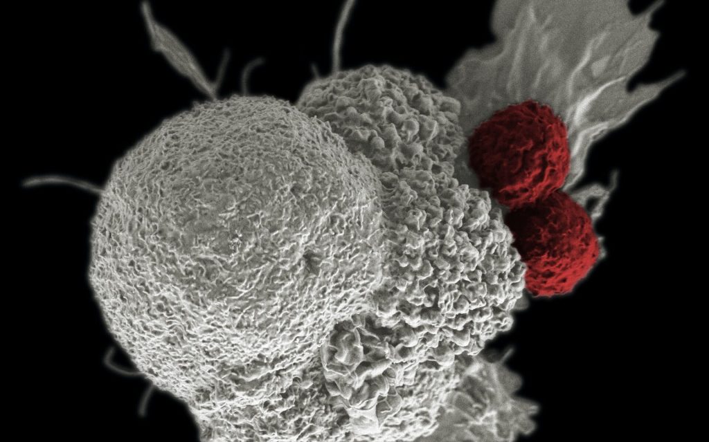
Pictured is a false-colour scanning electron micrograph of an oral squamous cancer cell (white) being attacked by two cytotoxic T cells (red), part of a natural immune response. Photo by National Cancer Institute on Unsplash

Scientists have discovered how natural killer (NK) cells fuel their activities when fighting infections, which will in turn help inform the development of immune therapies.

When it comes to dealing with infections and cancer, if T cells are like a team of specialist doctors in an emergency room, then NK cells are the paramedics: They arrive first on the scene and perform damage control until reinforcements arrive. Their existence was revealed in the 1970s when scientists were trying to characterise T cell cytotoxicity.

NK cells belong to our innate immune system, which dispatches these first responders, and they come with a built-in ability to recognise and respond to danger. Learning what powers NK cells is an ongoing area of immunology research, with important clinical implications.

“There’s a lot of interest right now in NK cells as a potential target of immunotherapy,” said Joseph Sun, an immunologist in the Sloan Kettering Institute. “The more we can understand what drives these cells, the better we can program them to fight disease.”

First responders

Previous studies have shown that aerobic glycolysis provides the energy for T cells to carry out their protective activities. But it was not known whether NK cells use this form of metabolism in performing their functions.

Dr Sun and his colleagues studied NK cells in animal models instead of in vitro, in order to find out, in a natural setting, what type of metabolism NK cells use and compare it to T cells. They discovered that NK cells increase aerobic glycolysis about five days before T cells respond with their own glycolytic surge.

“This fits with the idea that NK cells are innate immune cells that are really critical for mounting a rapid response,” said Research Fellow Sam Sheppard.

The findings are relevant to ongoing efforts to use NK cells as immunotherapy in people with cancer and other conditions. These are particularly relevant for procedures that make use of NK cells as a form of cell therapy—when cells are grown outside the body and then introduced back into the patient.

Finding a delicate balance

“If you’re growing these cells in a dish and you push them to divide too rapidly, they may not have as much potential to undergo aerobic glycolysis when you put them into a patient,” Dr Sheppard explained.

For researchers designing clinical trials, the goal is to find a balance between encouraging NK cells to multiply and preserving their stamina. These NK cells are the paramedics of our immune system, so it’s important to keep them speedy and responsive.

The findings were reported June 1, 2021, in the journal Cell Reports.

Source: Eureka Alert

US Supreme Court Snubs Johnson & Johnson Talc Lawsuit Appeal

Photo by Bill Oxford on Unsplash

On Tuesday, the US Supreme Court declined to hear Johnson & Johnson’s appeal challenging a $2.12 billion ruling in favour of 20 women who developed ovarian cancer which they alleged was linked to the company’s talcum powder. 

The company was appealing a 2018 court ruling in favor of 22 women who alleged asbestos-contaminated talcum powder was linked to their cases of ovarian cancer. The women had said the company did not provide adequate warning of the risks associated with using their products. The initial settlement amount had initially been over $4 billion before being cut down.

The judge in that case ruled Johnson & Johnson had “misrepresented the safety of these products for decades” and the evidence shown at the trial demonstrated “particularly reprehensible conduct on the part of Defendants.”
Johnsons & Johnson has dominated the talcum powder market for over a century.
While there is no established link between talcum powder exposure and cancer, but talcum powder is often mined close to asbestos, a known carcinogen for which there is no safe level of exposure, and which can have a long latency period between exposure and cancer development.
Some studies have shown an increase in lung cancer risk for miners working underground when exposed to raw talc, which can be contaminated with asbestos, while others have found no effect. Use of talcum powder in the genital or perineal area is thought to contribute to ovarian cancer risk, but results are also mixed.
Companies have been directed not to use asbestos in cosmetic products since the 1970s, according to the American Cancer Society. According to the National Cancer Institute, “the weight of evidence does not support an association between perineal talc exposure and an increased risk of ovarian cancer.”


A 2018 investigation by Reuters uncovered documents showing that Johnson & Johnson was not only aware of the asbestos contamination problem, the company covered it up. It even tried to influence US regulatory policy over asbestos in cosmetic products. Lawyers representing the company have argued in court that the tests were unreliable, although recent independent lab tests of samples obtained from various time periods detected asbestos contamination.

In response to queries from Reuters, Johnson & Johnson’s outside litigation counsel Peter Bicks wrote: “The scientific consensus is that the talc used in talc-based body powders does not cause cancer, regardless of what is in that talc. This is true even if – and it does not – Johnson & Johnson’s cosmetic talc had ever contained minute, undetectable amounts of asbestos.”

Source: Forbes

Surprising Mechanism of Action Discovered for Stem Cell Drugs

Two cytotoxic T cells (red) attacking an oral squamous cancer cell. Photo by National Cancer Institute on Unsplash

A new study revealed surprising insights into how specialised drugs that regenerate immune cells lost to chemotherapy actually work. 

In cancer patients following chemotherapy, there is a decrease in immune cells because chemotherapy also impacts the stem cells in bone marrow, which were meant to develop into new immune cells. This means that the immune system is then left short of immune cells to fight new infections.

Certain drugs exist, such as plerixafor, that can stimulate the release of stem cells from the bone marrow into the blood stream, so that they can be harvested and then reintroduced into the patients after treatment. These stem cells develop into new immune cells, bolstering the immune system. However, there was a lack of detailed knowledge of how these drugs actually worked.

Now, a study conducted in mice by researchers at the University of Copenhagen demonstrates how the medicine works at the cell level—and, surprisingly, how plerixafor, one of the two applied and tested drugs, is more effective than the other, despite the fact that the other drug, on paper, appears to be the most effective of the two. This discovery may not just help improve stem cell transplantation; it may also lead to improved drugs in the future.

“We have tested two drugs for stem cell transplantation which appear to have the same effect. What they do is block a receptor, causing the bone marrow to release stem cells into the blood. What the new study shows, though, is that they do not just block the receptor; one of the two drugs also affects other signaling pathways in the cell. And in short, that makes it more effective than the other of the two drugs,” explained PhD student Astrid Sissel Jørgensen from the Department of Biomedical Sciences at the University of Copenhagen.

“We used to believe that all we had to do was block the receptor, and that the two drugs had the same effect. It now appears that there is more to it,” she said.

The drugs tested by the researchers mobilise stem cells by acting as CXCR4 receptor antagonists. There are several drugs that target this receptor, including drugs inhibiting HIV replication.

“The drugs not only block the receptor’s normal signaling. One of the two drugs we have tested also affect some of the other cell pathways and even make the receptor withdraw into the cell and disappear from the surface,” explained corresponding author Professor Mette Rosenkilde. The study results revealed that one of the two drugs makes the bone marrow release more stem cells into the blood.

These findings on how the drugs affect cell pathways differently is also known as biased signalling. Mechanisms like these are what make the one drug more effective in practice than on paper, and they challenge the current view of these drugs.

“The results of our study directly influence our view of drugs used for stem cell transplantation. In the long term, though, it may also affect our view of future drugs, and how new drugs should be designed to have the best possible effect, both in connection with stem cell mobilisation, but also for treating HIV infections, where this particular receptor also plays a main role,” said Prof Rosenkilde.

Source: Medical Xpress

Journal information: Astrid S. Jørgensen et al, Biased action of the CXCR4-targeting drug plerixafor is essential for its superior hematopoietic stem cell mobilization, Communications Biology (2021). DOI: 10.1038/s42003-021-02070-9

New Study Finds Genetic Switch Role in Melanoma

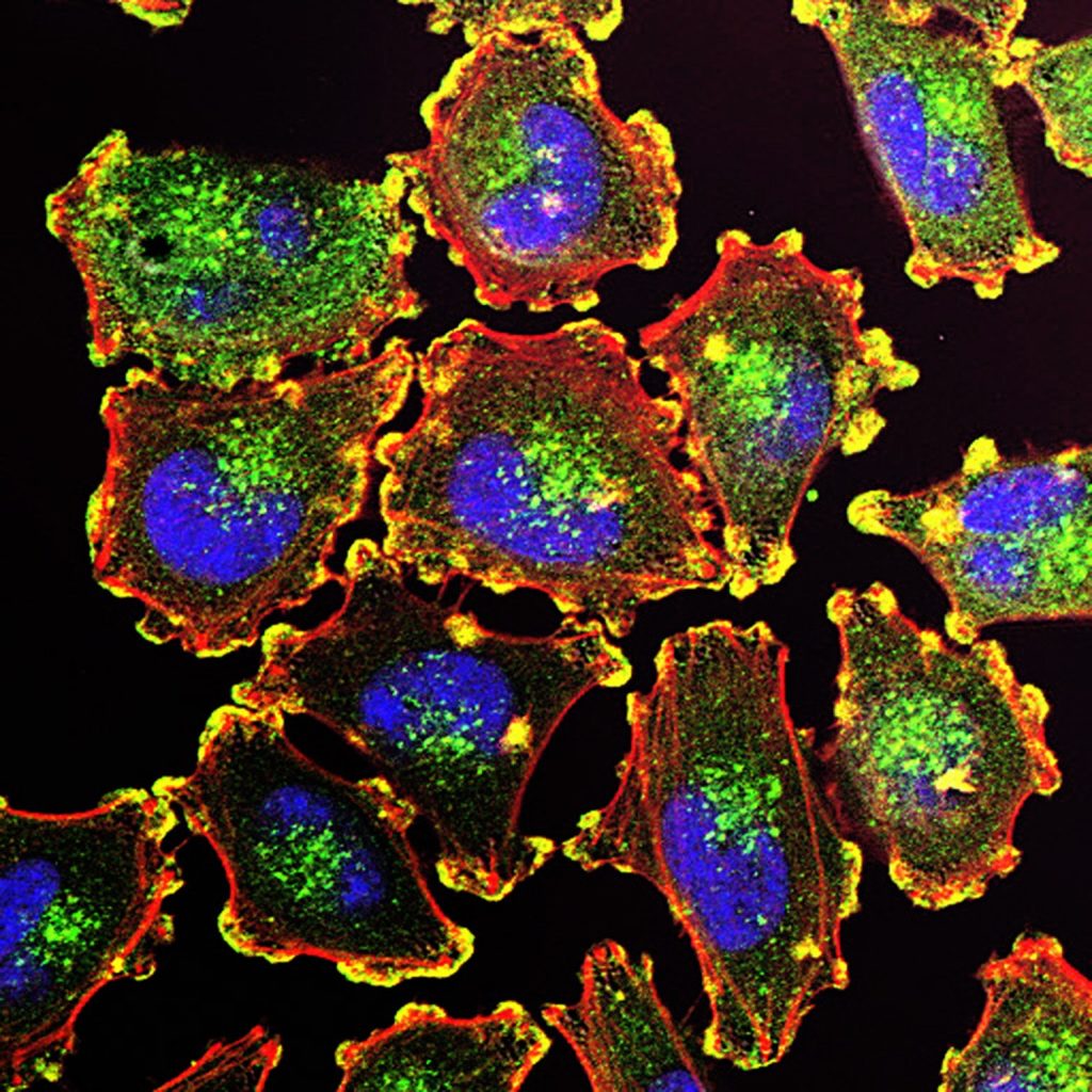
The ability of cancer cells to move and spread depends on actin-rich core structures such as the podosomes (yellow) shown here in melanoma cells. Cell nuclei (blue), actin (red), and an actin regulator (green) are also shown. Source: National Cancer Institute
Metastatic melanoma cells

A study published in the journal Cell Reports reveals that a genetic switch that could potentially be targeted to develop new treatments for melanoma by keeping the switch turned off.

Melanoma causes the majority of skin cancer-related deaths, despite only making up roughly 1 percent of skin cancers. The incidence of malignant melanoma is rapidly increasing around the world, and this increase is occurring at a faster rate than that of any other cancer except lung cancer in women. Treatments exist for this serious disease, but the effectiveness of these drugs can vary depending on the individual.

“We’ve been able to correlate the activity of this genetic switch to melanin production and cancer,” said Salk study corresponding author Marc Montminy, a professor in the Clayton Foundation Laboratories for Peptide Biology.

Melanoma develops when melanocytes, the pigment-producing skins in the cell, mutate and begin to multiply out of control. These mutations can cause proteins such as CRTC3 to prompt the cell to produce an abnormal amount of pigment, or to migrate and be more invasive.

While it was known that the CRTC family of proteins (CRTC1, CRTC2, and CRTC3) is involved in pigmentation and melanoma, obtaining precise details about the individual proteins has proven difficult. “This is a really interesting situation where different behaviours of these proteins, or genetic switches, can actually give us specificity when we start thinking about therapies down the road,” said first author Jelena Ostojic, a former Salk staff scientist and now a principal scientist at DermTech.

When the researchers deleted the CRTC3 gene in mice caused a color change in the animal’s coat color, demonstrating that the protein is needed for melanin production. They also found that when melanoma cells lacked the protein, they migrated and invaded less, meaning they were less aggressive, suggesting that inhibiting the protein could help treat the disease.

The team also described the connection between two cellular signalling systems that work on the CRTC3 protein in melanocytes. These two systems tell the cell to either proliferate or make the pigment melanin. Montminy likens this process to a relay race: essentially, a baton (chemical message) is passed from one protein to another until it reaches the CRTC3 switch, either turning it on or off.

“The fact that CRTC3 was an integration site for two signaling pathways—the relay race—was most surprising,” says Montminy, who holds the J.W. Kieckhefer Foundation Chair. “CRTC3 makes a point of contact between them that increases specificity of the signal.”

Next, the team plans to further investigate the mechanism of how CTRC3 impacts the balance of melanocyte differentiation to develop a better understanding of its role in cancer.

Source: Salk Institute