Using Fat Tissue, Researchers 3D-Print Skin that Contains Hair Precursors

Fat tissue holds the key to 3D printing layered living skin and potentially hair follicles, according to researchers who recently harnessed fat cells and supporting structures from clinically procured human tissue to precisely correct injuries in rats. The advancement could have implications for reconstructive facial surgery and even hair growth treatments for humans.
The team’s findings published in Bioactive Materials, and the team received a patent in February for the bioprinting technology it developed and used in this study.
“Reconstructive surgery to correct trauma to the face or head from injury or disease is usually imperfect, resulting in scarring or permanent hair loss,” said Ibrahim T. Ozbolat, professor of engineering science and mechanics, of biomedical engineering and of neurosurgery at Penn State, who led the international collaboration that conducted the work. “With this work, we demonstrate bioprinted, full thickness skin with the potential to grow hair in rats. That’s a step closer to being able to achieve more natural-looking and aesthetically pleasing head and face reconstruction in humans.”
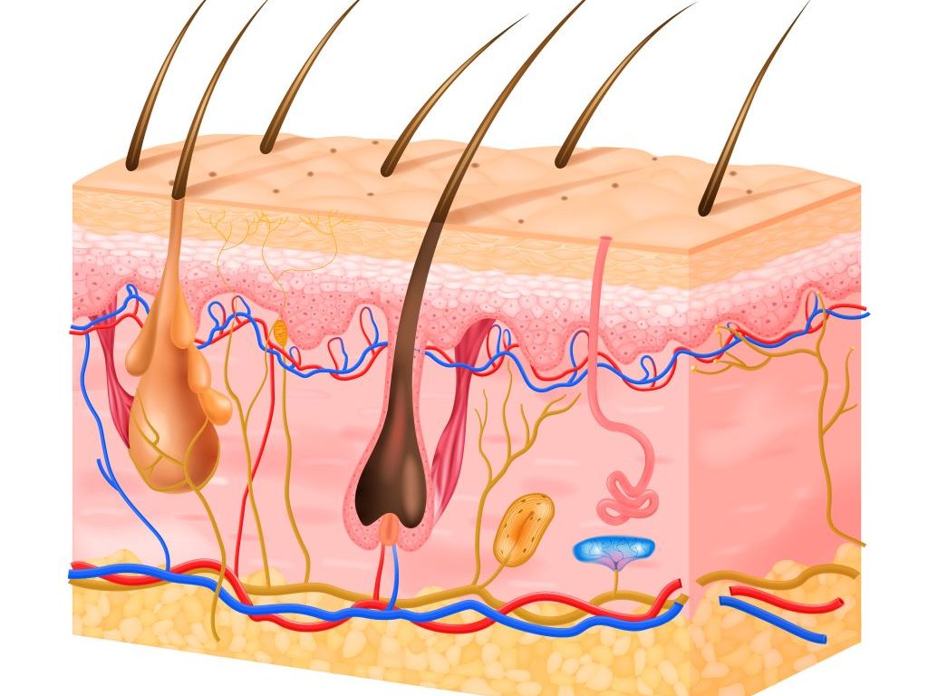
While scientists have previously 3D bioprinted thin layers of skin, Ozbolat and his team are the first to intraoperatively print a full, living system of multiple skin layers, including the bottom-most layer or hypodermis. Intraoperatively refers to the ability to print the tissue during surgery, meaning the approach may be used to more immediately and seamlessly repair damaged skin, the researchers said. The top layer — the epidermis that serves as visible skin — forms with support from the middle layer on its own, so it doesn’t require printing. The hypodermis, made of connective tissue and fat, provides structure and support over the skull.
“The hypodermis is directly involved in the process by which stem cells become fat,” Ozbolat said. “This process is critical to several vital processes, including wound-healing. It also has a role in hair follicle cycling, specifically in facilitating hair growth.”
The researchers started with human adipose, or fat, tissue obtained from patients undergoing surgery at Penn State Health Milton S. Hershey Medical Center. Collaborator Dino J. Ravnic, associate professor of surgery in the Division of Plastic Surgery at Penn State College of Medicine, led his lab in obtaining the fat for extraction of the extracellular matrix to make one component of the bioink.
Ravnic’s team also obtained stem cells, which have the potential to mature into several different cell types if provided the correct environment, from the adipose tissue to make another bioink component. Each component was loaded into one of three compartments in the bioprinter. The third compartment was filled with a clotting solution that helps the other components properly bind onto the injured site.
“The three compartments allow us to co-print the matrix-fibrinogen mixture along with the stem cells with precise control,” Ozbolat said. “We printed directly into the injury site with the target of forming the hypodermis, which helps with wound healing, hair follicle generation, temperature regulation and more.”
They achieved both the hypodermis and dermis layers, with the epidermis forming within two weeks by itself.
“We conducted three sets of studies in rats to better understand the role of the adipose matrix, and we found the co-delivery of the matrix and stem cells was crucial to hypodermal formation,” Ozbolat said. “It doesn’t work effectively with just the cells or just the matrix – it has to be at the same time.”
They also found that the hypodermis contained downgrowths, the initial stage of early hair follicle formation. According to the researchers, while fat cells do not directly contribute to the cellular structure of hair follicles, they are involved in their regulation and maintenance.
“In our experiments, the fat cells may have altered the extracellular matrix to be more supportive for downgrowth formation,” Ozbolat said. “We are working to advance this, to mature the hair follicles with controlled density, directionality and growth.”
According to Ozbolat, the ability to precisely grow hair in injured or diseased sites of trauma can limit how natural reconstructive surgery may appear. He said that this work offers a “hopeful path forward,” especially in combination with other projects from his lab involving printing bone and investigating how to match pigmentation across a range of skin tones.
Source: Penn State