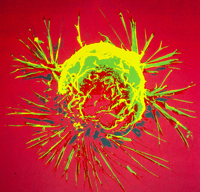
Radio DJ Mark Pilgrim has revealed on Twitter that his latest scan results showed a ‘significant shrinkage’ of all of the tumours present in his lung, spine, leg and lymph nodes.
Thank you for all the thoughts and prayers over the past 3 months. I received my scan results this morning… pic.twitter.com/rRcWx9XLRE
In March 2022, the radio star revealed that he had been diagnosed with stage 4 lung cancer, and that he was to start treatment “in a week”.
Speaking about his most recent diagnosis, he said: “I’m not Chuck Norris. Yes, I’m scared. I am also strong. Both emotions run parallel with each other. I’m under the care of incredible doctors and surrounded by love.”
It is not the first time the popular DJ has been through such difficult times. Pilgrim had already survived stage stage 4 testicular cancer in 1998 at the age of 18. The cancer had spread to his lungs and kidney, and he recalls that his oncologist said that his prognosis was “uncertain”. He endured 9 months of 4-hour chemotherapy sessions.
In 2008 he suffered heart damage from a massive myocardial infarction suffered in the doctor’s office, and last year he tested positive for COVID.
For patients with non-small cell lung cancer (NSLC), the most common form of lung cancer, half present at stage 4. In one Canadian study, only 14.9% of patients received chemotherapy as first-line treatment, with most patients receiving palliative radiotherapy.
While patients usually report any medications they are on, over-the-counter drugs and supplements are not reported as often to the medical team, according to a study on polypharmacy published in The Oncologist, an overlooked situation that complicates the problem of polypharmacy, especially in cancer.
Polypharmacy can lead to harmful drug interactions, especially dangerous for cancer patients about to undergo therapy.
Even for those without cancer, multiple medication use has risks and is tricky to navigate because of the emotions involved, said Erika Ramsdale, MD, study leader.
“As doctors, we tell people to take medications but we don’t always do a great job of following up,” she said. “From the patient perspective, if it’s determined that a medication is no longer needed, it’s hard to stop taking it. There’s a sense of, ‘What will happen if I stop?’ or ‘Are you giving up on me?’ A lot of uncertainty and emotions are tied up in this issue.”
The more drugs and supplements a person takes, the higher the risk of inappropriate use and serious drug interactions, she said.
The fragmentation of healthcare across specialties complicates the issue. “Sometimes, there is no quarterback,” Dr Ramsdale said, which can result in “prescribing cascades,” where additional drugs are given to offset the adverse side effects of the original medications.
Researchers analysed medication use in a sample of 718 adults with a mean age of 77 who had stage 3 or 4 cancer and other health conditions. They screened for potentially inappropriate medications that have risks higher than benefits (known as PIMS), drug-drug interactions (DDI), and drug-cancer treatment interactions (DCI). Drug interaction can have consequences such as falls, functional decline, and death. Patients on multiple medications are also more likely to have anxiety or depression.
Among the 718 patients, 70% were at risk of drug-drug interactions and 67% were taking at least one drug that was potentially inappropriate.
In fact, 61% of the patients were taking five or more medications before starting chemotherapy – and nearly 15% were taking 10 or more medications.
Other findings from the study include:
Nearly 68% of the patients had serious health issues besides cancer, requiring associated medications. Most common was cardiovascular disease. When a person has cancer combined with other ailments, there is a greater risk of toxicity from cancer treatments due to polypharmacy.
Approximately 10% of hospital admissions for older adults are associated with hazardous drug interactions. Among older adults with cancer receiving chemotherapy, polypharmacy is associated with dramatic increases (up to 114%) in unplanned hospitalisations.
Cholesterol-lowering medications, minerals, and thyroid therapy are most commonly involved in potential drug interactions.
More than 25% of the medications used by the patients in the study were non-prescription—and these accounted for 40% of the potentially inappropriate medications detected by investigators.
Common non-prescription remedies included vitamins and minerals, anti-anemic preparations such as ferrous sulfate, and drugs for acid-related disorders and constipation.
“Older adults may incorrectly assume that over-the-counter medications are safe for them,” the authors wrote. “This study helps delineate the size and shape of a problem under-recognised by both providers and patients.”
It’s also an understudied problem, Dr Ramsdale said, and including over-the-counter medications sets Wilmot’s data apart from previous research; most polypharmacy studies among cancer patients look only at prescription drugs.
The study highlights an opportunity for education and problem-solving, such as deprescribing some drugs.
Deprescribing is the planned reduction of medications to avoid harm. Doctors take into account the risks-versus-benefits of each medication and the patient’s life expectancy. For example, statins that are taken for high cholesterol do not have an immediate effect. They are meant to be preventive and can take 10 years to have an impact. Therefore, if a patient is old and has incurable cancer, he or she may not need to take statins. (Discontinuing statins in this setting is supported by a landmark study, according to the Ramsdale publication.)
However, these conversations can be quite delicate, Ramsdale said. The goal is to promote better quality of life, and she is conducting a clinical trial to test the best way to intervene in cases of polypharmacy among older people with cancer.
Research which appears in ACS Central Science has shown that a direct link exists between the amount of fat in the diet and bodily levels of nitric oxide, a naturally occurring signalling molecule that is related to inflammation and cancer development.
Nitric oxide (NO) is one of the critical components of the vasculature, regulating key signalling pathways in health. In macrovessels, NO functions to suppress cell inflammation as well as adhesion. In this way, it inhibits thrombosis and promotes blood flow. It also functions to limit vessel constriction and vessel wall remodelling. In microvessels and particularly capillaries, NO, along with growth factors, is important in promoting new vessel formation, a process termed angiogenesis. With age and cardiovascular disease, animal and human studies confirm that NO is dysregulated at multiple levels including decreased production, decreased tissue half-life, and decreased potency.
“We are trying to understand how subtle changes in the tumour microenvironment affect cancer progression at the molecular level. Cancer is a very complicated disease,” said Anuj Yadav, a senior research associate and the study’s lead coauthor.
Cancer is more than just a few tumour cells, rather the entire microenvironment of the tumour supporting the cells, Yadav explained.
“Inflammation can play a significant role in this environment. Certain inflammatory response comes from highly processed foods, which are high in calories and high in fat. We wanted to understand the links between food, inflammation, and tumors at a molecular level, so we had to develop advanced probes to be able to visualise these changes,” he said.
Yadav and coauthors are familiar with existing research linking increased NO levels to inflammation, and inflammation to cancer. Proving the connection between high-fat diets and NO levels on a molecular level required developing a highly sensitive molecular probe capable of deep-tissue imaging.
The team designed a molecular probe which is the first of its kind to be used in bioluminescence imaging of NO in cancer.
“Our group specialises in making designer molecules, which allows us to look at molecular features that are invisible to the naked eye,” said Jefferson Chan, an associate professor of chemistry at the University of Illinois Urbana-Champaign and the study’s principal investigator. “We design these custom-made molecules to discover things that weren’t previously known.”
With the probe, the researchers compared the tumourigenicity of the breast-cancer-carrying mice on a high-fat diet (60% of calories coming from fat) with mice on a low-fat diet (10% of calories coming from fat) by measuring the NO levels in both groups.
“As a result of the high-fat diet, we saw an increase in nitric oxide in the tumor microenvironment,” said Michael Lee, a student researcher in the Chan lab and a lead coauthor on this study. “The implication of this is that the tumor microenvironment is a very complex system, and we really need to understand it to understand how cancer progression works. A lot of factors can go into this from diet to exercise – external factors that we don’t really take into account that we should when we consider cancer treatments.”
The authors emphasised the importance of proving a direct link between a high-fat diet, NO levels, and cancer development. With this association now known, new implications exist for cancer diagnosis and treatment.
“Without this technology, you wouldn’t see this missing molecular link,” said Chan. “Now that we know that this is happening, how do we prevent it, and how do we improve the situation?”
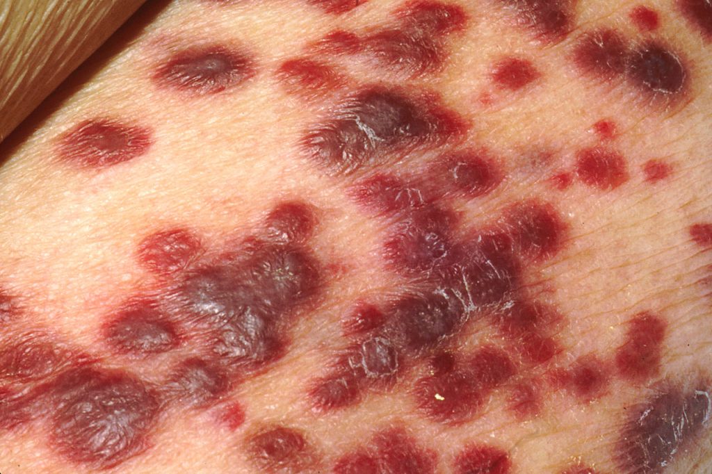
Kaposi sarcoma on the skin of an AIDS patient. Credit: National Cancer Institute
A study published in Cell Reportshas identified a protein in the cancer cell’s nucleus as a critical agent keeping Kaposi sarcoma-associated herpesvirus (KSHV) dormant and hidden from the immune system. The virus, in the same family as Epstein-Barr virus, is linked to AIDS-related Castleman’s disease and cancers such as Kaposi sarcoma.
Up to 50% of the population in some parts of Africa are affected with KSHV, though not everyone with KSHV will develop Kaposi sarcoma. Those who do typically have a weakened immune system due to HIV infection, organ transplant, being older or other factors.
The introduction of antiretroviral therapy significantly reduced AIDS-related Kaposi sarcoma prevalence in Western countries; however, in sub-Saharan Africa, the disease continues to have a poor prognosis.
On entry into a human cell the virus causes a hidden infection in the nucleus: the virus simply latches onto parts of the cell’s chromosomes without replicating.
Researchers studied KSHV’s latent-lytic switch, a process in which the virus exits its dormancy state to replicate in the host cell. This replication phase, called the lytic cycle, ends with the disintegration of the cell and the release of the viruses, infecting neighbouring cells.
“The virus likes to stay silent as long as possible to avoid being detected by the body’s immune system,” said Professor Yoshihiro Izumiya, the study’s senior author.
The team sought to understand the mechanisms behind this latent-lytic switch and the role the host cell environment played in this process.
“Where the virus latches onto the host cell, how it manages to stay dormant, and what triggers its activation were very exciting and important puzzles to solve,” Prof Izumiya said.
The study identified where the virus genome could be found on the host genome.
Izumiya and his team profiled and analysed chromosomal interactions on three cancer cell lines naturally infected with KSHV, locating the virus’s preferred chromosome docking sites. The binding patterns, similar among the three cancer cell lines, showed a nuclear ecosystem that can attract and help keep the virus in its silent form.
The team also found that CHD4 (chromodomain helicase DNA binding protein 4) binds to the virus’s genomic elements. CHD4, a protein in the host cell’s chromosomes, suppresses the work of the gene responsible for viral replication. The study showed that CHD4 is a key regulator of the KSHV latency-lytic switch.
“The location where the virus genome attaches to the host chromosome is not random,” said Ashish Kumar, a postdoctoral researcher in Izumiya Lab and the paper’s first author. “Without having enriched CHD4 protein, the virus starts to replicate, kicking in a cell destructive mode. For the virus to select CHD4 among many other host proteins, CHD4 must play a unique and important role in host cells.”
Virology can help identify cellular proteins essential for cell homeostasis. Over millions of years, the virus’s genome developed to encode or assemble a small number of very efficient proteins, which strategically connect to host cell proteins to keep viral chromatin dormant and impact the host cell’s tumour suppression function.
“We used virology as an entry point to shed light on the function of CHD4 in gene regulation in general. During virus-host co-evolution, KSHV cleverly learned to hijack host proteins that can help keep the gene responsible for viral replication dormant.”
The researchers found a viral protein which could serve as the basis for a replication inhibitor. Since CHD4 is critical for cancer cell growth in a variety of cancers, they hope this virus-host interaction could inform cancer treatment research.
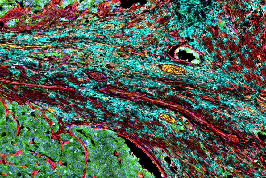
Cancer-associated fibroblasts surrounding a prostate tumour. Credit: Moscat/Diaz-Meco labs.
Some tumours can force neighbouring cells into supporting cancer growth by releasing lactate into their local environment, according to a study published in Cell Reports. The findings could lead to drugs that target that defence mechanism to help cancer patients ؘ– and also boost a current class of cancer drugs.
In the study, the researchers determined how developing tumours recruit nearby cells called fibroblasts to work as their enablers. Fibroblasts form part of the stroma, organs’ connective tissue, and normally have important repair and maintenance functions. But cancer-associated fibroblasts (CAFs) acquire properties that allow them to assist tumours in ways that make the tumours more malignant and harder to kill.
The researchers also discovered that PARP-1 inhibitors, a widely used class of cancer drugs, mimic one of the key steps in CAF recruitment, and can hamstring their own effectiveness by inadvertently switching local fibroblasts to this cancer-enabling mode.
“Future therapeutics that block this cancer-associated state of fibroblasts might be useful on their own or as a way to improve the effectiveness of PARP-1 inhibitors,” said study co-senior author Dr Maria Diaz-Meco.
Dr Diaz-Meco collaborated in the study with the laboratory of co-senior author Dr Jorge Moscat.
Developing tumours are known to often modify their local environments for their own survival and growth. Cancer-associated fibroblasts are a central component of the tumour microenvironment in prostate, lung, colon and other cancer types. Targeting these cells is therefore seen as a promising complementary approach to standard cancer treatment – and one that could work very broadly against cancers of different cellular and genetic origins.
“Cancer-associated fibroblasts support tumour growth by providing growth factors and essential metabolites to the tumour, by fending-off anti-tumour immune cells, and in many other ways,” Dr Moscat said. “The result is a tumour that is more malignant and treatment-resistant.”
Several years ago, the researchers discovered that a protein called p62, produced in fibroblasts, normally suppresses the CAF state, though many tumours find a way to restore this state by reducing fibroblast p62 production. In the new study, they showed that tumours achieve this by secreting high levels of an organic compound called lactate, also known as lactic acid.
Lactate is a normal by-product of certain energy-production processes in cells, which are often hyperactive in tumours. In experiments with prostate cancer cells, the researchers detailed the molecular chain of events by which tumour-secreted lactate disrupts the normal metabolism of fibroblasts, leading to reduced p62 gene activity and the activation of the tumour-enabling CAF state.
The finding is significant because it illuminates a major cancer-promoting pathway, which in principle can be targeted with future drugs as a standalone or add-on treatment strategy.
A second, surprising finding was that a key step leading from tumour lactate secretion to fibroblast p62 suppression turned out to be the inhibition of a DNA-repair enzyme called PARP-1, which has the same effect as PARP-1 inhibitors – suggesting that these drugs might be working partly against themselves by creating a more tumour-friendly microenvironment.
In vitro and animal testing confirmed that the PARP-1 inhibitor olaparib does reduce p62 in fibroblasts, and pushes them into the CAF state, in turn increase tumours’ resistance to the drug’s primary cancer-killing effect.
Thus, the researchers emphasised, future treatments that reprogram CAF cells to the non-cancer state or prevent their development might greatly enhance PARP-1 inhibitors’ anti-tumour effectiveness.
“We’re now studying several potential CAF-blocking therapeutics in our labs,” Dr Moscat said.
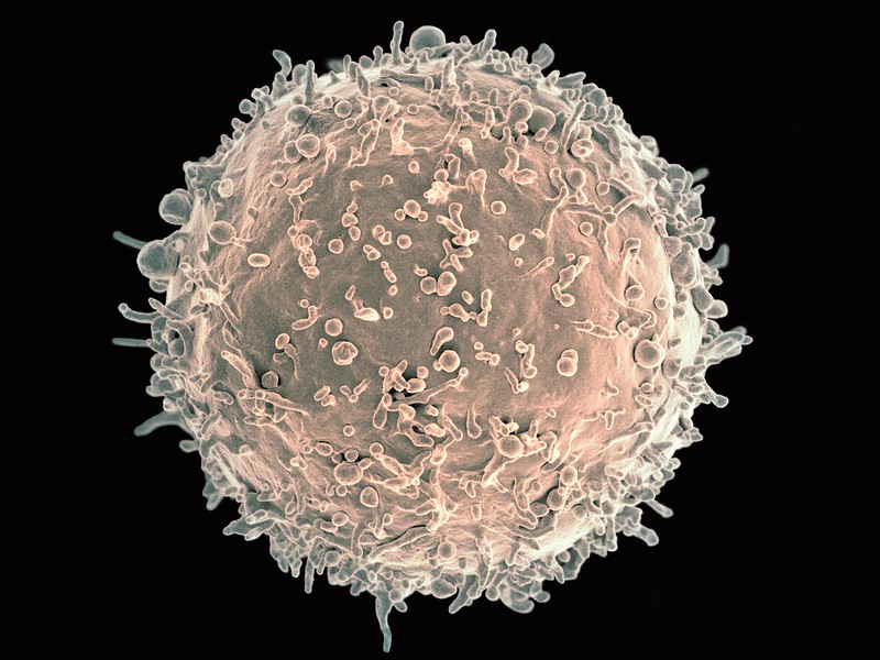
Scanning Electron Micrograph of a human B Cell. Credit: NIH
When patients in the UK showed adverse side effects during a cancer immunotherapy trial, researchers went back through the data and worked with patient samples to see what went wrong. Published in Nature, their findings provide clues into the dangerous side effects of many immunotherapies – and point to a better strategy for treating solid tumours.
“This work shows the importance of learning from early stage clinical trials,” says La Jolla Institute for Immunology (LJI) Professor Pandurangan Vijayanand, MD, PhD, who co-led the new research with Christian H. Ottensmeier, MD, PhD, FRCP, a professor with the University of Liverpool.
“In the oncology world, immunotherapy has revolutionised the way we think about treatment,” said Prof Ottensmeier. “We can give immunotherapies to patients even with metastatic and spreading disease, and then just three years later wave goodbye and tell them their cancer is cured. This is an astounding change.”
Unfortunately, only 20–30% of solid cancer patients given immunotherapies go into long-term remission. Some people see no change after immunotherapy, but others develop serious side effects, which can be debilitating, even fatal, and these patients are forced to discontinue the immunotherapy.
The researchers worked with samples from a recent UK clinical trial for head and neck cancers. The patients were given an oral cancer immunotherapy – a PI3Kδ inhibitor. At the time, PI3Kδ inhibitors had proven effective for B cell lymphomas but had not yet been tested in solid tumours.
PI3Kδ inhibitors are a new to cancer immunotherapy, but they hold promise for their ability to inhibit ‘regulatory’ T cells (Tregs). Tregs normally try to stop other T cells, called effector T cells, from targeting the body’s own tissues. Oncologists inhibit Tregs inside tumours so effector T cells can let loose and generate cancer-killing CD8+ T cells.
“Having an oral tablet that can take off the brakes – the Tregs– can be a great asset for oncologists,” said Prof Vijayanand.
Unfortunately, 12 of the 21 patients in the trial had to discontinue treatment early because they developed inflammation in the colon, a condition called colitis. “We thought this drug wouldn’t be toxic, so why was this happening?” said Prof Vijayanand.
Simon Eschweiler, PhD, an instructor at La Jolla Insitute, led the review into exactly how PI3Kδ inhibitor treatment affected immune cells in these patients. Gene sequencing showed that in the process of increasing tumour-fighting T cells in tumours, the PI3Kδ inhibitor also blocked a specific Treg cell subset from protecting the colon. Without Tregs patrolling there, pathogenic T cells, called Th17 and Tc17 cells, moved in and caused inflammation and colitis.
It was clear that a larger than needed PI3Kδ inhibitor dose had been given, and had disrupted the immune cell balance in the gut.
The pathway that leads to the toxicity seen in the new study may be broadly applicable to other organs harbouring similar Treg cells, and to other Treg cell-targeting immunotherapies like anti-CTLA-4, Eschweiler says.
The team found that intermittent dosing could be a valid treatment strategy that combines sustained anti-tumour immunity with reduced toxicity. The researchers are now designing a human clinical trial to test the intermittent dosing strategy in humans.
Why the lack of toxicity in trials for B cell lymphomas? Dr Eschweiler noted that in previous studies, lymphoma patients had been given several prior therapies leading to an overall immunocompromised state. This means the lymphoma patients didn’t have the same type or magnitude of immune response upon PI3Kδ inhibition. Meanwhile, the head and neck cancer patients were treatment-naive. Since their immune system was uncompromised, the immune-related adverse events were more rapid and pronounced.
Overall, the new study shows the importance of studying not just personalised therapies but personalised therapy doses and schedules.
A study published in Pharmacology & Therapeutics provides the first in-depth description of the anti-cancer activity of capsaicin sustained release formulations. Capsaicin, responsible for the hot and spicy taste of chili peppers, is being, explored in sustained release formulations of capsaicin for extended anti-cancer activity. However, how to get around its fiery taste remains a challenge.
The article chronicles the growth-suppressive activity of sustained release capsaicin drugs, including solid dispersion systems, liposomes, phospholipid complexes and nanoparticles. This marks the first publication to provide an in-depth description of the anti-cancer activity of capsaicin sustained release formulations. The research team was led by Associate Professor of Biomedical Sciences Piyali Dasgupta, PhD., and Professor of Biomedical Sciences Monica Valentovic, PhD.
“This review article is the first to provide a comprehensive overview of capsaicin formulations in human cancer,” said Prof Dasgupta, the study’s corresponding author. “Previous publications in the literature only briefly address sustained release formulations of capsaicin.”
The reviewers found that capsaicin displayed robust growth-inhibitory activity in a diverse array of human cancers. However, the clinical applications of capsaicin as a viable anti-cancer agent were hindered by three factors: poor solubility, low bioavailability and spicy flavour.
“Oral use of capsaicin is associated with unfavourable side effects such as stomach cramps, nausea, a burning sensation in the gut and gastrointestinal irritation,” said Prof Valentovic, a senior author on the publication. “A strategy to overcome these drawbacks is the development of different delivery systems, such as encapsulating capsaicin in long-acting sustained release drug delivery systems could allow for more consistent capsaicin levels that could be more efficient as anti-cancer agents.”
Killer T cells about to destroy cancer cell (centre). Credit: NIH
Though cancer is a genetic disease, the environment and metabolism must also be considered – and could narrow down avenues for cancer treatment, according to a research review by a leading expert which appears in Metabolites.
Nearly all the theories about the causes of cancer that have emerged over the past several centuries can be sorted into three larger groups, said Professor David Wishart at the University of Alberta. The first is cancer as a genetic disease, focusing on the genome. The second is cancer as an environmental disease, focusing on the exposome, which includes everything the body is exposed to throughout life. The third is cancer as a metabolic disease, focusing on the metabolome, which is all the chemical byproducts of the process of metabolism.
Until now, there has been little research on the metabolic perspective, but it’s gaining the interest of more scientists, who are beginning to understand the metabolome’s role in cancer.
The genome, exposome and metabolome operate together in a feedback loop as cancer develops and spreads.
According to the data, heritable cancers account for only 5–10%of all cancers, Wishart said. The remaining 90–95% are initiated by factors in the exposome, which in turn trigger genetic mutations.
“That’s an important thing to consider, because it says that cancer isn’t inevitable.”
The metabolome is a crucial part of the process, as those genetically mutated cancer cells are sustained by the cancer-specific metabolome.
“Cancer is genetic, but often the mutation itself isn’t enough,” said Wishart. As cancer develops and spreads in the body, it creates its own environment and introduces certain metabolites. “It becomes a self-fuelled disease. And that’s where cancer as a metabolic disorder becomes really important.”
The multi-omics perspective considers the genome, exposome and metabolome in unison when thinking about cancer. It is showing promise for finding treatments and for overcoming the limitations of looking at only one of these factors.
Researchers focusing only on the genetic perspective, for example, are looking to address particular mutations. The problem is, Prof Wishart said, is that there are around 1000 genes that can become cancerous when mutated, at least two different mutations within these cells are usually for cancer to grow. Thus, a there are a million possible mutation pairs, so “it becomes hopeless” to narrow down the possibilities when seeking new treatments.
From the metabolic perspective, there are just four major metabolic types, said Prof Wishart. Therefore instead of searching for a treatment plan for a one in a million mutation combination, determining the patient’s cancer metabolic type can immediately guide doctors in deciding on the best treatment for their specific cancer.
“It really doesn’t make a difference where the cancer is – it’s something you’ve got to get rid of. It’s how it thrives or grows that matters,” said Prof Wishart. “It becomes a question of, ‘What’s the fuel that powers this engine?'”
However, health-care providers still need a mix of therapeutics for cancer, and he noted that a deeper understanding of the metabolome and its role in the cancer feedback loop is also critical to preventing cancer.
“If we understand the causes of cancer, then we can start highlighting the known causes, the lifestyle issues that introduce or increase our risk,” he said.
“From the prevention side, changing our metabolism through lifestyle adjustments will make a huge difference in the incidence of cancer.”
Scanning Electron Micrograph of a breast cancer cell. Credit: NIH
A team of biologists has discovered a factor that increases cellular plasticity can explain why, in breast cancer, metastases spread to the bones. Their findings appear in the journal Nature Communications.
The organs affected by cancer metastases depend in part on their tissue of origin – in the case of breast cancer, they usually form in the bones. No cure for metastatic breast cancer exists yet, and it is associated with a poor prognosis with a 5-year survival rate of 26%. However, treatments can improve and extend the lives of patients.
From the primary site of a tumour, cancer cells can invade their microenvironment and then circulate via blood and lymphatic vessels to distant healthy tissue to form metastases. In the case of metastatic breast cancer, the cancer cells primarily colonise the bones, but can also be found in other organs such as the liver, lungs or brain.
Plasticity of tumour cells
Although the mechanisms behind the different stages of the metastatic process are not yet fully understood, cellular plasticity plays an important role: tumour cells that become metastatic change their shape and become mobile.
Using mice, the researchers investigated the potential role of the protein ZEB1, known to increase cell plasticity, in breast cancer cell migration.
“Unlike in women, mice transplanted with human breast cancer cells develop metastasis to the lungs, not the bones. We therefore sought to identify factors capable of inducing metastasis in bone tissue and in particular tested the effect of the factor ZEB1.,” explained researcher Nastaran Mohammadi Ghahhari, first author of the study
Directing metastasis to bone
In in vitro migration and invasion experiments, the scientists found that cancer cells expressing ZEB1 moved to bone tissue, unlike cancer cells that did not express it. These results were later confirmed when human breast cancer cells were transplanted into the mammary glands of mice. If the cancer cells did not express ZEB1, metastasis occurred primarily in the lungs. In contrast, when ZEB1 was present, metastases also developed in the bones, as is the case in women.
‘‘We can therefore assume that this factor is expressed during tumour formation and that it directs cells that have acquired metastatic characteristics to the bones,’’ explained Didier Picard, the study’s last author. These findings confirm the importance of plasticity in metastases, and could help lead to new therapies.
University of Michigan scientists have developed an ultrasound technology that uses high-powered pulses to break down liver tumours in rats, kill cancer cells and stimulate the immune system to destroy the remaining tumour and prevent metastasis.
The researchers reported in Cancers that, by destroying just 50% to 75% of liver tumour volume, the rats’ immune systems were able to clear away the rest, with no evidence of recurrence or metastases in more than 80% of animals.
“Even if we don’t target the entire tumour, we can still cause the tumour to regress and also reduce the risk of future metastasis,” said Professor Zhen Xu, corresponding author of the study.
The treatment was also found to spur the rats’ immune responses, possibly contributing to the eventual regression of the untargeted portion of the tumour and preventing further spread of the cancer.
The noninvasive technique, called histotripsy, focuses ultrasound waves to mechanically destroy target tissue with high precision. The relatively new technique is currently being used in a human liver cancer trial in the US and Europe.
Often, a tumour cannot be directly targeted for certain treatments due to the mass’ size, location or stage. To investigate the effects of partially destroying tumours with sound, this latest study targeted only a portion of each mass, leaving behind a viable intact tumour. It also allowed the team to demonstrate the technique in less ideal conditions.
“Histotripsy is a promising option that can overcome the limitations of currently available ablation modalities and provide safe and effective noninvasive liver tumour ablation,” said Tejaswi Worlikar, a doctoral student in biomedical engineering.
Liver cancer ranks among the top 10 causes of cancer related deaths worldwide, with poor prognosis despite multiple treatment options. Tumour recurrence and metastasis after initial treatment is common, demanding improved treatments.
The ultrasound approach comes without the side effects from present treatments such as radiation and chemotherapy.
“Our transducer, designed and built at U-M, delivers high amplitude microsecond-length ultrasound pulses – acoustic cavitation – to focus on the tumour specifically to break it up,” Prof Xu said. “Traditional ultrasound devices use lower amplitude pulses for imaging.”
The microsecond pulses create microbubbles within targeted tissues that rapidly expand and collapse, tearing up cancer cells and disrupting the tumour’s structure.