Published in Cancer Research, researchers have discovered why some patients with nonsmall cell lung cancer stop responding to treatment with tyrosine kinase inhibitors (TKIs): an epigenetic switch is flipped, reactivating genes that inhibit the effect of these drugs.
TKIs, specifically epidermal growth factor receptor inhibitors, are typically used to treat people with nonsmall cell lung cancer, a prevalent and usually incurable type of cancer that accounts for 80–85% of lung cancers. About 15–20% of these patients will become resistant to these standard treatments, resulting in their eventual death. This is because the cells develop a mutation that leads to resistance. But about half of the remaining resistant patients remain unexplained.
Cellular biologist Andrea Kasinski and her lab have found that the cause involves epigenetics. When cells lose a histone called methyltransferase (KMT5C), genes that KMT5C were repressing instead become expressed, leading to resistance to epidermal growth factor receptor inhibitors. This could open up development of new therapeutics and gives researchers and doctors a deeper insight into the biology and progression of cancers, especially the role that epigenetic-modifying proteins play in drug resistance, a poorly understood phenomenon.
“For the majority of genes that contribute to cancer, we’re not sure how they work yet,” Kasinski said. “And for many, we don’t have a way to therapeutically target them. Research like this, that helps us understand how those genes work to determine cancer outcomes, adds to our understanding of the network. This knowledge will ultimately lead us to better therapeutics.”
A drug screen on various cancer cell lines revealed hundreds of compounds with inhibitory effects on cancer, and one in particular, the leukaemia drug clofarabine, showed effectiveness in two specific types of bladder cancer. The study was published in the journal European Urology.
A joint study group conducted a drug screen which investigated the effects of over 1700 chemical compounds on 23 cell lines representing different stages and subtypes of bladder cancer. From this, the researchers identified more than 470 substances with inhibitory effects. These included a large number of drugs already used for cancers, but also medications for other diseases, such as malaria, parasitic diseases and various mental disorders.
One of these compounds, clofarabine, an antimetabolite drug currently used to treat childhood leukaemia, was studied in more detail. For this purpose, the researchers developed models from patient material representing different types of bladder cancer. Besides ‘conventional’ urothelial carcinoma, they were also able to establish an animal model for sarcomatoid carcinoma – a rare subtype of bladder cancer, for which there is currently no effective chemotherapy.
Describing the results, first author Iris Ertl said: “We found that clofarabine induced complete remission in mice with conventional urothelial carcinoma and massive, sustained tumour shrinkage in animals with sarcomatoid carcinoma, while not causing any apparent side effects.”
Next steps will be clinical trials in which patients with metastatic bladder cancer who cannot receive cisplatin-based therapy, will be treated with clofarabine prior to radical cystectomy. Shahrokh Shariat explains: “Our discovery was made possible by the close interdisciplinary collaboration with CeMM and the Center of Cancer Research. We very much look forward to continuing to work with our partners to incorporate our findings into clinical practice.”
Even though cigarette smoking is by far the main cause of lung cancer, only a minority of smokers develop the disease. A study reported in Nature Genetics suggests that some smokers may have robust mechanisms that protect them from lung cancer by keeping mutations in check. The findings could help identify those smokers who face an increased risk for the disease and therefore warrant especially close monitoring.
“This may prove to be an important step toward the prevention and early detection of lung cancer risk and away from the current herculean efforts needed to battle late-stage disease, where the majority of health expenditures and misery occur,” said Simon Spivack, MD, MPH, a co-senior author of the study.
For a long time, it has been assumed that smoking leads to lung cancer by triggering DNA mutations in normal lung cells. “But that could never be proven until our study, since there was no way to accurately quantify mutations in normal cells,” said Dr Jan Vijg, a study co-senior author. Dr Vijg overcame that obstacle a few years ago by developing an improved method for sequencing the entire genomes of individual cells.
Single-cell whole-genome sequencing methods can inadvertently introduce sequencing errors difficult to distinguish from true mutations – a major flaw when looking for rare and random mutations. To get around this, Dr Vijg developed a sequencing technique called single-cell multiple displacement amplification (SCMDA).
The researchers used SCMDA to compare the mutational landscape of normal lung epithelial cells from two types of people: 14 never-smokers, ages 11 to 86; and 19 smokers, ages 44 to 81, who had smoked a maximum of 116 pack-years. (One pack-year of smoking equals 1 pack of cigarettes smoked per day for one year.) The cells were collected from patients who were undergoing bronchoscopy for diagnostic tests unrelated to cancer. “These lung cells survive for years, even decades, and thus can accumulate mutations with both age and smoking,” said Dr Spivack. “Of all the lung’s cell types, these are among the most likely to become cancerous.”
The researchers found that mutations accumulated in the lung cells of non-smokers as they age, and significantly more mutations were found in the lung cells of the smokers. “This experimentally confirms that smoking increases lung cancer risk by increasing the frequency of mutations, as previously hypothesised,” said Dr Spivack. “This is likely one reason why so few non-smokers get lung cancer, while 10% to 20% of lifelong smokers do.” The study also revealed that the number of cell mutations detected in lung cells increased in a straight line with the number of pack years of smoking and, presumably, so did the lung cancer risk. However, the cell mutations stopped rising after 23 pack-years of exposure.
“The heaviest smokers did not have the highest mutation burden,” said Dr Spivack. “Our data suggest that these individuals may have survived for so long in spite of their heavy smoking because they managed to suppress further mutation accumulation. This leveling off of mutations could stem from these people having very proficient systems for repairing DNA damage or detoxifying cigarette smoke.”
The finding has led to a new research direction. “We now wish to develop new assays that can measure someone’s capacity for DNA repair or detoxification, which could offer a new way to assess one’s risk for lung cancer,” said Dr Vijg.
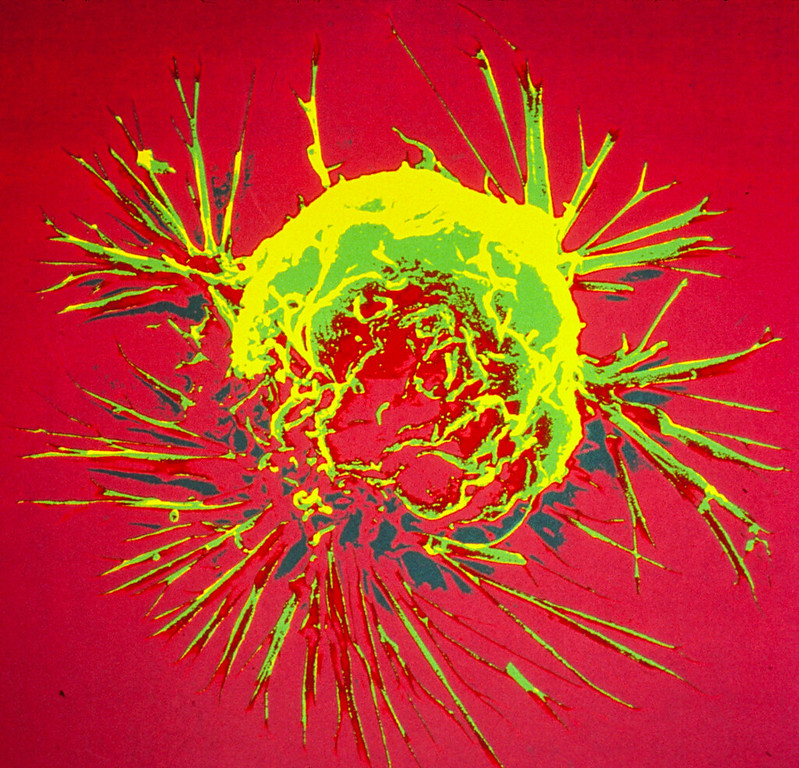
Scanning Electron Micrograph of a breast cancer cell. Credit: NIH
Researchers have found that bacteria lurking inside tumours promote cancer metastasis. They do so by enhancing the strength of host cells against mechanical stress in the bloodstream, promoting cell survival during tumour progression, researchers report in the journal Cell.
“Our study reveals that the cancer cell’s behaviour is also controlled by the microbes hiding inside tumours, the majority of which were originally thought to be sterile,” said senior author Shang Cai of the Westlake Laboratory of Life Sciences and Biomedicine. “This microbial involvement is distinct from the genetic, epigenetic, and metabolic components that most cancer drugs target.”
“However, our study does not mean that using antibiotics during cancer treatment will benefit patients,” he cautioned. “Therefore, it is still an important scientific question of how to manage the intratumor bacteria to improve cancer treatment in the future.”
It is known that microbes play a critical role in affecting cancer susceptibility and tumour progression, particularly in colorectal cancers. New evidence suggests however that, in a broad range of cancer types, they also form integral components of the tumour tissue itself, such as pancreatic cancer, lung cancer, and breast cancer. Microbial features are linked to cancer risk, prognosis, and treatment responses, yet the biological functions of tumour-resident microbes in tumour progression remain unclear.
Whether these microbes are actually drivers of tumour progression has been an intriguing question. “Tumour cells hijacked by microbes could be more common than previously thought, which underscores the broad clinical value of understanding the exact role of the tumour-resident microbial community in cancer progression,” Cai explained.
To find answers, Cai’s team utilised a mouse model of breast cancer with significant amounts of bacteria inside cells, similar to human breast cancer. The bacteria were found to be capable of travelling through the circulatory system with the cancer cells, playing critical roles in tumour metastasis. These passenger bacteria have the capacity to modulate the cellular actin network, promoting cell survival against mechanical stress in circulation.
“We were surprised initially at the fact that such a low abundance of bacteria could exert such a crucial role in cancer metastasis. What is even more astonishing is that only one shot of bacteria injection into the breast tumour can cause a tumour that originally rarely metastasises to start to metastasise,” Cai said. “Intracellular microbiota could be a potential target for preventing metastasis in broad cancer types at an early stage, which is much better than to have to treat it later on.”
While intratumour bacteria was found to have a clear role in promoting cancer cell metastatic colonisation, the authors did not exclude the possibility that the gut microbiome and immune system may act together with intratumour bacteria to determine cancer progression. Future in-depth analyses of how bacteria invade tumour cells, how intracellular bacteria are integrated into the host cell system, and how bacteria-containing tumor cells interact with the immune system will help inform how to properly deploy antibiotics in cancer treatment.
A recent analysis of published research found that more than half (59%) of cancer survivors and patients experience at least a moderate level of fear of cancer recurrence and that about one in five (19%) have a high level of fear. The findings, published in Psycho-Oncology, show women and younger people in particular have more fear of recurrence.
Cancer prevalence is increasing due to ageing populations, and more people are surviving cancer thanks to improved treatments. Managing fear of cancer recurrence (FCR) has been reported as one of the most important unmet needs for this growing group. FCR is defined as “fear, worry, or concern relating to the possibility that cancer will come back or progress”. Low levels of FCR can be helpful by promoting treatment compliance and healthy lifestyle adaptations. However, at clinical levels, FCR can limit quality of life and daily functioning and require professional help. Four features have been defined as key characteristics of clinical FCR: “(a) high levels of preoccupation; (b) high levels of worry; (c) that are persistent; and (d) hypervigilance to bodily symptoms”. It is important to address FCR, because FCR may also lead to increased healthcare costs14 and for most patients, it does not decrease over time without intervention.
The analysis, which is included 46 studies from 13 countries. Investigators found similar fear of cancer recurrence rates in survivors and patients. On average, younger people and women experienced more fear of cancer recurrence.
Additional research is needed to not only identify which patients desire support to address their fear of cancer recurrence but also to determine how to tailor interventions to different levels of fear and to individual needs and preferences.
“Knowing the prevalence and severity of fear of cancer recurrence for the general cancer population and for different subgroups is an important development, because it is essential for shaping healthcare provision, policy, and research on fear of cancer recurrence,” said lead author Yvonne Luigjes-Huizer, a PhD candidate at the Helen Dowling Institute and the University Medical Centre Utrecht, in the Netherlands.
Factors such as anaemia and anticoagulants have more impact on hospitalisation time after breast reconstruction than “common” risk factors according to a new study published in the Journal of Clinical Medicine.
The study investigated the impact of different factors on postoperative blood loss and drainage fluid volume, two factors which can lengthen hospitalisation time of patients after breast reconstruction after breast cancer surgery. The findings of the study allow for an improved risk assessment and planning of reconstructive breast surgery to offer patients personalised and improved treatment.
Partial or total mastectomy is often necessary in breast cancer surgery, and reconstructive breast surgery lessens the psychological stress on the patient. Fast wound healing after surgical breast reconstruction is crucial to not delay subsequent cancer treatments. Factors influencing the length of hospital stay (LOS) or wound healing are therefore particularly significant in cancer treatment. This study identified previously unrecognised risk factors.
Blood loss and drainage fluid volumes after breast reconstruction due to breast cancer were recorded, parameters which are closely linked to the healing process and LOS .Lower loss equals earlier patient discharge and early start of subsequent treatment. “We analysed factors that might affect blood loss and drainage fluid volumes after surgery – but can be identified before the surgery,” explained lead author Dr Tonatiuh Flores, plastic surgeon. “These factors included age, body mass index and smoking status – factors that are known to have a strong impact on the course of disease.” Additionally, haemoglobin levels and possible antithrombotic prophylaxis were reviewed – two parameters that are particularly significant in oncological treatment.
Surprising results emerged from the evaluation of a total of 257 breast reconstructions in 195 patients. Professor Konstantin Bergmeister, senior author of the study explained that “the classic risk factors did not significantly influence postoperative blood loss and drainage fluid output. Haemoglobin levels and anticoagulant concentration, however, did.” The analysis revealed a close relation between low haemoglobin values or anaemia and fluid loss after reconstructive breast surgery. Co-author Prof. Klaus Schroegendorfer, elaborated on this: “Especially breast cancer patients often show perioperative anaemia, caused by the frequently required neoadjuvant chemotherapy which can affect blood values, in particular haemoglobin.”
There were similar findings regarding low molecular heparin used in cancer patients as antithrombotic prophylaxis. Patients receiving heparin tended to have increased drainage fluid output after surgery, though the effect was not as strong as with perioperative anaemia.
The study authors recommend that, to cut LOS and continue the necessary cancer treatment after reconstructive breast surgery in cancer patients as early as possible, patients should preoperatively be screened for anaemia and administration of low molecular heparin should be adapted to the patients’ risk. In correlation to the results, follow-up treatment can be improved, patients can be discharged earlier and cancer treatment can be continued.
In mouse studies, researchers found that exercising prior to developing cancer was associated with slower tumour growth and helped reduce the effects of a cachexia, a cancer complication also known as wasting syndrome.
Cachexia is a metabolic wasting disorder that affects up to 80% of patients with advanced cancer and is associated with about a third of all deaths from cancer. Cachexia is characterised by severe progressive muscle wasting, a decline in heart structure and function and an overall poorer quality of life.
“Most exercise, especially aerobic exercise, is easily accessible and affordable,” said Louisa Tichy, a graduate student in Traci Parry’s lab at the University of North Carolina at Greensboro. “Therefore, engaging in consistent aerobic exercise such as running is a cost-effective way to reduce the risk of cancer and cancer complications.”
Tichy presented the research at the American Society for Investigative Pathology annual meeting.
Previous research has shown that exercise could have anti-inflammatory effects and might positively impact cancer cachexia by slowing its development and preserving cardiac structure and function. However, very few studies have focused on preconditioning.
“Our preclinical study indicated that preconditioning – or exercise prior to tumour bearing – appears to play an important cardioprotective role during cancer cachexia by preserving cardiac structure and function,” said Tichy. “It also helped stunt tumour growth, even when animals did not exercise during the tumour-bearing period.”
For the new study, the researchers studied mice that exercised on a treadmill for eight weeks and non-exercising mice. After the eight weeks, the researchers induced cancer in some of the exercised mice and some sedentary mice while keeping some mice from both groups cancer-free to act as controls.
Mice with cancer and a sedentary lifestyle were found to have poorer heart function – as measured with echocardiography – than the mice that exercised prior to cancer induction. Also, exercising mice had a smaller tumour volume and a 60% smaller tumour mass than the sedentary mice.
“This data is crucial in identifying the significance of exercise and the best timing of exercise as a protective and preventative measure against the detrimental effects of cancer cachexia,” said Tichy.
Understanding how the underlying proteins and pathways are affected by cancer and exercise can be used to inform exercise interventions. The researchers say that devising safe and effective exercise interventions for cancer patients would require evaluating the best intensity, duration and timing of exercise in preclinical models before human tests.
Breast cancer cells. Image source: National Cancer Institute on Unsplash
The natural compound cardamonin present in the spice cardamom and other plants could have therapeutic potential for triple-negative breast cancer, according to a new study using human cancer cells. The findings also show that the compound targets a gene that helps cancer cells elude the immune system.
Around 10–15% of breast cancers are triple-negative, which means they lack receptors for oestrogen or progesterone and don’t make excess amounts of a protein called HER2. These tumours are difficult to treat because they don’t respond to the hormone-based therapies used for other types of breast cancer. They also tend to be more aggressive and have a higher mortality rate than other breast cancers.
“It has been challenging to develop a targeted therapy for triple-negative breast cancer that is safe and effective at the same time,” said Assistant Professor Patricia Mendonca, PhD, of Florida A&M University. “Because of this, there is a critical need to investigate medicinal plants as a new way to combat this cancer.”
“The fact that cardamonin has been used for centuries as a spice and, more recently, as a supplement shows that its intake is safe and may bring health benefits,” said A/Prof Mendonca. “Our research shows that cardamonin holds potential for improving cancer therapy without as many side effects as other chemotherapeutic agents.”
For the new study, the researchers investigated how cardamonin affected the expression of the programmed cell death ligand 1 (PD-L1) gene, which is found in tumour cells. PD-L1 is overexpressed during breast cancer progression and plays a critical role in helping breast cancer cells evade the body’s immune system.
The researchers used two genetically different triple-negative breast cancer cell lines – one derived from women with African American ancestry and the other from women of European origin (Caucasian). They found that cardamonin treatment caused a dose-dependent decrease in cell viability in both cell lines. It also reduced PD-L1 expression in the Caucasian cell line but not the African American cell line, indicating that cells from different races may respond differently to cardamonin because of genetic variations among races.
“This is the first study to describe cardamonin’s inhibitory effect on the expression of PD-L1, which is relevant for the treatment of triple-negative breast cancer,” said Mendonca. “These findings add support to other research that has shown differences in the tumour microenvironment between African and non-African Americans.”
A recent study published in JAMA Surgery has demonstrated the viability of living-donor liver transplant for patients who have systemically controlled colorectal cancer and liver tumours that cannot be surgically removed.
“This study proves that transplant is an effective treatment to improve quality of life and survival for patients with colorectal cancer that metastasised to the liver,” said senior study author Dr Gonzalo Sapisochin.
The study focused on colorectal cancer partly for its tendency to spread to the liver. Nearly half of all patients with colorectal cancer develop liver metastases within a few years of diagnosis and 70% of liver tumours in these patients cannot be removed without removing the entire liver.
Unfortunately, most of these patients cannot get deceased-donor liver transplants because their liver function is fairly normal in spite of their tumours. This puts them near the bottom of the national organ transplant waiting list.
Thanks to recent advances in cancer treatments, many of these patients are able to get their cancer under systemic control, which means only their liver tumours prevent them getting a ‘cancer free’ label. It also increases the odds that these patients – and their new livers – will remain cancer free, which is crucial when balancing the benefit to the patient with the risk to a living donor.
“I’ve seen so many cancer patients, whose cancers were not spreading, but we couldn’t remove the tumours from their livers and we knew they would die,” said first study author Dr Roberto Hernandez-Alejandro. “We hoped living-donor liver transplant could give them another chance.”
Because it offered a last resort, the study attracted patients from near and far. All patients and donors went through a rigorous screening process to ensure they were good candidates for the procedure, and they were educated about the risks of the surgery and the possibility of cancer recurrence.
Patients and donors underwent staggered surgeries to fully remove patients’ diseased livers and replace them with half of their donors’ livers. Over time, both patients’ and donors’ livers regenerated and regain normal function.
Patient imaging and blood analysis was closely monitored for any signs of cancer recurrence and will continue to be followed for up to five years after their surgery. At the time of study publication, two patients had follow-up of two or more years and both remained alive and well, cancer-free.
“We have seen very good outcomes with this protocol, with 100 percent survival and 62 percent of patients remaining cancer free one year and a half after surgery,” said study author Dr Mark Cattral. “It is very strong data to support that we can offer this treatment safely and make appropriate use of scarce life-saving organs.”
An observational study with over 100 000 participants suggests that some artificial sweeteners are associated with increased cancer risk. The findings, published in PLOS Medicine, reflect other results from experimental studies.
The safety of artificial sweeteners has long been a subject of debate. To evaluate the potential carcinogenicity of artificial sweeteners, researchers analysed data from 102 865 French adults participating in the NutriNet-Santé study. The NutriNet-Santé study is an ongoing web-based cohort initiated in 2009 by the Nutritional Epidemiology Research Team (EREN). Participants enrolled voluntarily, and self-reported their medical history, sociodemographic, diet, lifestyle, and health data.
Researchers gathered data concerning artificial sweetener intake from 24-hour dietary records. After collecting cancer diagnosis information during follow-up, the researchers conducted statistical analyses to investigate the associations between artificial sweetener intakes and cancer risk. They also adjusted for a range of variables including age, sex, education, physical activity, medical history and dietary intake.
The researchers found that participants who consumed larger quantities of artificial sweeteners, particularly aspartame and acesulfame-K, had higher risk of overall cancer compared to non-consumers (hazard ratio 1.13). Higher risks were observed for breast cancer and obesity-related cancers.
In addition to being an observational study, there were a number of limitations; dietary intakes are self-reported. Selection bias may also have been a factor, as participants were more likely to be women, to have higher educational levels, and be more health-conscious. Additional research will be required to confirm the findings and clarify the underlying mechanisms.
According to the authors, “Our findings do not support the use of artificial sweeteners as safe alternatives for sugar in foods or beverages and provide important and novel information to address the controversies about their potential adverse health effects. While these results need to be replicated in other large-scale cohorts and underlying mechanisms clarified by experimental studies, they provide important and novel insights for the ongoing re-evaluation of food additive sweeteners by the European Food Safety Authority and other health agencies globally”.
First author Charlotte Debras added: “Results from the NutriNet-Santé cohort (n = 102 865) suggest that artificial sweeteners found in many food and beverage brands worldwide may be associated with increased cancer risk, in line with several experimental in vivo / in vitro studies. These findings provide novel information for the re-evaluation of these food additives by health agencies.”