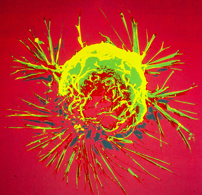
Human colon cancer cells. Credit: National Cancer Institute
Tumours often contain areas of oxygen-deficient tissue that frequently withstand conventional therapies. This is because the drugs applied in tumours require oxygen to be effective. An international research team has developed a novel mechanism of action that works without oxygen: polymeric incorporated nanocatalysts target the tumour tissue selectively and switch off the glutathione that the cells need to survive. The team published their findings in the journal Nature Communications.
Why tumours shrink but don’t disappear
Study leader Dr Johannes Karges from Ruhr University Bochum, Germany, explained: “As tumours grow very quickly, consume a lot of oxygen and their vascular growth can’t necessarily keep pace, they often contain areas that are poorly supplied with oxygen.” These areas, often in the centre of the tumour, frequently survive treatment with conventional drugs, so that the tumour initially shrinks but doesn’t disappear completely. This is because the therapeutic agents require oxygen to be effective.
The mechanism of action developed by Karges’ team works without oxygen. “It’s a catalyst based on the element ruthenium, which oxidises the naturally present glutathione in the cancer cells and switches it off,” explains Karges. Glutathione is essential for the survival of cells and protects them from a wide range of different factors. If it ceases to be effective, the cell deteriorates.
Compound accumulates in tumour tissue
All cells of the body need and contain glutathione. However, the catalyst has a selective effect on cancer cells as it is packaged in polymeric nanoparticles that accumulate specifically in the tumour tissue. Experiments on cancer cells and on mice with human tumours, that were considered incurable, proved successful. “These are encouraging results that need to be confirmed in further studies,” concludes Johannes Karges. “Still, there’s a lot of research work to be done before it can be used in humans.”
US county-level data point to specific pesticides that may increase prostate cancer incidence and death.
Photo by Arjun Mj on Unsplash
Researchers have identified 22 pesticides consistently associated with the incidence of prostate cancer in the United States, with four of the pesticides also linked with prostate cancer mortality. The findings are published by Wiley online in CANCER, a peer-reviewed journal of the American Cancer Society.
To assess county-level associations of 295 pesticides with prostate cancer across counties in the United States, investigators conducted an environment-wide association study, using a lag period between exposure and prostate cancer incidence of 10–18 years to account for the slow-growing nature of most prostate cancers. The years 1997–2001 were assessed for pesticide use and 2011–2015 for prostate cancer outcomes. Similarly, 2002–2006 were analysed for pesticide use and 2016–2020 for outcomes.
Among the 22 pesticides showing consistent direct associations with prostate cancer incidence across both time-based analyses were three that had previously been linked to prostate cancer, including 2,4-D, one of the most frequently used pesticides in the United States. The 19 candidate pesticides not previously linked to prostate cancer included 10 herbicides, several fungicides and insecticides, and a soil fumigant.
Four pesticides that were linked to prostate cancer incidence were also associated with prostate cancer mortality: three herbicides (trifluralin, cloransulam-methyl, and diflufenzopyr) and one insecticide (thiamethoxam). Only trifluralin is classed by the Environmental Protection Agency as a “possible human carcinogen,” whereas the other three are considered “not likely to be carcinogenic” or have evidence of “non-carcinogenicity.”
“This research demonstrates the importance of studying environmental exposures, such as pesticide use, to potentially explain some of the geographic variation we observe in prostate cancer incidence and deaths across the United States,” said lead author Simon John Christoph Soerensen, MD, of Stanford University School of Medicine. “By building on these findings, we can advance our efforts to pinpoint risk factors for prostate cancer and work towards reducing the number of men affected by this disease.”
A new study published in BJU International has found that the conventional, versatile open surgery approach to removal of the lymph nodes behind the intestines for patients whose testicular cancer has not advanced beyond the abdomen is the gold standard of care for men in this condition.
Open surgery involves making an incision on the abdomen for a direct view and access to the surgical area. In the appropriate patients, cancer cure rates are excellent with this surgery alone due to continued clinician experience and refinement of technique, particularly at high-volume centres.
The study, led by urologist and health services researcher Clint Cary, MD, MPH, MBA, of the Indiana University School of Medicine and the Regenstrief Institute, was conducted using information from the on 165 patients with clinical stage I or II testicular cancer and no prior chemotherapy. One of the highest-volume testicular cancer treatment groups in the US, IU School of Medicine’s Department of Urology is among those groups setting the bar for both better surgical results and fewer negative outcomes such as infertility. Study benchmarks included low blood loss, short hospital stay and rare major postoperative complications.
Dr Cary notes that the paper, which includes analysis of risk factors for complications, clearly indicates that prior to treatment, men and their families should have conversations with their local urologist about their experience level across risk factors, such as high body mass index (BMI), as well as have detailed discussions of complication rates. He adds that both patients and clinicians can use the findings of this paper as a bar for comparison as they make choices.
“In my clinical role at Indiana University in the operating room, I frequently perform lymph node dissections for men with a lower burden of metastatic cancer who have not had chemotherapy or may never receive it for this cancer. In my complementary role as a Regenstrief Institute health services researcher, I study the impact of clinical decisions,” said Dr Cary. “For this study, I wanted to know that this and other treatments I perform are making a difference and I want to know – whether we are continuing established treatments or introducing new approaches to surgery – how these decisions and the resulting care will impact patients’ quality of life and their cancer outcomes.
“This study is an example of how the benefit of providing clinical care and also being in a research environment enables us to learn from what we’re doing and to report it in a manner that is meaningful to both patients and physicians. We’re always looking for ways to improve upon surgical outcomes. Going beyond the scope of this paper, we are now expanding our work to begin a randomised trial comparing two surgical techniques for removing lymph nodes to understand if one is better than the other regarding postoperative recovery.”
The paper did not compare robot-assisted surgery with traditional open surgery, which may allow a more complete lymph node dissection. While robotic surgery in general offers potential benefits to both surgeons and patients, the authors note that the head to head comparison between these two approaches is limited due to small experiences with the robotic approach in testicular cancer.
The authors conclude that for patients undergoing removal of lymph nodes for testicular cancer and certainly more complex and challenging patients at higher risk of post-operative complications, the traditional open surgery remains the most effective approach and the gold standard.
Obesity and type 2 diabetes are risk factors for various malignancies, including pancreatic cancer, which has a high death rate. A new analysis in Diabetes/Metabolism Research and Reviews suggests that metabolic-bariatric surgery may lower the risk of developing pancreatic cancer in people with obesity, especially in those who also have type 2 diabetes.
In the systematic review and meta-analysis, investigators identified 12 relevant studies that explored the effects of metabolic-bariatric surgery on pancreatic cancer incidence, with a total of 3 711 243 adults with obesity. Surgery was associated with a 44% reduction in pancreatic cancer risk among individuals with obesity but without type 2 diabetes and a 79% risk reduction in those with both obesity and type 2 diabetes.
“Metabolic-bariatric surgery not only has beneficial effects on obesity and type 2 diabetes but also may play a crucial role in reducing the risk of pancreatic cancer in these individuals,” said corresponding author Angeliki M. Angelidi, PhD, of the Broad Institute of MIT and Harvard. “These findings underscore the need for further research to elucidate the underlying mechanisms and understand the full spectrum of health benefits of metabolic-bariatric surgery beyond weight loss.”
Clinical trial reveals improved self-reported cognitive function in women with breast cancer who started an exercise program when initiating chemotherapy.
Photo by Ketut Subiyanto on Pexels
Many women who receive chemotherapy experience a decreased ability to remember, concentrate, and/or think – commonly referred to as “chemo-brain” or “brain fog” – both short- and long-term. In a recent clinical trial of women initiating chemotherapy for breast cancer, those who simultaneously started an aerobic exercise program self-reported greater improvements in cognitive function and quality of life compared with those receiving standard care. The findings are published by Wiley online in CANCER, a peer-reviewed journal of the American Cancer Society.
The study, called the Aerobic exercise and CogniTIVe functioning in women with breAsT cancEr (ACTIVATE) trial, included 57 Canadian women in Ottawa and Vancouver who were diagnosed with stage I–III breast cancer and beginning chemotherapy. All women participated in 12–24 weeks of aerobic exercise: 28 started this exercise when initiating chemotherapy and 29 started after chemotherapy completion. Cognitive function assessments were conducted before chemotherapy initiation and after chemotherapy completion (therefore, before the latter group started the exercise program).
Women who participated in the aerobic exercise program during chemotherapy self-reported better cognitive functioning and felt their mental abilities improved compared with those who received standard care without exercise. Neuropsychological testing – a performance-based method used to measure a range of mental functions – revealed similar cognitive performance in the two groups after chemotherapy completion, however.
“Our findings strengthen the case for making exercise assessment, recommendation, and referral a routine part of cancer care; this may help empower women living with and beyond cancer to actively manage both their physical and mental health during and after treatment,” said lead author Jennifer Brunet, PhD, of the University of Ottawa.
Dr Brunet noted that many women undergoing chemotherapy for breast cancer remain insufficiently active, and there are limited exercise programs tailored to their needs. “To address this, we advocate for collaboration across various sectors – academic, healthcare, fitness, and community – to develop exercise programs specifically designed for women with breast cancer,” she said. “These programs should be easy to adopt and implement widely, helping to make the benefits of exercise more accessible to all women facing the challenges of cancer treatment and recovery.”
Credit: Darryl Leja National Human Genome Research Institute National Institutes Of Health
In a peer-reviewed study believed to be the first of its kind published, a research team led by Johns Hopkins Medicine provides scientific evidence that a healthy diet may reduce the chance of low risk prostate cancer progressing to a more aggressive state in men undergoing active surveillance – a clinical option in which men with lower risk cancer are carefully monitored for progression in lieu of treatments that could have undesired side effects or complications.
The findings are reported in the journal JAMA Oncology.
“Many men diagnosed with low grade prostate cancer are interested in changes they can make to reduce the risk of their tumour becoming more aggressive, and the role of diet and nutrition is one of the most commonly asked questions,” says study co-senior author Bruce Trock, PhD, a professor of urology, epidemiology and oncology at the Johns Hopkins University School of Medicine, and director of the Brady Urological Institute’s epidemiology division. “These men are motivated to make changes that may improve their prognosis, which is why we began collecting data on their diets, lifestyles and exposures 20 years ago. Hopefully, these latest findings will enable us to develop some concrete steps they can take to reduce the risk of cancer progression.”
When a patient is found after a biopsy to have developed prostate cancer, the sampled cells are assigned to a grade group based on comparison normal prostate tissue. Grade groups range from 1 to 5, with grade group 1 indicating indolent cancer cells that don’t look very different than normal tissue and do not metastasise. At the other end of the scale, grade group 5 indicates cancer cells that are quite abnormal in appearance, and can grow and metastasise if untreated
During active surveillance, biopsies are performed at regular intervals to see if prostate cancer should be move it to a higher grade group. Called grade reclassification, this often leads to a recommendation for treatment. It also is a common way for researchers to evaluate the effectiveness of therapies and lifestyle modifications.
“While there have been previous research studies looking at diet and its relationship to prostate cancer, we believe that ours is the first to provide statistically significant evidence that a healthy diet is associated with a reduction in risk of prostate cancer progressing to a higher grade group, as shown by a reduction in the percentage of men on active surveillance experiencing grade reclassifications over time,” says study co-senior author Christian Pavlovich, MD, a professor in urologic oncology at the Johns Hopkins University School of Medicine and director of the Brady Urological Institute’s prostate cancer active surveillance program.
In the newly published study, the researchers prospectively evaluated the histories of 886 men (median age at diagnosis: 66) diagnosed with grade group 1 prostate cancer from January 2005 to February 2017, all of whom were in the Johns Hopkins Medicine active surveillance program and whom, at the time of enrolment, completed a validated food frequency survey regarding their usual dietary patterns. Of the participants, 55 were Black (6.2%), 803 (90.6%) were white and 28 (3.2%) identified as other races and ethnicities.
Based on their responses to the questionnaire, a Healthy Eating Index (HEI) score was calculated for each patient. The HEI ranges from 0 to 100.
“The HEI is a validated measure of overall diet quality, quantifying how well an individual’s dietary pattern adheres to the recommendations of the U.S. Department of Agriculture’s Dietary Guidelines for Americans,” says study lead author Zhuo Tony Su, MD. “We looked at each patient’s HEI score – as calculated from their dietary information recorded at enrolment in our active surveillance programme – and assessed whether men with a higher quality diet were less likely to experience grade reclassification in the years afterward.”
Su says the researchers also evaluated the patients using an energy-adjusted HEI (E-HEI) score that takes into account a person’s daily caloric intake.
Along with those two metrics, Su says, the researchers calculated scores for each study participant using the Dietary Inflammatory Index (DII) and the energy-adjusted DII (E-DII).
“The DII and E-DII scores assess the inflammatory or anti-inflammatory potential of any diet, so higher scores indicate a diet that may cause more inflammation, which in turn, may contribute to the development and progression of prostate cancer,” says Su. “We evaluated whether higher inflammatory potential was associated with increased risk of grade reclassification.”
By a follow-up assessment at 6.5 years after diagnosis, 187 men (21%) had been reclassified as grade group 2 or greater, of whom 55 (6%) had extreme grade reclassification to grade group 3 or greater.
“When our team looked at the HEI and E-HEI scores in relation to the grade reclassification rates, we found a statistically significant inverse association between adherence to a high quality diet – as indicated by high HEI and E-HEI scores – and the risk of grade reclassification during active surveillance,” says Trock. “In other words, the higher the HEI and E-HEI scores, the more reduced the risk that a low grade prostate cancer had progressed to a higher grade disease that mandated curative treatment.”
Pavlovich says for patients adhering to a high quality diet, every increase of 12.5 points in the HEI score was associated with an approximately 15% reduction in reclassification to grade group 2 or greater, and a 30% reduction in reclassification to grade group 3 or greater.
The researchers say their findings also indicate that lower inflammation potential is among several possible risk lowering mechanisms as a result of a higher quality diet. However, they did not find an association between grade reclassification and baseline DII/E-DII scores.
“This lack of association with DII/E-DII may indicate that inflammation plays a role in driving the progression from a healthy prostate to one with cancer,” says Trock. “Whereas, in men who already have prostate cancer, the more subtle biological change from a lower to higher grade may reflect other mechanisms potentially influenced by diet.”
The researchers report several limitations in their study, including diet data based on patient self-reporting, results subject to potential nonresponse bias (bias occurring when respondents and nonrespondents differ in ways that impact the research, making the sample population less representative of the whole population) and not accounting for dietary changes over time. Additionally, they say the study population, consisting predominantly of white men with grade group 1 disease at diagnosis, may not be generalisable to all patients.
“Our findings-to-date should be helpful for the counselling of men who choose to pursue active surveillance and are motivated to modify their behaviours, including quality of diet,” says Pavlovich. “However, to truly validate the association between higher quality diet and reduced risk of prostate cancer progression, future studies with more diverse populations are needed.”
Colourised scanning electron micrograph of a breast cancer cell. Credit: NIH
Researchers from Tampere University, Finland, and Izmir Institute of Technology, Turkey, have developed an in vitro cancer model to investigate why breast cancer spreads to bone. Their findings, published in PLOS One, hold promise for advancing the development of preclinical tools to predict breast cancer bone metastasis.
Breast canceris a significant global public health challenge, with 2.3 million new cases and 700 000 deaths every year. Approximately 80% of patients with primary breast cancer can be cured, if they are diagnosed and treated promptly. However, in many cases, the cancer has already metastasised at the time of diagnosis.
Metastatic cancer is incurable and accounts for more than 90% of cancer-related deaths. Currently, there are no reliable in vitro models to study how breast cancer spreads to secondary organs such as bone, lung, liver or brain. Now, researchers from the Precision Nanomaterials Group at Tampere University in Finland, and the Cancer Molecular Biology Lab at Izmir Institute of Technology in Turkey, have used lab-on-a-chip platforms to create a physiologically relevant metastasis model to study the factors controlling breast cancer bone metastasis.
“Breast cancer most frequently spreads to bone, with an estimated rate of 53%, resulting in severe symptoms such as pain, pathological bone fractures, and spinal cord compressions. Our research provides a laboratory model that estimates the likelihood and mechanism of bone metastasis occurring within a living organism. This advances the understanding of molecular mechanisms in breast cancer bone metastasis and provides the groundwork for developing preclinical tools for predicting bone metastasis risk,” says Burcu Firatligil-Yildirir, postdoctoral researcher at Tampere University and the first author of the paper.
According to Nonappa, Associate Professor and leader of the Precision Nanomaterials Group at Tampere University, developing sustainable in vitro models that mimic the complexity of the native breast and bone microenvironment is a multidisciplinary challenge.
“Our work shows that physiologically relevant in vitro models can be generated by combining cancer biology, microfluidics and soft materials. The results open new possibilities for developing predictive disease, diagnostic and treatment models,” he says.
Plant fungus provides new drug with a new cellular target
Human colon cancer cells. Credit: National Cancer Institute
Novel chemical compounds from a fungus could provide new perspectives for treating colorectal cancer, one of the most common and deadliest cancers worldwide. The fungus, Bipolaris victoriae, is otherwise known as a fungal plant pathogen which in the 1940s caused the “Victoria blight”, decimating oats and similar grains in the US.
In the journal Angewandte Chemie, researchers reported on the isolation and characterisation of a previously unknown class of metabolites (terpene-nonadride heterodimers). One of these compounds effectively kills colorectal cancer cells by attacking the enzyme DCTPP1, which thus may serve as a potential biomarker for colorectal cancer and a therapeutic target.
Rather than using conventional cytostatic drugs, which have many side effects, modern cancer treatment frequently involves targeted tumour therapies directed at specific target molecules in the tumour cells. The prognosis for colorectal cancer patients remains grim however, demanding new targets and novel drugs.
Targeted tumour therapies are mostly based on small molecules from plants, fungi, bacteria, and marine organisms. About half of current cancer medications were developed from natural substances. A team led by Ninghua Tan, Yi Ma, and Zhe Wang at the China Pharmaceutical University (Nanjing, China) chose to use Bipolaris victoriae S27, a fungus that lives on plants, as the starting point in their search for new drugs.
The team first analysed metabolic products by cultivating the fungus under many different conditions (OSMAC method, one strain, many compounds). They discovered twelve unusual chemical structures belonging to a previously unknown class of compounds: terpene-nonadride heterodimers, molecules made from one terpene and one nonadride unit. Widely found in nature, terpenes are a large group of compounds with very varied carbon frameworks based on isoprene units. Nonadrides are nine-membered carbon rings with maleic anhydride groups. The monomers making up this class of dimers termed “bipoterprides” were also identified and were found to contain additional structural novelties (bicyclic 5/6-nonadrides with carbon rearrangements).
Nine of the bipoterprides were effective against colorectal cancer cells. The most effective was bipoterpride No. 2, which killed tumour cells as effectively as the classic cytostatic drug Cisplatin. In mouse models, it caused tumours to shrink with no toxic side effects.
The team used a variety of methods to analyse the drug’s mechanism: bipoterpride 2 inhibits dCTP-pyrophosphatase 1 (DCTPP1), an enzyme that regulates the cellular nucleotide pool. The heterodimer binds significantly more tightly than each of its individual monomers. The activity of DCTPP1 is elevated in certain types of tumours, promoting the invasion, migration, and proliferation of the cancer cells while also inhibiting programmed cell death. It can also help cancer cells to resist treatment. Bipoterpride 2 inhibits this enzymatic activity and disrupts the now pathologically altered amino acid metabolism in the tumour cells.
The team was thus able to identify DCTPP1 as a new target for the treatment of colorectal cancer and bipoterprides as new potential drug candidates.
Pretoria, 16 October 2024: A group of African women leaders have come together to form the Africa Breast Cancer Council, with the support of Roche, one of the world’s largest biotech companies.
The Africa Breast Cancer Council is a response to the growing burden of breast cancer on the African continent. Breast cancer is the most commonly diagnosed cancer in women in Africa and causes the most cancer-related deaths. Only one in two women in sub-Saharan Africa are currently expected to survive for five years after receiving a diagnosis.
The Council will leverage their extensive, diverse and complementary experience to guide and drive policy change in their home countries and across the continent. They will encourage increased collaboration, better data collection and improved patient outcomes. The Council’s work will seek to foster systemic improvements in healthcare infrastructure, access, and funding, driving sustainable, scalable impact for breast cancer care. The Council will focus initially on African healthcare systems which are open to partnership and innovation, then move to scale this work across the continent.
Wendy Cupido, Africa Breast Cancer Council Co-chair and General Manager at Roche South Africa and Sub Region said: “Every woman on this Council has a day job that plays an important role in the fight against breast cancer. Our aim in coming together is to channel our knowledge, expertise, relationships and energy into a collective voice, and a collective force, to focus on significant areas of concern.”
Dr Magda Robalo, Africa Breast Cancer Council member, and President and Co-founder of the Institute for Global Health and Development said: “Most African women with breast cancer are diagnosed too late and, even after a diagnosis, many do not receive the treatment they need. This egregious injustice is utterly preventable. In response, the Africa Breast Cancer Council will work to shape policies and advocate for governments to urgently commit the resources needed.”
It can take more than six months for women in Africa to receive a breast cancer diagnosis after noticing symptoms, due in part to healthcare system inefficiencies and limited access to specialised care. This contributes to 60-70% of African women receiving a diagnosis in the late stage, reducing chances of survival and increasing cancer care costs with more expensive therapies and longer hospital stays. This is exacerbated by a lack of awareness of the importance of breast self-examinations, along with widespread stigma surrounding breast cancer treatment. In light of this need, the Council will work at a national level to reduce the time to diagnosis from six months to 60 days.
In South Africa, the average three-year survival rate for breast cancer is below 45%. This is due in part to poor uptake of early screening and detection for breast cancer, exacerbated by significant stigma, leading to late-stage diagnosis and lower survival rates. Lengthy regulatory pathways and a lack of ring-fenced budget for cancer care also present barriers to progress.
Dr Miriam Mutebi, Africa Breast Cancer Council member and Breast Cancer Surgical Oncologist said: “This Council has chosen to launch during Breast Cancer Awareness Month, a recognised annual global movement to drive awareness, early detection and improved outcomes. We are using this month to start raising our collective voice, alongside others committed to reducing the unacceptable toll of breast cancer on African women.”
I was diagnosed with breast cancer on an ordinary Thursday afternoon in February 2023. I was 34 years old. The December before, my GP had performed a breast exam as part of a general check-up and was concerned that with my dense breast tissue she might be missing something. She wanted me to have an ultrasound, but there was no rush. Her exact words to me were something like: “Don’t worry, it can wait until you have medical aid savings again in January.”
The ultrasound turned up a small shadow, just a centimetre in diameter – something that could be a cyst, but the radiologist thought we should do a mammogram “just in case”. Would I mind waiting? No, I wouldn’t mind. The mammogram was worrying enough that she got approval to do a biopsy the next day. “Just in case”. The results came in the following week.
I had none of the risk factors for breast cancer. I didn’t drink, didn’t smoke, didn’t have any family members with a history of breast cancer, was nowhere near the age of 50. A few months later, I would find out I had none of the genetic markers which can predict risk either – not only did I test negative for the genes associated with breast cancer called BRCA 1 and 2, I didn’t have any of the genes connected with any kind of cancer at all.
As I say, I was diagnosed on a Thursday afternoon. I had my first appointment with an oncologist that Friday morning. I had my first set of scans two days later on Monday and my initial surgery the following Friday. I started chemotherapy treatment within three weeks of first having the word “cancer” used in relation to my body. My doctors moved quickly because they had to. On a scale of 1 to 9 on something called the Bloom and Richardson classification, my cancer was a 9. So, even though I was only stage 1, I was also a grade 3. “Aggressive” doesn’t begin to cover it.
During this time, I held onto five facts. First, we had caught the tumour at exactly the right time. Had I gone in for screening any earlier, we might not have found the cancer yet. Had I gone any later, it likely would have grown and spread to my lymph nodes and other parts of my body and I might have needed more radical treatment and surgeries. Second, it was treatable. My particular kind of cancer ought to respond well to a combination of chemotherapy and radiation. Third, I was otherwise very healthy, aside from the cancer. Fourth, I had a medical aid which was covering almost everything I needed. And, most importantly, fifth, I had a wonderful support system of my partner and his family and our close friends to rely on.
From the beginning, I had an incredible standard of care. To the point where the doctors I saw had heated examination beds – they didn’t want their patients to experience any additional discomfort and distress during such a difficult time. And it was difficult. Chemotherapy and immunotherapy left me feeling battered and broken. Nausea, intense muscular pain, fatigue, vomiting, diarrhoea, constipation, weight gain, hair loss, brain fog, depression – some of the awful side effects it’s impossible to really prepare for. In fact, I had such a hard time mentally during treatment that at one point I had to be hospitalised.
The same day I received my diagnosis, I overheard a woman in my doctor’s office asking if it was possible to make a payment plan for her treatment. The administrators replied that treatment was likely to cost in excess of R300 000 at a minimum. I cannot even begin to imagine having to go into debt to fight off cancer. For treatment that makes you feel more than just sick, more like you’re dying. For treatment that may not necessarily work.
But this is the choice that faces most people with cancer in our country. With a relatively small number of people on comprehensive medical aids with screening benefits and prescribed minimum benefits, many face waiting for treatment in government facilities or running up huge bills at private clinics.
According to the most recent report by Statistics SA, breast cancer is the most commonly diagnosed cancer in women in South Africa, accounting for 23% of all cancers. It is also one of the most deadly, representing 17% of cancer deaths in women, just behind cervical cancer.
The Stats SA report lists “awareness of the symptoms and need for screening” as the main intervention to reduce the risk of death by breast cancer. The report also draws attention to the discrepancy in mortality rates in different population groups. For example, Coloured women have a relatively low incidence of breast cancer, but a high mortality rate – meaning that they are dying of breast cancer after being diagnosed too late. Stats SA points out that this is likely due to “poor access to cancer treatment facilities” as well as a lack of medical aid coverage. It is perhaps unsurprising that Black and Coloured women are the groups least likely to have medical aid in South Africa.
There are also some NGOs trying to step in to fill the gaps, like the aptly named I Love Boobies or the PinkDrive. These organisations make it their mission to give women a fighting chance to beat breast cancer. They provide free screenings to women around the country who would otherwise not be able to afford this necessary medical care.
I am one of the lucky ones. I officially went into remission on 30 August 2023 when I had a lumpectomy to remove the tumour in my right breast. Remission means that the cancer can no longer be detected in your body through scans and blood tests. It doesn’t mean you’re “cured”. There could still be cancerous cells in the body, which is why cancer is also often treated with radiation like mine was. Some people prefer not to use the term “survivor” until they have been in remission for over five years.
Five years is an important milestone for many people diagnosed with cancer. It’s often the period in which someone is most likely to suffer a relapse. I live with the possibility that my cancer will come back every day; I am reminded by my scars and by the fact that I am still recovering physically and mentally from a traumatic year. I still battle with periods of fatigue and depression and I will never be the same person I was before falling ill.
Still, remission is better than relapse. So far, so good. I continue to see my myriad of doctors every few months for scans and tests and examinations to check that nothing has come back yet and I feel like I’m getting stronger.
Almost a year to the day after I went into remission, my fiancé and I ran the Johannesburg Women’s Race in support of the PinkDrive. A mobile health unit was parked on the field in Mark’s Park offering free screenings all morning, which women were queuing up to access after the run. The festive atmosphere was bittersweet to me. Certainly, some of the women in that line would not know that they were starting on a long and painful journey, a journey which sometimes feels like it has no end. Hopefully, they would be starting early enough to be given a chance to become a survivor.
There’s another meaning of “remission” I wasn’t aware of until I looked it up. It can also be defined as “a cancellation of debt”. No-one with cancer should have to crowdfund in order to get treatment, but that is the reality we are faced with in our country. This October, I encourage everyone to contribute in whatever way they can to a cancer survivor’s remission. Join the Imagine Challenge, try a secret swim, pick up a pink bottle of milk or a scrunchie, support someone raising funds on GivenGain, get yourself examined. Every one of us can join the fight against breast cancer.