In a new study published in the American Journal of Transplantation, researchers report that they successfully transplanted two kidneys from a genetically modified pig into a human recipient who had suffered brain death.
The use of pig organs, genetically modified to enable transplantation into humans, could ease the shortage of available donor organs for transplantation and prevent thousands of deaths that result each year due to a shortage of organs. Recently, a US man became the first human recipient of a genetically modified pig heart.
This study used a novel preclinical human model to answer numerous critical safety questions. No hyperacute rejection was seen for the 74 hours until termination. No chimerism or transmission of porcine retroviruses was detected. Longitudinal biopsies revealed thrombotic microangiopathy that did not progress in severity, without evidence of cellular rejection or deposition of antibody or complement proteins. Although the xenografts produced variable amounts of urine, creatinine clearance did not recover. Whether renal recovery was impacted by the milieu of brain death and/or microvascular injury remains unknown.
The study provides important insights and identifies several areas where additional research is needed before xenotransplantation can be used to help address the current organ shortage.
“This study provides knowledge that could not be generated in animal models and moves us closer to a future where organ supply meets the tremendous need,” said senior author Jayme E. Locke, MD, MPH, of the University of Alabama at Birmingham.
Among 25 183 women aged 50 to 79 years, there were 1455 cases of hospitalisation for heart failure during a median follow-up of 16.9 years. Compared with women who walked at a casual pace, those who walked at an average pace or fast pace had 27% and 34% lower risks of heart failure, respectively.
Fast walking for less than 1 hour per week was associated with the same risk reduction of heart failure as average or casual walking for more than 2 hours per week.
“This study confirms other studies demonstrating the importance of walking speed on mortality and other cardiovascular outcomes,” said senior author Charles B. Eaton, MD, MS, of the Warren Alpert Medical School of Brown University. “Given that limited time for exercise is frequently given as a barrier to regular physical activity, walking faster but for less time might provide similar health benefits as the recommended 150 minutes per week of moderate physical activity.”
Further study is warranted to determine whether interventions to increase the walking pace in older adults will reduce heart failure risk and whether fast pace will compensate for the short duration of walking.
A five year-long randomised, placebo-controlled study found that in older adults taking vitamin D supplements, alone or with omega-3 fatty acids, the risk of developing autoimmune disease was reduced.
Autoimmune diseases (AD) such as rheumatoid arthritis, polymyalgia rheumatica, autoimmune thyroid disease and psoriasis, are a leading cause of morbidity and mortality as people age. Few effective treatments are available for AD, but some research has hinted that supplements, including vitamin D and omega-3 fatty acids, could have beneficial effects.
In a new study published in the BMJ, investigators from Brigham and Women’s Hospital evaluated whether taking vitamin D and/or omega fatty acid supplements could affect rates of AD. The large-scale vitamin D and Omega-3 Trial (VITAL), a randomised study which followed participants for approximately five years. Taking vitamin D, or vitamin D and omega-3 fatty acids had a significantly lower rate of AD than placebo.
“It is exciting to have these new and positive results for non-toxic vitamins and supplements preventing potentially highly morbid diseases,” said senior author Karen Costenbader, MD, MPH. “This is the first direct evidence we have that daily supplementation may reduce AD incidence, and what looks like a more pronounced effect after two years of supplementation for vitamin D.”
“Now, when my patients, colleagues, or friends ask me which vitamins or supplements I’d recommend they take to reduce risk of autoimmune disease, I have new evidence-based recommendations for women aged 55 years and older and men 50 years and older,” said Dr Costenbader. “I suggest vitamin D 2000 IU a day and marine omega-3 fatty acids (fish oil), 1000 mg a day – the doses used in VITAL.”
VITAL included 25 871 participants, with men aged 50 and older and women aged 55 and older, conducted to investigate whether taking vitamin D3 and/or omega-3 supplements could reduce the risk for developing cancer, heart disease and stroke in people who do not have a prior history of these illnesses. Prior to the launch of VITAL, investigators determined that they would also look at rates of AD among participants, as part of an ancillary study.
Participants answered questionnaires about new diagnoses of diseases, including rheumatoid arthritis, polymyalgia rheumatica, autoimmune thyroid disease, psoriasis and inflammatory bowel disease, with space to write in all other new onset ADs. Medical records were reviewed to confirm reported diagnoses.
“Autoimmune diseases are common in older adults and negatively affect health and life expectancy. Until now, we have had no proven way of preventing them, and now, for the first time, we do,” said first author Jill Hahn, ScD, post-doctoral fellow at the Brigham.
Among patients randomised to vitamin D, 123 participants in the treatment group and 155 in the placebo group were diagnosed with confirmed AD (22 percent reduction). Among those in the fatty acid arm, confirmed AD occurred in 130 participants in the treatment group and 148 in the placebo group. Omega-3 fatty acids alone did not significantly lower incidence of AD, but there was some evidence of a decrease over long periods.
Using a mix of drugs and a regenerative seal, scientists were able to successfully regrow frog legs, as reported in Science Advances. This represents an eventual step towards possibly regrowing limbs in humans.
On adult frogs, which are naturally unable to regenerate limbs, the researchers were able to trigger regrowth of a lost leg using a five-drug cocktail applied in a silicone wearable bioreactor dome that seals in the treatment over the stump for just 24 hours. That brief treatment sets in motion an 18-month period of regrowth that restores a functional, near-complete leg.
In humans and mammals loss of a large and structurally complex limb cannot be restored by any natural process of regeneration in humans or mammals. In fact, we tend to cover major injuries with an amorphous mass of scar tissue, protecting it from further blood loss and infection and preventing further growth.
The Tufts University researchers triggered the regenerative process in African clawed frogs by enclosing the wound in a silicone cap, which they call a BioDome, containing a silk protein gel loaded with the five-drug cocktail.
Each drug fulfilled a different purpose, including tamping down inflammation, inhibiting collagen production which would lead to scarring, and encouraging the new growth of nerve fibres, blood vessels, and muscle. The combination and the bioreactor provided a local environment and signals that tipped the scales away from the natural tendency to close off the stump, and toward the regenerative process.
A dramatic growth of tissue was observed in many of the treated frogs, re-creating an almost fully functional leg which was able to respond to stimuli, though the “toes” grown had no bones.
“It’s exciting to see that the drugs we selected were helping to create an almost complete limb,” said Nirosha Murugan, research affiliate at the Allen Discovery Center at Tufts and first author of the paper. “The fact that it required only a brief exposure to the drugs to set in motion a months-long regeneration process suggests that frogs and perhaps other animals may have dormant regenerative capabilities that can be triggered into action.”
Within the first few days after treatment, they detected the activation of known molecular pathways that are normally used in a developing embryo. Activation of these pathways could allow the burden of growth and organisation of tissue to be handled by the limb itself, similar to that in an embryo, rather than require ongoing therapeutic intervention over the many months it takes to grow the limb.
Animals naturally capable of regeneration live mostly in an aquatic environment. The first stage of growth after loss of a limb is the formation of a blastema, a mass of stem cells at the end of the stump, which is used to gradually reconstruct the lost body part. The wound is rapidly covered by skin cells within the first 24 hours after the injury, protecting the reconstructing tissue underneath.
“Mammals and other regenerating animals will usually have their injuries exposed to air or making contact with the ground, and they can take days to weeks to close up with scar tissue,” said Tufts University Professor David Kaplan, co-author of the study. “Using the BioDome cap in the first 24 hours helps mimic an amniotic-like environment which, along with the right drugs, allows the rebuilding process to proceed without the interference of scar tissue.”
Previous work using just progesterone with the BioDome resulted in a spike-like limb.
The five-drug cocktail is a major step toward the restoration of fully functional frog limbs and suggests further exploration of drug and growth factor combinations could lead to regrown limbs that are even more functionally complete.
“We’ll be testing how this treatment could apply to mammals next,” said corresponding author Professor Michael Levin at Tufts University.
“Covering the open wound with a liquid environment under the BioDome, with the right drug cocktail, could provide the necessary first signals to set the regenerative process in motion,” he said. “It’s a strategy focused on triggering dormant, inherent anatomical patterning programs, not micromanaging complex growth, since adult animals still have the information needed to make their body structures.”
A new meta-analysis has established an association between alcohol and epilepsy, in contrast to previous studies which reported conflicting results on the relationship.
Epilepsy is one of the most common neurological conditions, with an annual incidence of 40–70 per 100 000 people in industrialised countries. It is also a disease that is highly stigmatised.
A number of studies have focused on how alcohol consumption leads to provoked seizures, commonly resulting from alcohol withdrawal, or heavy intoxication. Very few of these however focused on the link between alcohol consumption and unprovoked seizures. A 2010 meta-analysis found that alcohol users were more prone to developing unprovoked seizures – but data from recent cohort studies contradict these findings. A 2018 meta-analysis suggested that the relationship may only hold true for heavy drinkers.
Now, using more accurate diagnostic methods and recent data, a team of scientists from Pusan National University, South Korea, conducted an updated meta-analysis to conclusively clarify the relationship between alcohol consumption and unprovoked seizures and epilepsy.
For this meta-analysis, appearing in Drug and Alcohol Dependence, the researchers included a total of eight studies, of which five were case-control studies and three were cohort studies. They analysed the data to assess the dose-response relationship between alcohol intake and epilepsy. The results suggested that overall, compared to non-drinkers, alcohol drinkers were at a significantly higher risk of developing epilepsy, which increased with alcohol intake. These findings are consistent with previous meta-analyses.
An important finding was that cohort studies did not show a positive association between alcohol intake and epilepsy. In fact, 2 out of 3 cohort studies suggested that alcohol intake reduces the risk of epilepsy.
More large cohort studies are needed to prove a causal relationship between alcohol drinking and epilepsy, as well as a threshold of onset, said second author Professor Yun Hak Kim.
The National Institute for Communicable Diseases has warned that, as South Africa enters its peak malaria season, cases of malaria are being misdiagnosed as COVID. Both malaria and COVID have similar non-specific early symptoms such as fever, chills, headaches, fatigue and muscle pain. Undiagnosed and untreated malaria rapidly progresses to severe illness and can be fatal.
Speaking at a media briefing on Wednesday, principal NICD medical scientist Dr Jaishree Raman said that Gauteng has seen a slight increase of malaria cases recently.
Dr Raman noted that COVID “has pulled resources from the malaria programmes, reducing active surveillance and case investigation, which is reducing the ability [to] classify cases accurately.”
However, the NICD does not know the exact source of the malaria. “Data cleaning and case classification is ongoing, so at the moment, we cannot say whether the uptick in cases is due to locally-acquired or imported malaria,” she said.
The NICD advises that any individual that prevents with fever or ‘flu-like illness, if they reside in a malaria-risk area in Limpopo, KwaZulu-Natal and Mpumalanga or have travelled to a malaria-risk area, especially Mozambique, in the past six weeks, must be tested for malaria by blood smear microscopy or malaria rapid diagnostic test. If they test positive for malaria, the patient must be started on malaria treatment, immediately.
The NICD also advises patients to remember to inform their healthcare provider of their recent travel, especially to neighbouring countries and malaria risk areas in South Africa.
‘Taxi malaria’, transmitted by hitch-hiking mosquitoes, should be considered in a patient with unexplained fever who has not travelled to a malaria-endemic area, but is getting progressively sicker, with a low platelet count.
New guidelines for managing and treating brain metastases have been published in the Journal of Clinical Oncologyand are set to improve care for cancer patients and extend and improve the quality of their lives.
The new guidelines come from an expert panel assembled by the American Society of Clinical Oncology (ASCO). The panel included a diverse range of top cancer doctors, as well as a patient representative.
The guidelines reflect the enormous advances in care for brain metastases over the last few decades. In the 1970s, early attempts to develop guidelines largely emphasised steroids and whole-brain radiation therapy, without controlled, randomised studies to guide the use of surgery and chemotherapy.
Far more encompassing and far more evidence-based, the new guidelines will help doctors and patients make the best treatment decisions and achieve the best outcomes.
“When I started in this field 30 years ago, the average survival with brain metastases was 4 months, and most patients died from the brain disease. With improvements in therapies, fewer than one-quarter of patients die from the brain metastases, and some patients live years or are even cured,” said David Schiff, MD, a co-chair of the ASCO panel and the co-director of UVA Cancer Center’s Neuro-Oncology Center. “Equally importantly, the use of advanced localised radiation techniques and new targeted chemotherapies and immunotherapies have improved the quality of survival for most patients suffering from brain metastases.”
Up to 30% of cancer patients will have it spread to the brain, where it can be extremely difficult to treat. In the United States, approximately 200 000 new brain metastases are diagnosed each year.
The likelihood of a solid tumour spreading to the brain varies by cancer type, with approximately 20% of lung cancers spreading to the brain within a year after diagnosis. For patients with breast cancer, renal cell cancer or melanoma, that number is up to 7%. That is in addition to the patients found to have brain metastases at the time of their initial diagnosis.
Bringing together a diverse range of disciplines, the ASCO panel incorporated the results of more than 30 randomised trials published since 2008. The resulting guidelines cover everything from when surgery is appropriate and when and in what form radiation should be used to those circumstances in which medication alone may be employed.
The guidelines emphasise the importance of local therapies (surgery or stereotactic radiosurgery) for symptomatic brain metastases and lay out when these options are feasible. They highlight situations in which local therapy or whole brain radiotherapy can be deferred in place of chemotherapy, targeted therapy or immunotherapy depending on tumour type and molecular features. They also lay out how, in many cases, doctors can avoid the cognitive toxicity of whole brain radiotherapy by using either stereotactic radiosurgery or hippocampal-avoidant whole brain radiotherapy with the drug memantine.
“Patients with brain metastases may initially see a neurosurgeon, radiation or medical oncologist. The rigorous analysis underpinning these guidelines will provide each subspecialist a comprehensive picture of the treatment options appropriate for a given patient,” Dr Schiff said. “The result will allow patients the optimal personalised approach to maximise long-term control of brain metastases with good functional outcome.”
Additional information is available at the ASCO website.
Chronic use of marijuana could greatly impact male fertility and reproductive outcomes, according to a new study appearing in Fertility & Sterility. This is the first study to assess the impacts of typical substance use on testicular function via a mode of delivery, and dosage.
Robust research in animal models has shown that cannabis negatively impacts male fertility. Researchers monitored the reproductive systems of healthy male nonhuman primates following exposure to THC, the main psychoactive ingredient in marijuana.
The nonhuman primates in the study received a THC edible once daily over the course of seven months. Their THC dose was based on published medical marijuana acclimation recommendations for humans, and was increased every 70 days in alignment with the animals’ sperm development cycle. Dose adjustments were made until it reached the equivalent of a heavy medical marijuana dose in humans. Semen samples were collected at baseline before initiation of THC, and again at the end of each THC dosing timepoint.
The study found that THC use was linked to significant adverse impacts to the animals’ reproductive hormones, including decreased levels of testosterone and severe testicular shrinkage. “Specifically, we observed a greater than 50% decrease in testicular size,” said senior author Jamie Lo, MD, MCR. “Unfortunately, these effects appeared to worsen as the THC dose was increased, suggesting a possible dose-dependent effect.”
These results align with Dr Lo’s previous studies indicating similar THC-associated impacts to female reproductive hormones and the menstrual cycle.
“These early findings are concerning from a clinical standpoint,” said study lead author Jason Hedges, MD, PhD. “Even moderate doses could have a profound impact on fertility outcomes. While family planning may not be top of mind for those in their late teens and early 20s, the longer-term effects of THC on male reproductive health are not well-defined; it is possible that THC could cause lasting impacts that may alter family planning later in life.”
The researchers will continue to explore the relationship between THC and reproductive health in both male and female nonhuman primates, examining longer term THC exposure impacts, including changes to sperm count and motility, and whether discontinuing use of THC products could reverse adverse effects.
Vaccination against COVID did not affect fertility outcomes in patients undergoing in-vitro fertilisation (IVF), according to a new study. The findings, which were published in Obstetrics & Gynecology, add to the growing body of evidence providing reassurance that COVID vaccination does not affect fertility.
Investigators compared rates of fertilisation, pregnancy, and early miscarriage in IVF patients who had received two doses of vaccines manufactured by Pfizer or Moderna with the same outcomes in unvaccinated patients.
“The study found no significant differences in response to ovarian stimulation, egg quality, embryo development, or pregnancy outcomes between the vaccinated compared to unvaccinated patients.” said first author Devora Aharon, MD.
The study involved patients whose eggs were frozen and then thawed for in vitro fertilisation and womb transfer, and patients who underwent medical treatment to stimulate the development of eggs. The two groups of patients who underwent frozen-thawed embryo transfer (214 vaccinated and 733 unvaccinated) had similar rates of pregnancy and early pregnancy loss. The two groups of patients who underwent ovarian stimulation (222 vaccinated and 983 unvaccinated) had similar rates of eggs retrieved, fertilisation, and embryos with normal numbers of chromosomes, among several other measures.
The authors of the study anticipate that the findings will ease the anxiety of people considering pregnancy.
Patients undergoing IVF treatment are closely tracked, enabling the researchers to capture early data on the implantation of embryos in addition to pregnancy losses that might be undercounted in other studies.
Previous studies have found that COVID vaccination helped protect pregnant persons (already at greater risk from severe illness and death from COVID) from severe illness, conferred antibodies to their infants, and did not raise the risk of preterm birth or foetal growth problems.
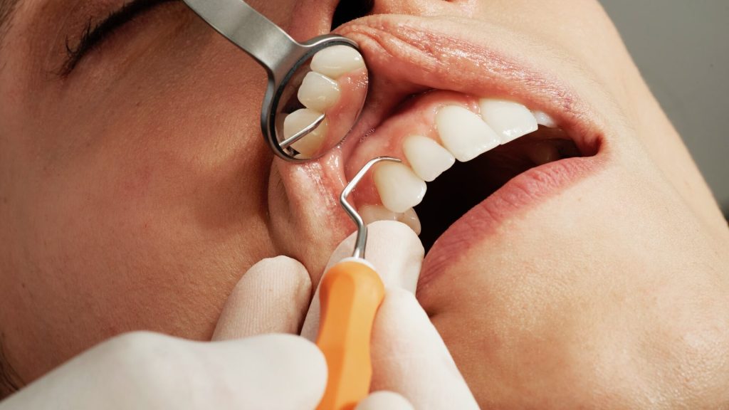
A new treatment approach for tissue inflammation around the dental root and root canal has been implemented, which uses ozone therapy. When used in a complex clinical case, the method helped save a patient’s tooth.
Inflammation of the dental pulp and the periodontium, if not localised only on the inside or the outside but affects both areas, can be extremely difficult to completely cure a tooth. Such cases are difficult to diagnose, therefore, professional literature contains only a few descriptions of them, and there are no general treatment recommendations. Some dentists prefer to focus on the internal canals first, while others suggest treating the pulp and the periodontium simultaneously. A dentist from RUDN University implemented a single protocol for comprehensive pulp and periodontium treatment.
Explaining the problem, RUDN Associate Professor Maria Makeeva, PhD, said: “Inflammation of periodontal tissues often leads to tooth loss. In some cases, a lost tooth cannot be replaced with an implant, because the consequences of the infection worsen the implantation prognosis. Therefore, keeping the tooth is a more desirable outcome in this scenario.”
As described in described in Clinical, Cosmetic and Investigational Dentistry, the new approach was carried out in a patient (aged 44) with significant inflammation of the right mandibular canine. The patient was initially diagnosed with periodontium inflammation; the tooth was loose, and a flow of pus was seen. The first steps were typical for a periodontal inflammation treatment protocol: the patient was administered an antibiotic and then was instructed to rinse the tooth with chlorhexidine. The dentist also scaled the tooth. After that, the patient remained under observation for six months. Although the bleeding and pyorrhoea stopped, the dentist discovered tissue injury around the root of the tooth, which had caused bone destruction. It turned out that one narrow area had an 8mm deep periodontal pocket, and that the initial inflammation transferred to the internal tissues of the tooth.
A periodontologist and an endodontist together implemented a comprehensive treatment approach. After removing the dead pulp from the dental canals, which were then dried and treated with ozone for 24 seconds for better disinfection. After a week of dressing, the treatment was repeated, and the cavity was covered with a permanent filling made of a light-cured composite material. After cleaning, the periodontal pocket was rinsed, dried, and treated with ozone for 18 seconds. A recall examination in six months showed no inflammation of the tooth. Additionally, the bone tissue was recovering, and the periodontal pocket reduced to 4mm.
“Simultaneous inflammation of the pulp and periodontium tissues is very difficult to treat. It can be caused by several types of pathogens at the same time that migrate between tissues and worsen the prognosis. Our experience shows that such an infected tooth can be saved, but it requires close collaboration of a periodontologist and an endodontist, as well as a patient’s complete compliance with oral hygiene recommendations. One should also take into consideration possible bacterial resistance to antibiotics and therefore use additional antibacterial treatment, such as ozone,” explained Dr Makeeva.