Year: 2021

Pink Drinks Make You Run Faster

A new study led by the shows that sweetened pink drinks — purely as a result of their colour — can help people run faster and further compared to clear sweetened drinks.

The study, led by Centre for Nutraceuticals in the University of Westminster, found that a pink drink can raise exercise performance by 4.4% and can also bolster a ‘feel good’ effect, possibly making exercise seem less difficult. The findings were published in the journal Frontiers in Nutrition.

This marks the first investigation to assess the effect of drink colour on exercise performance and could open up a new avenue of future research in the field of sports drinks and exercise. Interest in colour and exercise performance had already resulted in studies, such as wearing red-coloured glasses during exercise which were found to raise testosterone but not performance.

The study involved asking participants to run on a treadmill for 30 minutes at a self-selected pace, ensuring a consistent exertion. Throughout the exercise they rinsed their mouths with either a pink artificially sweetened low-calorie drink or a clear drink which was also artificially sweetened and low-calorie.

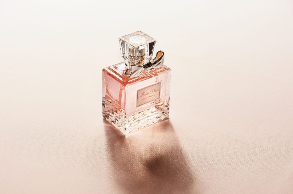
The drinks were identical in every respect aside from the addition of pink colouration to the one. Pink was selected as it is associated with perceived sweetness, therefore increasing expectations of sugar and carbohydrate intake.

In prior research, it was found that rinsing the mouth with carbohydrates can improve exercise performance by reducing the perceived exercise intensity, so the researchers wanted to see whether rinsing with a pink drink with no carbohydrate stimulus could create a similar result through the placebo effect.

The results show that the participants ran an average 212 metres further with the pink drink while their mean speed during the exercise test also increased by 4.4 %. Feelings of pleasure were also improved, meaning participants found running more enjoyable.

Additional investigations will be needed to understand whether the proposed placebo effect causes a similar activation to the reward areas of the brain that are commonly reported when rinsing the mouth with carbohydrates. 

“The influence of colour on athletic performance has received interest previously, from its effect on a sportsperson’s kit to its impact on testosterone and muscular power,”  said corresponding author Dr Sanjoy Deb, University of Westminster. “Similarly, the role of colour in gastronomy has received widespread interest, with research published on how visual cues or colour can affect subsequent flavour perception when eating and drinking.

“The findings from our study combine the art of gastronomy with performance nutrition, as adding a pink colourant to an artificially sweetened solution not only enhanced the perception of sweetness, but also enhanced feelings of pleasure, self-selected running speed and distance covered during a run.”

Source: News-Medical.Net

Journal information: Brown, D. R., et al. (2021) Mouth Rinsing With a Pink Non-caloric, Artificially-Sweetened Solution Improves Self-Paced Running Performance and Feelings of Pleasure in Habitually Active Individuals. Frontiers in Nutrition. doi.org/10.3389/fnut.2021.678105.

Sepsis Leaves a Dangerous Imprint in Immune System

E. Coli bacteria. Image by Gerd Altmann from Pixabay

New research suggests that sepsis can cause alterations in the functioning of defence cells that persist even after the patient is discharged from hospital.

This cellular reprogramming creates a disorder the authors term ‘post-sepsis syndrome’, symptoms of which include frequent reinfections, cardiovascular alterations, cognitive disabilities, declining physical functions, and poor quality of life.This explains why so many patients who survive sepsis die sooner after hospital discharge than patients with other diseases or suffer from post-sepsis syndrome, immunosuppression and chronic inflammation.

The article reviews studies done to investigate cases of septic patients who died up to five years after hospital discharge.

Sepsis is one of the main causes of death in intensive care units, sepsis is a life-threatening systemic organ dysfunction triggered by the body’s dysregulated response to a pathogen, usually a bacterium or fungus. While fighting the pathogen, the defence system injures the body’s own tissues and organs.

If not caught and treated in time, the condition can lead to septic shock and multiple organ failure. Patients with severe COVID and other infectious diseases have an increased risk of developing and dying from sepsis.

Worldwide, new sepsis cases are estimated to reach some 49 million per year. Hospital mortality from septic shock exceeds 40% globally, up to 55% in Brazil, according to the Sepsis Prevalence Assessment Database (SPREAD) study, conducted with support from FAPESP.

“The massive infection and the accompanying intense immune response with a cytokine outpouring during sepsis may promote irreversible cell metabolic reprogramming. Cell reprogramming is unlikely to occur in leukocytes or bone marrow only. This might happen in several tissues and cells that prompt systemic organ dysfunctions […] Bacteria can transfer genetic material to host cell DNA as eukaryotic cells develop tools to protect themselves against the microorganism invasion. The latter may induce cell biology and metabolic reprogramming that remains even after the infection’s elimination,” the investigators wrote in the article.

According to Raquel Bragante Gritte, joint first author with Talita Souza-Siqueira, one of the hypotheses was that metabolic reprogramming begins in the bone marrow, whose cells acquire a pro-inflammatory profile.

“Our analysis of blood samples from patients even three years after ICU discharge showed that monocytes [a type of defense cell] were activated and ready for battle. They should have been neutral. Monocytes are normally activated only when they are ‘recruited’ to the tissue,” Gritte told Agência FAPESP. Both Gritte and Souza-Siqueira are researchers at Cruzeiro do Sul University (UNICSUL) in the state of São Paulo, Brazil.

The researchers conducted a follow-up study of 62 patients for three years after discharge from the ICU at USP’s University Hospital, analysing alterations in monocytes, neutrophils and lymphocytes, as well as microRNAs, in order to identify prognostic markers and factors associated with post-sepsis syndrome.

“Our hypothesis is that white blood cells conserve a memory of sepsis, which helps explain why patients remain sick after they leave hospital,” said co-author Rui Curi, Professor at UNICSUL, and Director of Butantan Institute.

The investigators suggest that sepsis may create a specific macrophage phenotype that stays active even after hospital discharge. “Cell metabolism reprogramming is also involved in the functions and even generation of the different lymphocyte subsets. Several stimuli and conditions change lymphocyte metabolism, including microenvironment nutrient availability,” they wrote.

The next stage of research will be bone marrow studies to understand how cells are reprogrammed by sepsis. “We think the key to this alteration is in bone marrow,” she said. “However, another possibility is that activation occurs in the blood. We’ll need to do more in-depth research to find answers.”

Source:
News-Medical.Net

Distraction a Big Problem in Teletherapy Sessions

Photo by Tracy le Blanc from Pexels

A small survey has found that although therapists appear to prefer virtual sessions over in-person meetings, a significant proportion admit to being distracted while delivering care.

A third of respondents admitted to providing lower-quality care to clients during online sessions. Overall, 39% admitted to checking emails and social media while providing virtual care.

These were the results of a survey of 600 therapists conducted and published by OnlineTherapy.com, a virtual directory for teletherapists and counselors. It’s also an affiliate of the controversial app BetterHelp, stating on their website that the company “may receive compensation from BetterHelp if you purchase products or services through the links provided.”

Online care is generally well received by therapists: nearly half said they prefer virtual sessions over in-person meetings. They appreciated many advantages of virtual therapy, such as working from home which allows therapists to keep a flexible schedule and increases their availability. Video sessions also provide therapists with a uniquely intimate look into their clients’ daily lives, making assessing their mental health easier.

However, teletherapy has introduced its own challenges. Besides the struggle to remain for therapists to stay focused themselves, 56% of those surveyed said their clients are more easily distracted during virtual sessions, and 48% reported technological issues as a major impediment to their practice.

Peter Yellowlees, MD, of UC Davis Health in Sacramento, California, noted with concern and confusion that 16% of therapists reported substance use before or during their sessions.

“There are all sorts of people in this world who call themselves therapists, most of whom have very reasonable training, but quite a number don’t,” Dr Yellowlees told MedPage Today, expressing serious doubts about whether these rates of social media distraction and substance use during virtual sessions would ring true for mental health clinicians with PhDs and MDs.

According to a spokesperson from OnlineTherapy.com, the survey only asked if respondents were currently practicing mental health professionals and did not ask for their credentials or certifications.

The results of the survey did, however, echo Dr Yellowlees’s own concerns of mental health problems on the rise of among therapists in general. The vast majority (90%) said that during the last year they suffered mental health issues, including anxiety disorders (50%) and depressive disorder (48%). This would likely impact the level of care that a therapist able to provide to their clients.

A further concern is that 17% of respondents reported seriously considered suicide since the start of the pandemic.

But  Dr Yellowlees also sees teletherapy as a way for therapists to start getting the help that they need, rather than simply treating themselves in fear of stigma or possible repercussions for their licences.

“It’s undoubtedly helped significantly,” Dr Yellowlees said. “We know that certain teletherapies are actually good for the mental health of providers, as well as the patients.”

Source: MedPage Today

Muscle Atrophy Gene Identified from Mice Sent into Space

Dragon cargo capsule arriving at the International Space Station. Image by SpaceX-Imagery from Pixabay

Researchers from the University of Tsukuba have found a new gene involved in muscle atrophy when they sent mice into space to explore effects of weightlessness on skeletal muscles.

Extended periods of skeletal muscle inactivity or mechanical unloading (bed rest, immobilisation, spaceflight and reduced step) can result in a significant loss of muscle mass and strength which ultimately lead to muscle atrophy. Spaceflight is one of the leading models of understanding muscle atrophy from disuse.

As the molecular and cellular mechanisms involved in disuse skeletal muscle atrophy have been studied, several different signaling pathways have been studied to understand their regulatory role in this process. However, large gaps exist in the understanding of the regulatory mechanisms involved, as well as their functional significance.

Prior studies examining the effects of reduced gravity on muscle mass and function have used a ground control group which cannot be directly compared to the space experimental group. Researchers from the University of Tsukuba set out to explore the effects of gravity in mice subjected to the same housing conditions, such as the stresses of launch, landing and cosmic radiation. “In humans, spaceflight causes muscle atrophy and can lead to serious medical problems after return to Earth” says senior author Professor Satoru Takahashi. “This study was designed based on the critical need to understand the molecular mechanisms through which muscle atrophy occurs in conditions of microgravity and artificial gravity.”

Two groups of six mice each were housed onboard the International Space Station for 35 days. One group was subjected to artificial gravity (1g) and the other was left in microgravity. All mice were returned to Earth aboard a Dragon capsule and the team compared the effects of the different onboard environments on skeletal muscles. “To understand what was happening inside the muscles and cells, at the molecular level, we examined the muscle fibers. Our results show that artificial gravity prevents the changes observed in mice subjected to microgravity, including muscle atrophy and changes in gene expression,” explained Prof Takahashi. 

Transcriptional analysis of gene expression showed that the artificial gravity environment prevented altered expression of atrophy-related genes, and also identified other genes possibly associated with atrophy. Specifically, a gene called Cacng1 was identified as possibly having a functional role in myotube atrophy, which previously had no known function, and was shown to have increased activity when muscle atrophy was present.

When muscle fibres were cultured in vitro, ones which had Cacng1 expression upregulated were decreased in diameter by 27.5%. A similar effect was seen in newborn mice with upregulated Cacng1.

This work validated the use of 1g artificial gravity environments in spaceflight for examining the effects of microgravity in muscles. These studies add to the body of knowledge surrounding the mechanisms of muscle atrophy, possibly improving the treatment of related diseases.

Source: Tsukuba University

Journal information: Okada, R., et al. (2021) Transcriptome analysis of gravitational effects on mouse skeletal muscles under microgravity and artificial 1 g onboard environment. Scientific Reports. doi.org/10.1038/s41598-021-88392-4.

HIV Increases Risk of COVID Infection and Mortality

Man with red HIV ribbon on shirt. Photo by Anna Shvets from Pexels

New research shows that individuals living with HIV and AIDS have an increased risk of SARS-CoV-2 infection and death from COVID.

An estimated 38 million people around the world are living with HIV/AIDS, according to the World Health Organization, 7.5 million of whom are in South Africa, according to UNAIDS.

In their review, researchers at  Penn State College of Medicine found that people living with HIV had a 24% higher risk of SARS-CoV-2 infection and a 78% higher risk of death from COVID than people without HIV. They analysed data from 22 prior studies with nearly 21 million participants in North America, Africa, Europe and Asia to determine to what extent people living with HIV/AIDS are susceptible to SARS-CoV-2 infection and death from COVID.

Participants were mostly male (66%) and the median age was 56. The most common comorbidities among the HIV-positive population were hypertension, diabetes, chronic obstructive pulmonary disease and chronic kidney disease. Most patients (96%) were on antiretroviral therapy (ART).

“Previous studies were inconclusive on whether or not HIV is a risk factor for susceptibility to SARS-CoV-2 infection and poor outcomes in populations with COVID-19,” said Dr Paddy Ssentongo, lead researcher and assistant professor at the Penn State Center for Neural Engineering. “This is because a vast majority of people living with HIV/AIDS are on ART, some of which have been used experimentally to treat COVID-19.”

Pre-existing conditions common among people living with HIV/AIDS, may contribute to the severity of their COVID infections, noted the investigators. It remains inconclusive as to whether antiviral drugs, such as tenofovir and protease-inhibitors, reduce the risk of SARS-CoV-2 infection and death from COVID in people with living with HIV/AIDS.

“As the pandemic has evolved, we’ve obtained sufficient information to characterize the epidemiology of HIV/SARS-CoV-2 coinfection, which could not be done at the beginning of the pandemic due to scarcity of data,” said Vernon Chinchilli, fellow researcher and chair of the Department of Public Health Sciences. “Our findings support the current Centers for Disease Control and Prevention guidance to prioritize persons living with HIV to receive a COVID-19 vaccine.”

Source: Penn State University

South African Perfume Expert’s Smell Recovery Breakthrough

A perfume bottle. Image by StockSnap from Pixabay

A South African perfume expert has developed a treatment to help people who have lost their sense of smell due to COVID.

Loss of smell and taste is one of the most prominent symptoms and after-effects of COVID infection. A meta-analysis of 27 studies showed that 48.47% of COVID patients reported loss of sense of smell, and loss of taste was reported in 20 studies with a prevalence of 41.47%.

While some people only experience a minimal loss of smell, others, especially those who have experienced prolonged COVID infection, may wait months for their sense of smell to return, if it all.

For those patients who battle with their sense of smell after recovering from COVID-19, it is a disheartening experience. Luckily Scenterprises Inc founder, Sue Phillips, has found an amazing way to help these patients smell all the wonderful things in life again.

Phillips is a renowned South African fragrance expert and a self-proclaimed ‘scentrepreneur’, with over 40 years’ experience in the fragrance industry. She is a vocal advocate for the amazing qualities of fragrances and is the author of a book, titled The Power of Perfume.

In an interview with Cape Talk, Phillips described her breakthrough discovery – the role perfume can play in helping those who have had COVID regain their sense of smell.

A perfume is made up of a complex blend of notes – the top, middle and base notes, explained Phillips. She said that she met with a woman who had lost her sense of smell for over a year due to COVID. The woman was unable to identify the lighter, or top notes of the perfume. However, the woman was able to pick up on the stronger notes.

“Suddenly we had a breakthrough and she was quite emotional. She finally said, and she was crying, ‘Oh my goodness, I can finally smell something beautiful’,” Phillips told Cape Talk.

Phillips observed that as they continue with the process, more and more fragrances can be identified by patients.

The perfume expert is now offering Zoom sessions educating people about this process and is producing ‘scent kits’ to help patients battling with their sense of smell.

Source: The South African

How Legionnaire’s Disease Digs In

A bunker from World War II, emulating how Legionella makes a protective shelter. Image by herb1979 from Pixabay

Scientists have discovered how the bacteria that causes Legionnaires’ disease digs in and makes a tiny shelter inside the cells of humans and other hosts. 

The findings, published in Science, could offer insights into how other bacteria are able to survive inside cells, knowledge that could lead to new treatments for a wide variety of infections.

Discovered in 1976, Legionella, an aerobic gram-negative bacillus is responsible for Legionnaires’ disease, a condition of severe pneumonia. Spread through aerosolised water particles, it is a common cause of hospital and community-acquired pneumonia.

“Many infectious bacteria, from listeria to chlamydia to salmonella, use systems that allow them to dwell within their host’s cells,” explained study leader Vincent Tagliabracci, Ph.D., assistant professor of molecular biology at UTSW and member of the Harold C Simmons Comprehensive Cancer Center. “Better understanding the tools they use to make this happen is teaching us some interesting biochemistry and could eventually lead to new targets for therapy.”

Dr Tagliabracci’s lab studies atypical kinases, unusual forms of enzymes that put phosphates onto proteins or lipids, changing their function. Legionella is a particularly rich source of these noncanonical kinases. According to the Centers for Disease Control and Prevention, nearly 10 000 cases of Legionnaires’ disease were reported in the US in 2018, though the true incidence is believed to be higher.      

After identifying a new Legionella atypical kinase named MavQ, Dr Tagliabracci and colleagues used a live-cell imaging technique in concert with a relatively new molecular tagging method to see where MavQ is found in infected human cells. However, rather than residing in a specific location, the researchers were surprised to see that the protein moved back and forth between the endoplasmic reticulum – a network of membranes important for protein and lipid synthesis – and bubble- or tube-shaped structures within the cell.

Further research suggests that MavQ, along with a partner molecule called SidP, remodels the endoplasmic reticulum so that Legionella can strip off sections of the membrane to help create and sustain the vacuole, a structure that the parasitic bacteria uses to shelter inside cells, protecting it from immune attack.

Dr Tagliabracci said that he suspects other bacterial pathogens may use similar mechanisms to co-opt existing host cell structures to create their own protective shelters. 

 Source: University of Texas

Journal information: Ting-Sung Hsieh, et al. Dynamic remodeling of host membranes by self-organizing bacterial effectors. Science, 2021; eaay8118 DOI: 10.1126/science.aay8118

Manganese Sharpens MRI Scans of Heart Attacks

Clinician prepares an MRI scanner. Image by Michal Jarmoluk from Pixabay

Manganese, a common trace mineral, could improve MRI scans of hearts after a heart attack and guide therapy, according to a new study.

By far the most widely used contrast agent for MRI is gadolinium, which improves the visibility of different organs and tissue types in MRI scans. However, it is taken up equally by cells regardless of their activity, and spreads out in damaged tissue. Furthermore, there are also extremely rare instances of serious kidney damage from its use. 

Manganese, besides being less toxic, has a useful property in that it competes with calcium uptake. Calcium handling is highly sensitive to altered heart muscle viability and changes rapidly after damage. Manganese ions enter heart muscle cells through calcium channels, and thus give a useful surrogate for heart tissue viability.

The contrast agent was tested first in vitro with heart muscle cells, and then in mice which had a myocardial infarction (heart attack) induced. The manganese contrast agent was administered with a calcium supplement or administered slowly to negate the effects of manganese interfering with the heart’s calcium channel. Findings were evaluated by examining the infarct size and blood supply at three key intervals: one hour, one day and 14 days after a myocardial infarction was induced. Overall, the manganese contrast agent was superior to gadolinium.

These findings could have major implications for heart attack treatment, if confirmed. They could also be greatly useful in preclinical evaluation of treatments for patients with cardiac ischaemia – where blood supply to the heart muscle is reduced, possibly leading to cardiac arrest.

Furthermore, if manganese-enhanced MRI is performed within the first few hours of a heart attack it could be used to determine the optimal treatment regime for individual patients – helping to regulate changes in the cardiac muscle and thereby further improving survival chances. 

“Magnetic resonance imaging (MRI) is increasingly used to diagnose and give information on heart conditions,” said lead researcher Dr Patrizia Camelliti, Senior Lecturer in Cardiovascular Science, University of Surrey. “This research using mice allows us to measure the health status of the heart muscle rapidly after a heart attack and could provide important information for optimizing treatments in patients.”

Source: News-Medical.Net

Journal reference: Jasmin, N.H., et al. (2021) Myocardial Viability Imaging using Manganese‐Enhanced MRI in the First Hours after Myocardial Infarction. Advanced Science. doi.org/10.1002/advs.202003987.

Oesophageal Cancer Unleashes ‘Fossil Viruses’ Hidden in Human DNA

Colorectal cancer cells. Photo by National Cancer Institute on Unsplash.

Scientists have discovered that many oesophageal cancers turn on ancient viral DNA that was embedded in our genome hundreds of millions of years ago.

“It was surprising,” said principal investigator Adam Bass, MD, the Herbert and Florence Irving Professor of Medicine at Columbia University Vagelos College of Physicians and Surgeons and Herbert Irving Comprehensive Cancer Center. 

“We weren’t specifically searching for the viral elements, but the finding opens up a huge new array of potential cancer targets that I think will be extremely exciting as ways to enhance immunotherapy.”

The idea that bits of ancient retroviruses within the human genome—known as endogenous retroviral elements, or ERVs—play a role in cancer is not new. About 8% of our genome is made up of these remnants of viral infections that have accumulated over the last 100 million years. While ERV sequences have degraded over the aeons and cannot produce viral particles, the viral fossils sometimes wind up inside other genes, disrupting their normal activities, or act as switches that turn on cancer-causing genes.

Recent research however shows that ERVs may also have a cancer fighting role if they are transcribed into strands of RNA.

“When cells activate lots of ERVs, a lot of double-stranded RNA is made and gets into the cell cytoplasm,” Dr Bass explained. “That creates a state that’s like a viral infection and can cause an inflammatory response. In that way, ERVs may make the cancer more susceptible to immunotherapy, and many researchers are working on ways to trick cancer cells into activating ERVs.”

In the new study, Bass and colleagues created organoids of oesophageal mouse tissue to follow the development of cancer from normal cells to malignancy.

With these organoids, Bass discovered that a specific cancer-promoting gene in oesophageal cancers called SOX2 triggers the expression of many ERVs.  

As ERV expression and the accumulation of double-stranded RNAs that can result can be toxic to cells, the researchers found that there is a specific enzyme called ADAR1 that rapidly degrades these double-stranded RNAs.

ADAR1 has been implicated in oesophageal cancer by other researchers, although its role had been unclear. Levels of ADAR1 are known to correlate with poor survival. “The cancers are dependent on ADAR1 to prevent an immune reaction that can be very toxic to the cells,” Bass says.

Some patients with oesophageal cancer are currently treated with immunotherapy, which has been shown to increase survival by several months. “We have a lot of enthusiasm that blocking ADAR1 may both have direct efficacy for oesophageal cancers and that ADAR1 inhibition may have even great effects by enhancing the efficacy of cancer immunotherapy in patients with oesophageal cancer,” Bass said.

Beyond the results regarding ADAR1 and ERVs, the process of modelling the development of oesophageal cancer via genomic engineering of organoids also uncovered a variety of other processes in oesophageal cancer that could help develop new therapies.

“The way we used organoids to build cancers up from the normal cell is a powerful system for uncovering cancer-causing activities and testing therapeutic targets,” said Bass. “By making individual genome alterations in these models one at a time, we can see which combinations of genetic alterations lead to cancer and then determine specific mechanisms of tumor formation.”

In this study, the organoids started with overexpression of the SOX2 gene, a commonly amplified factor that promotes the development of squamous cancers.

In the study, Dr Bass’ team built a panel of organoids emulating conditions ranging from normal oesophagus to fully transformed cancer.

By comparing normal and cancerous organoids, the team could dissect how the activity of SOX2 differs in normal and cancerous tissues. “It’s important to understand the difference, since potential treatments need to target the cancer functions but have lesser impact upon normal tissue,” he says. “It’s relatively easy to kill cancer cells. The problem is, how do you kill cancer cells but spare other cells?”

Results from the organoids showed that when SOX2 is overactive—and two tumour suppressors are inactivated—SOX2 interacts with other factors, activating an array of cancer-causing genes in addition to their effects upon induction of ERVs.  

“These findings reveal new vulnerabilities in SOX2 oesophageal cancers,” Bass said, “that will now allow us to begin developing therapies that can precisely target the cancer cell and improve the treatment of patients.”

Source: Columbia University Irving Medical Center

Journal information: Zhong Wu, et al. Reprogramming of the esophageal squamous carcinoma epigenome by SOX2 promotes ADAR1 dependence. Nature Genetics, 2021; DOI: 10.1038/s41588-021-00859-2

Lenvatinib Produces Impressive Results Against Tough Tumours

Image by doodlartdotcom from Pixabay

Lenvatinib, a multitargeted tyrosine kinase inhibitor (TKI) induced a strong tumour response in patients with advanced gastrointestinal or pancreatic tumours, according to results from a phase II trial.

The study focused on previously treated advanced gastroenteropancreatic neuroendocrine tumors (GEP-NETs). An overall response rate (ORR) of 29.9% was seen in the trial, with a particularly high ORR — 44.2% — in patients with pancreatic NETs. 

“This study provides novel evidence for the efficacy of lenvatinib in patients with disease progression following treatment with other targeted agents, suggesting the potential value in the treatment of advanced GEP-NETs,” wrote Jaume Capdevila, MD, PhD, of Vall Hebron University Hospital in Barcelona, and colleagues.

TKIs are a group of pharmacologic agents that disrupt the signal transduction pathways of protein kinases by several modes of inhibition. Since sunitinib maleate (Sutent), another multitargeted TKI, was approved ten years ago, investigators have been evaluating newer-generation TKIs that target VEGF receptors (VEGFRs), among other receptors, both in pancreatic and non-pancreatic NETs.

Lenvatinib targets VEGFR 1-3, fibroblast growth factor receptors (FGFR) 1-4, and platelet-derived growth factor receptor alpha.

The researchers noted that studies have demonstrated its particular effectiveness against FGFR-1, which is a key driver of resistance to antiangiogenic drugs, “suggesting that it could potentially also reverse primary and acquired resistance to anti-VEGFR treatments or to other targeted agents.”

A total of 111 patients were enrolled in the study; 55 had histologically confirmed grade 1-2 pancreatic NETs, while 56 had gastrointestinal NETs. Patients were administered 24-mg lenvatinib once daily until disease progression or treatment intolerance. Median follow-up was 23 months.

The ORR was 16.4% for patients with gastrointestinal NETs, and median duration of response was 19.9 months for patients with pancreatic NETs and 33 months for gastrointestinal NETs. The median progression-free survival (PFS) for both groups was 15.7 months.

These results compare well with PFS outcomes reported in phase III trials, including those evaluating sunitinib and surufatinib, the authors noted.

“Interestingly, the ORR in pancreatic NETs was 44%, a rate not seen before with targeted agents,” Jonathan Strosberg, MD, head of the neuroendocrine tumor division at Moffitt Cancer Center in Tampa, told MedPage Today.

Dr Strosberg, who was not involved with this research, noted that the study group had been heavily treated beforehand, and that 29% had received prior sunitinib. “In contrast, the response rates with other TKIs have been <20% in this population, even in less heavily treated populations. The ORR for gastrointestinal NETs was more modest, but still impressive,” he added.

The most common grade 3/4 adverse events was hypertension (22.7%), while a majority of patients needed either a dose reduction or a pause.

“This suggests that lower starting doses might be considered in this population, and that particularly close monitoring of blood pressure is necessary,” said Dr Strosberg.

The study results “suggest that lenvatinib is more than just a ‘me-too’ competitor to sunitinib,” he noted. “It actually seems to have superior activity, potentially due to its ability to target both the VEGF and FGF receptors. Moreover, it appears to have activity in patients who have progressed on sunitinib. Randomized phase III studies with this drug are warranted, both for pancreatic and GI/lung NETs.”

Source: MedPage Today

Journal information: Capdevila J, et al “Lenvatinib in patients with advanced grade 1/2 pancreatic and gastrointestinal neuroendocrine tumors: results of the phase II TALENT trial (GETNE1509)” J Clin Oncol 2021; DOI: 10.1200/JCO.20.03368.