Tag: stem cells

A New Understanding of Graft-versus-host Disease

T cell
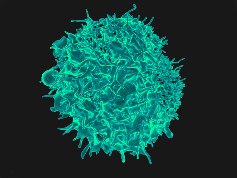
Scanning Electron Micrograph image of a human T cell. Credit: NIH/NIAID

New research published in the journal Immunity challenges the prevailing hypothesis for how donor stem cell grafts cause graft-versus-host disease (GVHD) and offers an alternative model that could guide development of novel therapies.

The study showed in a mouse model that GVHD, which often affects the skin, gut and liver, is maintained by donor T cells that seed those tissues soon after transplant and not by the continual recruitment of T cells from the blood as previously thought.

“This study changes the paradigm of how people think about GVHD,” said co-senior author Warren Shlomchik, MD, professor of medicine and immunology at the University of Pittsburgh School of Medicine. “It provides important mechanistic detail about what’s going on in the tissues affected by GVHD, which could ultimately inform the development of better therapeutics and lead to better outcomes for stem cell recipients.”

Allogeneic stem cell transplantation involves infusion of stem cells from a healthy donor’s blood or bone marrow to a recipient. While often lifesaving for patients with leukaemia and other blood disorders, the treatment also comes with a risk of developing GVHD, a life-threatening disease that occurs when donor alloreactive T cells attack the recipient’s healthy tissues.

According to a widely held theory, GVHD is maintained by T cells that continually migrate from secondary lymphoid organs throughout the body, including the spleen and lymph nodes, to affected tissues via the blood.

However, a different model posits that the disease is maintained locally by T cells in the tissues with little input from the blood. In the new study, Shlomchik, lead author Faruk Sacirbegovic, PhD, research assistant professor of surgery at Pitt, and their team investigated the two hypotheses for how GVHD is sustained in tissues.

The researchers developed a system to track alloreactive T cells in a mouse model of GVHD by labelling individual cells with unique tags to create different T cell “flavours.” By measuring the tags over time, they monitored where the T cells travelled and replicated.

The analysis showed that each tissue affected by GVHD had unique T cell populations with varying frequencies of each T cell flavour.

“This finding is strong evidence that the disease is locally maintained by T cells in each of the tissues,” explained Shlomchik. “If tissues were constantly getting T cells from circulating blood, then the frequencies of T cell flavors in each tissue should become more and more alike over time — but we didn’t see that.”

The team used mathematical models to predict that progenitor T cells seed out into recipient tissues early after transplant, differentiating there into disease-causing cells.

Next a series of experiments was conducted to confirm this prediction and identified these progenitors as T cells expressing a gene called Tcf7.

“We think that progenitor T cells are long-lived in target tissues and are critical for maintaining GVHD,” said co-senior author Thomas Höfer, PhD, professor of theoretical systems biology at the University of Heidelberg. “After the initial seeding phase, the disease is mostly sustained within the tissue itself without a lot of input from new T cells in the blood.”

Stem cell recipients are typically treated with immunosuppressants to prevent and treat GVHD. As these powerful drugs act systemically to suppress the immune system, they also lower immunity to infections and have other side effects.

According to the researchers, the study’s insights could eventually lead to new, targeted therapies for GVHD.

“Now that we know the identity of progenitor cells, we might be able to prevent them forming early post-transplant or target them directly after they’ve formed,” said Shlomchik. “The findings also suggest that treating GVHD in the tissues themselves would be effective – although targeting tissues beyond the skin remains a challenge.”

With better ways to minimise the risk of GVHD after stem cell transplantation, the procedure could become more widely used to treat a broader range of diseases, including blood disorders such as sickle cell anaemia and autoimmune diseases such as lupus and multiple sclerosis.

Source: University of Pittsburgh

World First Trial of Lab-grown Red Blood Cells for Transfusion

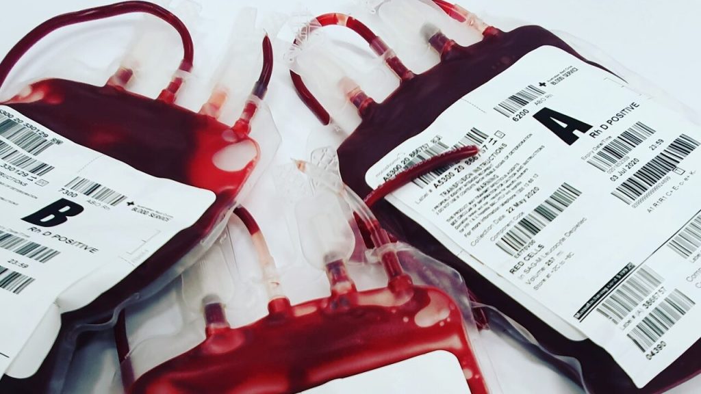
https://www.pexels.com/photo/a-close-up-shot-of-bags-of-blood-4531306/
Photo by Charlie-Helen Robinson on Pexels

In a world first, researchers have launched a clinical trial of lab-grown red blood cells for transfusion into another person. These manufactured blood cells were grown from stem cells from donors, for transfusion into volunteers in the RESTORE randomised controlled clinical trial.

If our trial is successful, it will mean that patients who currently require regular long-term blood transfusions will need fewer transfusions in future, helping transform their care

Professor Cedric Ghevaert, chief investigator

If the technique is proven safe and effective, manufactured blood cells could in time revolutionise treatments for people with blood disorders such as sickle cell and rare blood types. It can be difficult to find enough well-matched donated blood for some people with these disorders.

To produce the lab-grown blood cells, stem cells are first magnetically extracted from a normal 470ml blood donation. These stem cells are then coaxed into becoming red blood cells. Over the three week process, an initial pool of about half a million stem cells generates 50 billion red blood cells.

Chief Investigator Professor Cedric Ghevaert, Professor in Transfusion Medicine and Consultant Haematologist at the University of Cambridge and NHS Blood and Transplant, said: “We hope our lab grown red blood cells will last longer than those that come from blood donors. If our trial, the first such in the world, is successful, it will mean that patients who currently require regular long-term blood transfusions will need fewer transfusions in future, helping transform their care.”

Professor Ashley Toye, Professor of Cell Biology at the University of Bristol and Director of the NIHR Blood and Transplant Unit in red cell products, said: “This challenging and exciting trial is a huge stepping stone for manufacturing blood from stem cells. This is the first-time lab grown blood from an allogeneic donor has been transfused and we are excited to see how well the cells perform at the end of the clinical trial.”

The trial is studying the lifespan of the lab grown cells compared with infusions of standard red blood cells from the same donor. The lab-grown blood cells are all fresh, so the trial team expect them to perform better than a similar transfusion of standard donated red cells, which contains cells of varying ages.

Additionally, if manufactured cells last longer in the body, patients who regularly need blood may not need transfusions as often. That would reduce iron overload from frequent blood transfusions, which can lead to serious complications.

The trial is the first step towards making lab grown red blood cells available as a future clinical product. For the foreseeable future, manufactured cells could only be used for a very small number of patients with very complex transfusions needs. NHSBT continues to rely on the generosity of donors.

Co-Chief Investigator Dr Rebecca Cardigan, Head of Component Development NHS Blood and Transplant and Affiliated Lecturer at the University of Cambridge, said: “It’s really fantastic that we are now able to grow enough red cells to medical grade to allow this trial to commence. We are really looking forward to seeing the results and whether they perform better than standard red cells.”

Thus far, two people have been transfused with the lab grown red cells. They are well and healthy, and were closely monitored with no untoward side effects were reported. The amount of lab grown cells being infused varies but is around 5-10mls.

Donors were recruited from NHSBT’s blood donor base. They donated blood to the trial and stem cells were separated out from their blood. These stem cells were then grown to produce red blood cells in a laboratory at NHS Blood and Transplant’s Advanced Therapies Unit in Bristol. The recipients of the blood were recruited from healthy members of the NIHR BioResource.

A minimum of 10 participants will receive two mini transfusions at least four months apart, one of standard donated red cells and one of lab grown red cells, to see if the young lab-made red blood cells last longer than cells made in the body.

Further trials are needed before clinical use, but this research marks a significant step in using lab grown red blood cells to improve treatment for patients with rare blood types or people with complex transfusion needs.

Source: University of Cambridge

Fixing Spinal Cord Injuries with Stem Cell Grafts and Rehabilitation

Photo by Cottonbro on Pexels

In recent years, researchers have made strides in promoting tissue regeneration in spinal cord injuries (SCI) through implanted neural stem cells or grafts in animal models. Separate efforts have shown that intensive physical rehabilitation can improve function after SCI by promoting greater or new roles for undamaged cells and neural circuits.

University of California San Diego researchers tested whether rehabilitation can pair with pro-regenerative therapies, such as stem cell grafting. They published their findings in in JCI Insight, 

The researchers induced a cervical lesion in rats that impaired the animals’ ability to grasp with its forelimbs. The animals were divided into four groups: animals who underwent the lesion alone; animals who received a subsequent grafting of neural stem cells designed to grow and connect with existing nerves; animals who received rehabilitation only; and animals who received both stem cell therapy and rehabilitation.

Rehabilitation therapy for some animals began one month after initial injury, a time point that approximates when most human patients are admitted to SCI rehabilitation centers. Rehabilitation consisted of daily activities that rewarded them with food pellets if they performed grasping skills.

The researchers found that rehabilitation enhanced regeneration of injured corticospinal axons at the lesion site in rats, and that a combination of rehabilitation and grafting produced significant recovery in forelimb grasping when both treatments occurred one month after injury.

“These new findings indicate that rehabilitation plays a critically important role in amplifying functional recovery when combined with a pro-regenerative therapy, such as a neural stem cell transplant,” said first author Paul Lu, PhD, associate adjunct professor of neuroscience at UC San Diego School of Medicine and research health science specialist at the Veterans Administration San Diego Healthcare System.

“Indeed, we found a surprisingly potent benefit of intensive physical rehabilitation when administered as a daily regimen that substantially exceeds what humans are now provided after SCI.”

Senior author Mark H. Tuszynski, MD, PhD, professor of neurosciences and director of the Translational Neuroscience Institute at UC San Diego School of Medicine, and colleagues have long worked to address the complex challenges of repairing SCIs and restoring function.

In 2020, for example, they reported on the observed benefits of neural stem cell grafts in mice and in 2019, described 3D-printed implantable scaffolding that would promote nerve cell growth.

“There is a great unmet need to improve regenerative therapies after SCI,” said Tuszynski. “We hope that our findings point the way to a new potential combination treatment consisting of neural stem cell grafts plus rehabilitation, a strategy that we hope to move to human clinical trials over the next two years.”

Source: University of California – San Diego

Experiment Turns Back the Age of Human Skin Cells by 30 Years

This normal human skin cell was treated with a growth factor that triggered the formation of specialised protein structures that enable the cell to move.
Credit: Torsten Wittmann, University of California, San Francisco

In a finding which could revolutionise regenerative medicine, researchers have found a way to reverse the age of human skin cells by 30 years, reversing genetic ageing measures for cells without losing their specialised function. The function of older cells was partly restored, as well as rejuvenating the molecular measures of biological age. The research was published in the journal eLife.

One of the ways regenerative medicine aims to replace damaged or old cells is by creating ‘induced’ stem cells, which differentiate into specialised cells. Currently the process is not reversible.

The new method, based on stem cell production, overcomes the problem of entirely erasing cell identity by halting reprogramming part of the way through the process. This let researchers find the precise balance between reprogramming cells, making them biologically younger, while still being able to regain their specialised cell function.

Currently, cell reprogramming takes around 50 days using four key molecules called the Yamanaka factors. The new method, called ‘maturation phase transient reprogramming’, exposes cells to Yamanaka factors for just 13 days. At this point, age-related changes are removed and the cells have temporarily lost their identity. The partly reprogrammed cells were given time to grow under normal conditions, to observe whether their specific skin cell function returned. Genome analysis showed that cells had regained markers characteristic of skin cells (fibroblasts), and this was confirmed by observing collagen production in the reprogrammed cells.

To show that the cells had been rejuvenated, the researchers looked for changes in ageing indicators. Dr Diljeet Gill, who conducted the work as a PhD student explained: “Our understanding of ageing on a molecular level has progressed over the last decade, giving rise to techniques that allow researchers to measure age-related biological changes in human cells. We were able to apply this to our experiment to determine the extent of reprogramming our new method achieved.”

Cellular ages examined included the epigenetic clock, where chemical tags present throughout the genome indicate age. Another is the transcriptome, all the gene readouts produced by the cell. According to these two measures, the reprogrammed cells matched the profile of cells that were 30 years younger compared to reference data sets.

However, ‘rejuvenated’ cells need to function as if they were younger as well as looking younger. The rejuvenated fibroblasts were able to produce more collagen proteins compared to control cells that did not undergo the reprogramming process. Fibroblasts also move into areas that need repairing. Researchers tested the partially rejuvenated cells in vitro, and the treated fibroblasts moved into the gap faster than older cells – a sign that these could be used to improve wound healing,

The method also had an effect on other genes linked to age-related diseases and symptoms, the researchers saw, indicating possible future therapies. The APBA2 gene, associated with Alzheimer’s disease, and the MAF gene with a role in the development of cataracts, both showed changes towards youthful levels of transcription.

The researchers plan to explore the mechanism behind the successful transient programming, which is not yet completely understood. It is speculated that key areas of the genome involved in shaping cell identity might escape the reprogramming process.

Dr Diljeet concluded: “Our results represent a big step forward in our understanding of cell reprogramming. We have proved that cells can be rejuvenated without losing their function and that rejuvenation looks to restore some function to old cells. The fact that we also saw a reverse of ageing indicators in genes associated with diseases is particularly promising for the future of this work.”

Professor Wolf Reik, a group leader in the Epigenetics research programme who has recently moved to lead the Altos Labs Cambridge Institute, said: “This work has very exciting implications. Eventually, we may be able to identify genes that rejuvenate without reprogramming, and specifically target those to reduce the effects of ageing. This approach holds promise for valuable discoveries that could open up an amazing therapeutic horizon.”

Source: Babraham Institute

Space Could be Ideal Place for Stem Cell Production

Astronaut Raja Chari sequences DNA from bacteria samples to understand the microbial environment on the International Space Station. Credit: NASA

The lack of gravity in outer space could be the key to the efficient production of large quantities of stem cells. Scientists at Cedars-Sinai have found that the microgravity environment in space stations can potentially aid life-saving advances on Earth by facilitating the rapid mass production of stem cells.

A new paper in Stem Cell Reports outlines key opportunities discussed at a space biomanufacturing symposium to expand the manufacture of stem cells in space.

With new rocket technology, the cost of access to space has plummeted, opening up new opportunities for research and industry, as well as spaceflight by private citizens. Biomanufacturing of therapeutic and research biomaterials can be more productive in microgravity conditions.

“We are finding that spaceflight and microgravity is a desirable place for biomanufacturing because it confers a number of very special properties to biological tissues and biological processes that can help mass produce cells or other products in a way that you wouldn’t be able to do on Earth,” said stem cell biologist Arun Sharma, PhD, head of a new Cedars-Sinai research laboratory.

“The last two decades have seen remarkable advances in regenerative medicine and exponential advancement in space technologies enabling new opportunities to access and commercialise space,” he said.

Attendees at the virtual space symposium in December identified more than 50 potential commercial opportunities for conducting biomanufacturing work in space, according to the Cedars-Sinai paper. The most promising fell into three categories: Disease modelling, biofabrication, and stem-cell-derived products.

Scientists use disease modelling, to study diseases and possible treatments by replicating full-function structures – whether using stem cells, organoids or other tissues.

Decades of spaceflight experience has shown that when the body is exposed to low-gravity conditions for extended periods of time, it experiences accelerated bone loss and ageing. By developing disease models based on this accelerated ageing process, research scientists can better understand the mechanisms of the ageing process and disease progression.

“Not only can this work help astronauts, but it can also lead to us manufacturing bone constructs or skeletal muscle constructs that could be applied to diseases like osteoporosis and other forms of accelerated bone ageing and muscle wasting that people experience on Earth,” explained Dr Sharma.

Biofabrication, another major topic of discussion at the symposium, produces materials like tissues and organs with 3D printing a core technology.

A major issue with biofabrication on Earth involves gravity-induced density, which makes it hard for cells to expand and grow. This requires the use of scaffolding structures, but it generally cannot support the small, complex shapes found in vascular and lymphatic pathways. With the lack of gravity in space, scientists are hopeful that they can use 3D printing to print unique shapes and products, like organoids or cardiac tissues, in a way that can’t be replicated on Earth. This technology is being tested on the International Space Station.

The third category has to do with the production of stem cells and understanding how some of their fundamental properties are influenced by microgravity. Some of these properties include potency, or the ability of a stem cell to renew itself, and differentiation, the ability for stem cells to turn into other cell types.

Understanding some of the effects of spaceflight on stem cells can potentially lead to better ways to manufacture large numbers of cells in the absence of gravity. In coming months, Cedars-Sinai scientists will send stem cells into space to test whether it is possible to produce large batches in a low gravity environment.

“While we are still in the exploratory phase of some of this research, this is no longer in the realm of science fiction,” Dr Sharma said. “Within the next five years we may see a scenario where we find cells or tissues that can be made in a way that is simply not possible here on Earth. And I think that’s extremely exciting.”

Source: Cedars-Sinai Medical Center

Rapidly Correcting Genetic Disorders

Image source: Pixabay

Researchers have developed a new method to precisely and rapidly correct genetic alterations in cultured patient cells.

The genetically corrected stem cells are produced from a 2–3 mm skin biopsy taken from patients with different genetic diseases. The corrected stem cells are essential in the research and for the development of new therapies for the diseases in question.

The scientists based the new method on previous groundbreaking research in the fields of stem cells and gene editing; the first technique is the invention of induced pluripotent stem cells, iPSCs from differentiated cells, which won the Nobel in 2012. The other technique is the CRISPR-Cas9 ‘gene scissors’, which got the prize in 2020. The new method combines these techniques to correct gene alterations that cause inherited diseases, creating fully functional new stem cells.

The researchers aim to eventually produce autologous cells with therapeutic properties. The use of the patient’s own corrected cells could help in avoiding the immunological challenges hampering the organ and tissue transplantation from a donor. The new method was developed by PhD student Sami Jalil  and is published in Stem Cell Reports.

More than 6000 inherited diseases are known to exist, which are caused by various gene alterations. Currently, some are treated with a cell or organ transplant from a healthy donor, if available.

“Our new system is much faster and more precise than the older methods in correcting the DNA errors, and the speed makes it easier and diminishes also the risk of unwanted changes,” commented adjunct professor Kirmo Wartiovaara, who supervised the work.

“In perfect conditions, we have reached up to 100 percent efficacy, although one has to remember that the correction of cultured cells is still far away from proven therapeutic applications. But it is a very positive start” Prof Wartiovaara added.

Source: University of Helsinki

A New Mechanism Explains Hair Loss in Men and Women

Bald man
Photo by Brett Sayles on Pexels

Studies of balding male mice have uncovered a possible cause of hair loss in men and women as well. The findings, published in Nature Aging, provide new insight into how hair and tissues age.

The study shows as hair stem cells age, they lose the adhesion that keeps them lodged inside the hair follicle. As their adhesiveness wanes, the stem cells escape from their location, called the bulge, into the dermis. Once outside their delicate microenvironment, they generally can’t survive.

“The result is fewer and fewer stem cells in the hair follicle to produce hair,” said lead author Rui Yi, the Paul E. Steiner Research Professor of Pathology at Northwestern University Feinberg School of Medicine. “This results in thinning hair and ultimately baldness during ageing.”

This finding could be applicable to older men and women with thinning hair as mice and humans share hair and stem cell similarities, Prof Yi said.

By labelling individual stem cells with a fluorescent marker, the researchers were able, for the first time, to track hair follicle ageing in real time in live animals. Scientists also discovered two key genes responsible for enhancing adhesiveness of the stem cells. They are now trying to reinstate these genes to see if that will reverse hair loss.

During follicles’ normal cycles of life and death, a large number of stem cells remain permanently lodged in the stem cell compartment of hair follicles to keep producing hair follicle cells.

“We believe this stem cell escape mechanism has never been reported before, because nobody could track the aging process in live animals,” Yi said.

Though scientists knew hair follicles become miniaturised during aging, how it happened was unclear. Many thought it was due to cell death or the inability of cells to divide as they age.

“We discovered, at least in part, it is due to hair follicle stem cells migrating away from their niche,” Prof Yi said. “Cell death also occurs during our observation. So, our discovery doesn’t dispute existing theories but provides a new mechanism.”

Source: Northwestern University

The Rise of Phony Stem Cell COVID Treatments

SARS-CoV-2 virus. Source: Fusion Medical Animation on Unsplash

The global race to develop new stem cell-based COVID treatments during the pandemic was filled with violations of government regulations, inflated medical claims and distorted public communication, according to an article appearing in Stem Cell Reports.

While stem cell therapy has treatment applications for a limited range of diseases and conditions, at present no clinically tested or government-approved cell therapies are available for the treatment or prevention of COVID or long COVID.

Despite this, some clinics have started offering unproven and unsafe “stem cell” therapies that promise to prevent COVID by strengthening the immune system or improving overall health, according to lead author Laertis Ikonomou, PhD, associate professor of oral biology in the University at Buffalo School of Dental Medicine.

The article explores the negative effects that misinformation about cell therapies has on public health, as well as the roles that researchers, science communicators and regulatory agencies should play in curbing the spread of inaccurate information and in promoting responsible, accurate communication of research findings.

“Efforts to rapidly develop therapeutic interventions should never occur at the expense of the ethical and scientific standards that are at the heart of responsible clinical research and innovation,” said Prof Ikonomou.

Other investigators include Megan Munsie, PhD, professor of ethics, education and policy in stem cell science at the University of Melbourne; and 

Many of the studies on possible stem cell-based COVID treatments are at an early stage of investigation and further evaluation on larger sample sizes is required, says Munsie. However, the findings from preliminary studies are frequently exaggerated through press releases, social media and uncritical news media reports.

“Given the urgency of the ongoing pandemic, even the smallest morsel of COVID science is often deemed newsworthy and rapidly enters a social media landscape where—regardless of its accuracy – it can be widely shared with a global audience,” said Aaron Levine, PhD, associate professor of public policy at Georgia Institute of Technology..

Clinics selling such treatments sometimes use these findings and news reports to exploit the fears of vulnerable patients by unethically advertising unproven stem cell treatments benefits of boosting the immune system, regenerating lung tissue and preventing transmission of COVID, said co-author Leigh Turner, PhD, professor of health, society and behaviour at the University of California, Irvine.

Reportedly some harm to patients resulted from unproven stem cell therapies, including blindness and death. Patients suffer financially as well, said Prof Ikonomou, as the products range in price from a few thousand to tens of thousands of dollars, and people are often encouraged to receive the expensive treatments every few months.

Patients who COVID may decline vaccines, stop wearing masks and stop other COVID safety measures, Prof Turner warned. They may also be less likely to participate in ethically conducted clinical trials.

“The premature commercialisation of cell-based therapeutics will inevitably harm the field of regenerative medicine, increase risks to patients and erode the public’s trust,” said Prof Ikonomou.

Despite warnings, many offending companies continue to make false claims. The authors recommend that regulatory agencies consider implementing stronger measures.

They also suggest that scientific and professional societies lobby regulatory agencies to increase enforcement of laws and regulations. The authors recommended that science communicators and journalists can combat misinformation by not engaging in hyperbolic coverage of research results and conveying study limitations.

Source: University at Buffalo

Mice Born From Stem Cell-derived Gametes

Photo by Kanashi on Unsplash

For the first time, mice have been born from gametes that have been created entirely from stem cells, marking the beginning of a revolutionary new reproductive option.

The experiment is the brainchild of Dr Katsuhiko Hayashi of Kyushu University, who has led the pursuit of making gametes outside of a living body. If adapted for humans, these wild reproductive pursuits are bound to shake up our entire conception of the beginning of life, similar to the way “test-tube” babies did when in vitro fertilisation (IVF) was first introduced.

Dr Hayashi dreams of even bigger possibilities; since stem cells can be rapidly created from skin or other cells, they are an endless source of raw material to make sperm and egg cells. These gametes, if fully functional, can merge into a zygote inside a test tube, be transplanted into a surrogate, and birth a new generation without ever seeing testes or ovaries.

Though still far off for humans, in vitro gametogenesis, or IVG, has great potential. Researchers can use these lab-grown models to better understand how reproductive cells form and mature. For couples struggling to conceive, or people who’ve lost reproductive function due to diseases like cancer, IVG would offer a new route towards pregnancy. Same-sex couples could also potentially conceive children with their own genetic makeup. There are many possibilities, and a wide range of ethical problems.

The basis of the technology uses induced pluripotent stem cells (iPSCs), which can be nudged in any direction, including sperm and egg. Back in 2011, Dr Hayashi showed that by bathing stem cells in a particular chemical soup, his team was able to produce sperm cell precursors, with the capacity to turn into functional sperm.

In 2016, the team achieved the same with eggs in mice, mimicking the entire process of how ovaries make eggs – which were used to produce healthy pups. However, eggs made in a test tube couldn’t develop naturally outside the ovary. Fresh ovarian tissue from mice was needed, creating an obvious challenge for fertility treatments in humans.

In the current study, the team focused on the support cells that normally encapsulate a developing egg. These support cells thrive inside the ovary, secreting hormones and nutrients that help support the metabolic needs of an egg  – a crucial step, which includes forming ovarian follicles for the eggs to mature in.

These ovary-supporting cells can also be made from stem cells if the right chemical keys are used, and so after five years Dr Hayashi figured out those keys. Many of them sport fanciful names like ‘sonic hedgehog‘ (SHH), but most of these proteins belong to the morphogen family, in that they can morph the physical structure and identity of a tissue.

After dousing stem cells with this soup, the cells differentiated into foetal ovary supporting cells, with a gene expression profile closely mimicking that of their natural counterparts.

Next, the researchers added precursor immature egg cells, also made from stem cells. Together, the cells coalesced into tiny ovarian follicles, with support cells forming a bubble wrapping the developing egg. The eggs were then fertilised with sperm, transplanted into surrogate mouse mothers, and after normal pregnancies, resulted in about a dozen healthy pups. Those mice eventually gave birth to babies of their own.

The artificial ovary produces mature eggs less effectively than its natural counterpart, suggesting there’s still much to be learned about this stage of reproduction.

Application of this technology to assisted reproduction in humans is still decades away: human reproductive cells take far longer to mature than those in mice, and likely require different supporting nutrients for the sperm, egg, and surrounding tissue.

The team is now testing their chemical soup in marmosets, to be followed by primates.

Currently no laws or ethical frameworks deal with IVG, since the technology is so new.

Dr Hayashi is taking it step by step, and welcoming public discourse before even considering any clinical use. The first step, he said, is verifying the quality of the stem-cell derived eggs, adding, “That could take a long, long time.”

Source: SingularityHub

Chemical Fingerprints Improve Stem Cell Production

Photo by Louis Reed on Unsplash

Researchers in Japan have developed a new, noninvasive way to monitor the tricky art of stem cell production.

The current era of ethical stem cell research was ushered in by the 2012 Nobel prize-winning discovery that ordinary cells could be coaxed to revert to their earliest pluripotent stage ushered in. Suddenly, scientists could have an ethical, near-inexhaustible supply of pluripotent stem cells — the most versatile of stem cells — that can become any type of cell much like how embryonic stem cells function.

These reprogrammed cells called induced pluripotent stem cells (or iPS cells) hold great promise for regenerative medicine, where they can be used to develop tissue or organ replacement-based treatments for life-threatening diseases.

One key challenge is that it is a lengthy and delicate process to artificially induce ordinary cells to reset back to pluripotency. Obtaining iPS cells therefore is a matter of chance. However, knowing all they can about the complex chemical changes happening inside during reprogramming can help scientists increase the chances of successfully obtaining viable iPS cells for clinical applications. Current methods that track reprogramming status, however, use destructive and costly techniques.

A study led by Dr Tomonobu Watanabe, professor at Hiroshima University’s Research Institute for Radiation Biology and Medicine, showed that Raman spectroscopy could be a low-cost, simpler, and non-intrusive technique to monitor the cell’s internal environment as it transitions.

Dr Watanabe explained: “The quality evaluation and sorting of existing cells have been carried out by investigating the presence or absence of expression of surface marker genes. However, since this method requires a fluorescent antibody, it is expensive and causes a problem of bringing the antibody into the cells.”

He added that the “solution of these problems can accelerate the spread of safe and low-cost regenerative medicine using artificial tissues. Through our method, we provide a technique for evaluating and sorting the quality of iPS cells inexpensively and safely, based on scattering spectroscopy.”

Raman spectroscopy is an alternative to invasive approaches that require dyes or labels to extract biochemical information. It instead makes use of vibration signatures produced when light beams interact with chemical bonds in the cell. Since each chemical has its own distinct vibration frequency, scientists can use it to identify the cell’s molecular makeup.

The team used this spectroscopic technique to get the “chemical fingerprints” of mouse embryonic stem cells, the neuronal cells they specialised into, and the iPS cells formed from those neuronal cells. These data were then used to train an AI model to can track the reprogramming is progressing, and verify iPS cell quality by checking for a “fingerprint” match with the embryonic stem cell.

To measure the progress, they assigned the “chemical fingerprint” of neuronal cells as the transformation starting point and the embryonic stem cell’s patterns as the desired end goal. Along the axis, they used “fingerprint” samples collected on days 5, 10, and 20 of the neuronal cells’ reprogramming as reference points on how the process is advancing.

“The Raman scattering spectrum contains comprehensive information on molecular vibrations, and the amount of information may be sufficient to define cells. If so, unlike gene profiling, it allows for a more expressive definition of cell function,” Dr Watanabe said.

“We aim to study stem cells from a different perspective than traditional life sciences.”

Source: Hiroshima University

Journal information: Germond, A., et al. (2020) Following Embryonic Stem Cells, Their Differentiated Progeny, and Cell-State Changes During iPS Reprogramming by Raman Spectroscopy. Analytical Chemistry doi.org/10.1021/acs.analchem.0c01800.