Tag: spinal cord injury

MIT Engineers Turn Skin Cells Directly into Neurons for Cell Therapy

A new, highly efficient process for performing this conversion could make it easier to develop therapies for spinal cord injuries or diseases like ALS.

Anne Trafton | MIT News
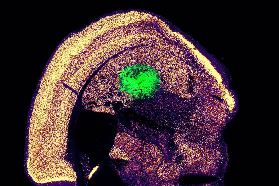
Researchers at MIT have devised a simplified process to convert a skin cell directly into a neuron. This image shows converted neurons (green) that have integrated with neurons in the brain’s striatum after implantation. Credits :Image: Courtesy of the researchers

Converting one type of cell to another – for example, a skin cell to a neuron – can be done through a process that requires the skin cell to be induced into a “pluripotent” stem cell, then differentiated into a neuron. Researchers at MIT have now devised a simplified process that bypasses the stem cell stage, converting a skin cell directly into a neuron.

Working with mouse cells, the researchers developed a conversion method that is highly efficient and can produce more than 10 neurons from a single skin cell. If replicated in human cells, this approach could enable the generation of large quantities of motor neurons, which could potentially be used to treat patients with spinal cord injuries or diseases that impair mobility.

“We were able to get to yields where we could ask questions about whether these cells can be viable candidates for the cell replacement therapies, which we hope they could be. That’s where these types of reprogramming technologies can take us,” says Katie Galloway, the W. M. Keck Career Development Professor in Biomedical Engineering and Chemical Engineering.

As a first step toward developing these cells as a therapy, the researchers showed that they could generate motor neurons and engraft them into the brains of mice, where they integrated with host tissue.

Galloway is the senior author of two papers describing the new method, which appear today in Cell Systems. MIT graduate student Nathan Wang is the lead author of both papers.

From skin to neurons

Nearly 20 years ago, scientists in Japan showed that by delivering four transcription factors to skin cells, they could coax them to become induced pluripotent stem cells (iPSCs). Similar to embryonic stem cells, iPSCs can be differentiated into many other cell types. This technique works well, but it takes several weeks, and many of the cells don’t end up fully transitioning to mature cell types.

“Oftentimes, one of the challenges in reprogramming is that cells can get stuck in intermediate states,” Galloway says. “So, we’re using direct conversion, where instead of going through an iPSC intermediate, we’re going directly from a somatic cell to a motor neuron.”

Galloway’s research group and others have demonstrated this type of direct conversion before, but with very low yields – fewer than 1 percent. In Galloway’s previous work, she used a combination of six transcription factors plus two other proteins that stimulate cell proliferation. Each of those eight genes was delivered using a separate viral vector, making it difficult to ensure that each was expressed at the correct level in each cell.

In the first of the new Cell Systems papers, Galloway and her students reported a way to streamline the process so that skin cells can be converted to motor neurons using just three transcription factors, plus the two genes that drive cells into a highly proliferative state.

Using mouse cells, the researchers started with the original six transcription factors and experimented with dropping them out, one at a time, until they reached a combination of three – NGN2, ISL1, and LHX3 — that could successfully complete the conversion to neurons.

Once the number of genes was down to three, the researchers could use a single modified virus to deliver all three of them, allowing them to ensure that each cell expresses each gene at the correct levels.

Using a separate virus, the researchers also delivered genes encoding p53DD and a mutated version of HRAS. These genes drive the skin cells to divide many times before they start converting to neurons, allowing for a much higher yield of neurons, about 1100 percent.

“If you were to express the transcription factors at really high levels in nonproliferative cells, the reprogramming rates would be really low, but hyperproliferative cells are more receptive. It’s like they’ve been potentiated for conversion, and then they become much more receptive to the levels of the transcription factors,” Galloway says.

The researchers also developed a slightly different combination of transcription factors that allowed them to perform the same direct conversion using human cells, but with a lower efficiency rate – between 10 and 30 percent, the researchers estimate. This process takes about five weeks, which is slightly faster than converting the cells to iPSCs first and then turning them into neurons.

Implanting cells

Once the researchers identified the optimal combination of genes to deliver, they began working on the best ways to deliver them, which was the focus of the second Cell Systems paper.

They tried out three different delivery viruses and found that a retrovirus achieved the most efficient rate of conversion. Reducing the density of cells grown in the dish also helped to improve the overall yield of motor neurons. This optimised process, which takes about two weeks in mouse cells, achieved a yield of more than 1000 percent.

Working with colleagues at Boston University, the researchers then tested whether these motor neurons could be successfully engrafted into mice. They delivered the cells to a part of the brain known as the striatum, which is involved in motor control and other functions.

After two weeks, the researchers found that many of the neurons had survived and seemed to be forming connections with other brain cells. When grown in a dish, these cells showed measurable electrical activity and calcium signaling, suggesting the ability to communicate with other neurons. The researchers now hope to explore the possibility of implanting these neurons into the spinal cord.

The MIT team also hopes to increase the efficiency of this process for human cell conversion, which could allow for the generation of large quantities of neurons that could be used to treat spinal cord injuries or diseases that affect motor control, such as ALS. Clinical trials using neurons derived from iPSCs to treat ALS are now underway, but expanding the number of cells available for such treatments could make it easier to test and develop them for more widespread use in humans, Galloway says.

The research was funded by the National Institute of General Medical Sciences and the National Science Foundation Graduate Research Fellowship Program.

Reprinted with permission of MIT News

Person with Tetraplegia Pilots Drone with Brain-computer Interface

Photo by Thomas Bjornstad on Unsplash

A brain-computer interface, surgically placed in a research participant with tetraplegia, paralysis in all four limbs, provided an unprecedented level of control over a virtual quadcopter – just by thinking about moving his unresponsive fingers.

The technology divides the hand into three parts: the thumb and two pairs of fingers (index and middle, ring and small). Each part can move both vertically and horizontally. As the participant thinks about moving the three groups, at times simultaneously, the virtual quadcopter responds, manoeuvring through a virtual obstacle course.

It’s an exciting next step in providing those with paralysis the chance to enjoy games with friends while also demonstrating the potential for performing remote work.

“This is a greater degree of functionality than anything previously based on finger movements,” said Matthew Willsey, U-M assistant professor of neurosurgery and biomedical engineering, and first author of a new research paper in Nature Medicine. The testing that produced the paper was conducted while Willsey was a researcher at Stanford University, where most of his collaborators are located.

While there are noninvasive approaches to allow enhanced video gaming such as using electroencephalography to take signals from the surface of the user’s head, EEG signals combine contributions from large regions of the brain. The authors believe that to restore highly functional fine motor control, electrodes need to be placed closer to the neurons. The study notes a sixfold improvement in the user’s quadcopter flight performance by reading signals directly from motor neurons vs. EEG.

To prepare the interface, patients undergo a surgical procedure in which electrodes are placed in the brain’s motor cortex. The electrodes are wired to a pedestal that is anchored to the skull and exits the skin, which allows a connection to a computer.

“It takes the signals created in the motor cortex that occur simply when the participant tries to move their fingers and uses an artificial neural network to interpret what the intentions are to control virtual fingers in the simulation,” Willsey said. “Then we send a signal to control a virtual quadcopter.”

The quadcopter is on a serpentine path around rings that hang in midair over a virtual basketball court. The fingers of the hand are curled in with a line indicating a neutral point for the fingers. Four vectors point away from the thumb: up, down, right and left.
A screenshot of the game display shows the quadcopter following a green path around the rings. The inset shows a hand avatar. The neural implant records from nearby neurons and algorithms determine the intended movements for the hand avatar. The finger positions are then used to control the virtual quadcopter. Image credit: Nature Medicine

The research, conducted as part of the BrainGate2 clinical trials, focused on how these neural signals could be coupled with machine learning to provide new options for external device control for people with neurological injuries or disease. The participant first began working with the research team at Stanford in 2016, several years after a spinal cord injury left him unable to use his arms or legs. He was interested in contributing to the work and had a particular interest in flying.

“The quadcopter simulation was not an arbitrary choice, the research participant had a passion for flying,” said Donald Avansino, co-author and computer scientist at Stanford University. “While also fulfilling the participant’s desire for flight, the platform also showcased the control of multiple fingers.”

Co-author Nishal Shah, incoming professor of electrical and computer engineering at Rice University, explained, “controlling fingers is a stepping stone; the ultimate goal is whole body movement restoration.”

Jaimie Henderson, a Stanford professor of neurosurgery and co-author of the study, said the work’s importance goes beyond games. It allows for human connection.

“People tend to focus on restoration of the sorts of functions that are basic necessities – eating, dressing, mobility – and those are all important,” he said. “But oftentimes, other equally important aspects of life get short shrift, like recreation or connection with peers. People want to play games and interact with their friends.”

A person who can connect with a computer and manipulate a virtual vehicle simply by thinking, he says, could eventually be capable of much more.

“Being able to move multiple virtual fingers with brain control, you can have multifactor control schemes for all kinds of things,” Henderson said. “That could mean anything, from operating CAD software to composing music.”

Source: University of Michigan

Engineered T Cells aid the Recovery of Spinal Cord Injury

View of the spinal cord. Credit: Scientific Animations CC4.0

In a recent study published in Nature, researchers prevented T cells from causing the normal autoimmune damage that comes with spinal cord injury, sparing neurons and successfully aiding recovery in mouse models.

In spinal cord injury, the wound site attracts a whole host of peripheral immune cells, including T cells, which result in both beneficial and deleterious effects. Notably, antigen-presenting cells activate CD4+ T cells to release cytokines, ultimately leading to neuroinflammation and tissue destruction. This neuroinflammation is notably most pronounced during the acute phase of spinal cord injury. The problem is that these same T cells have a neuroprotective effect initially, only later developing autoimmunity and attacking the injury site.

Using single cell RNA sequencing, the researchers found that CD4+ T cell clones in mice showed antigen specificity towards self-peptides of myelin and neuronal proteins. Self-peptides have been implicated in a wide range of autoimmune conditions.

Using mRNA techniques, the researchers edited the T cell receptor, so that they shut off after a few days. In mouse models of spinal cord injury, they showed notable neuroprotective efficacy, partly as a result of modulating myeloid cells via interferon-γ.

Their findings provided insights into the mechanisms behind the neuroprotective function of injury-responsive T cells. This will help pave the way for the future development of T cell therapies for central nervous system injuries, and perhaps treatments for neurodegenerative diseases such as Alzheimer’s.

Meet Kamogelo – The Teen with the Can-do Attitude

Spinal cord injury survivor is a capable and helpful big brother

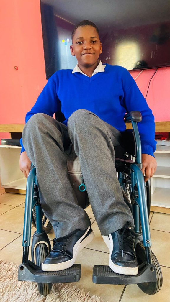
Kamogelo Sodi, who was injured in a car crash when he was just six years old, says he learned valuable skills on how to regain his independence at the Netcare Rehabilitation Hospital. The teenager enjoys cooking for himself, taking care of his three younger brothers, and playing basketball when he’s not studying hard to achieve his dream of being a medical practitioner one day.

5 September 2024: At 14 years old, Kamogelo Sodi of Alberton enjoys listening to music, chatting with his friends on social media and working hard at school towards his dream of becoming a neurosurgeon one day. He cooks for himself when he’s hungry and loves looking after his three little brothers. He also likes playing basketball. The difference between him and most other teenagers is that he does all this from his wheelchair.

“Since I’ve been in a wheelchair, I’ve become more confident,” says the vivacious teenager. “I was extremely shy, and I didn’t have a lot of friends, but now I have loads of friends.”

In 2016, when he was just six years old, Kamogelo’s life changed forever. He was in a devastating car crash, which left him with fractures in the lumbar region of his spine, resulting in complete paraplegia.

Once discharged from the hospital, where he had emergency surgery, Kamogelo was sent to the Netcare Rehabilitation Hospital to learn how to cope with, as his mother Reshoketswe Sodi calls it, his new normal. He was to stay there for almost six months.

Mrs Sodi, a radiation therapist, says the enduring care of the doctors, occupational therapists and physiotherapists there helped support Kamogelo and their family on their journey towards accepting and learning to cope with this difficult transition in his life. “It was important for me that he continued his schoolwork while there. When the social worker asked me what I wanted to happen, the first thing I said was that I didn’t want to break the routine of what he had been doing and that I wanted him to continue with school.

“It’s been a struggle, but with the help of the occupational therapists and physiotherapists, it has been an easier journey. We saw real progress when they taught Kamogelo something, and he grasped it, putting all his energy into it by thinking positively about it. It’s been hard, but with the support of the team from Netcare Rehabilitation Hospital, we managed it,” she says.

“After he was discharged, initially, we lived in a flat on the seventh floor. When the lifts weren’t working, like during load shedding, I’d have to carry him upstairs on my back – there was no other way to take him up. I’m so fortunate that I had a lot of support from my family and friends who’ve been pillars of strength for us.”

Kamogelo remembers his first visit to the Netcare Rehabilitation Hospital in Auckland Park. “When I first got to the hospital, I was lost. I didn’t know how to use a wheelchair. I was still so young. But they were so kind and taught me everything I needed to know. 

“At first, I struggled to move around. I battled to transfer myself from place to place, but they showed me what to do, and over time, I started getting used to it. I managed to start moving myself around, and I began to enjoy it. From that day forward, I didn’t like people pushing me around. The staff also taught me how to transfer myself from my wheelchair to the car. It was a bit difficult at first, but I learned to push myself up properly so my bottom wouldn’t scrape on the wheelchair.

“It does help you become more independent, but you must be consistent. You don’t need to complain about things,  you just need to listen to the people who want to help you learn to be independent.”

Later, in 2022, when he was 12 years old, Kamogelo returned to the Netcare Rehabilitation Hospital after he developed a severe pressure sore.

Dr Anrie Carstens, a doctor at the Netcare Rehabilitation Hospital, said Kamogelo was operated on at Netcare Milpark Hospital under the care of a plastic surgeon who did a flap to close the wound. “When the doctor was happy with his progress, Kamogelo came to us to help him because you get weak after surgery. The wound had healed, but the skin was delicate, so we had a graded seating approach for him to build up his strength and so that the areas of the skin didn’t break down. Another area of focus for Kamogelo was spasticity at the ankles. We worked on relaxing the ankles to get to a ninety-degree angle so he could sit better in his chair with his feet positioned well in the footrest.”

When homesickness inevitably struck, the staff comforted Kamogelo. “I began to miss home, and I cried and said I wanted to go home. They spoke nicely to me and said they first had to help me so I could go back home with no problems so my parents wouldn’t have to worry about me because of the pressure sore.”

Kamogelo said the staff also taught him valuable techniques to help him empty his bladder and bowels and assisted him in his journey to independence. “I was worried it would be painful and was a bit hesitant to try them out. But, doing it daily helped my routine and helped me become independent.”

Charne Cox, a physiotherapist at Netcare Rehabilitation Hospital, describes Kamogelo as bubbly, intelligent and with lovely manners. “He’s so motivated and tried so hard in therapy. He manages to go to school each day, not because of us, but because of his character.”

She says as children grow, their needs change. “The pressure sore developed because his seating in his wheelchair was not adequate because he had grown so much. We collaborated with the wheelchair manufacturer to re-evaluate and reassess the wheelchair seating, and they made him a new wheelchair. He was getting heavier, and his feet weren’t in alignment, so it was trickier for him to safely transfer from the wheelchair to the bed, for instance. It was good to re-educate him on pressure relief and pressure sores. It’s vital that adolescents are taught to take responsibility for themselves.”

Cox also helped Kamogelo work towards getting his feet in a better position.

“Children are so good about learning to use a wheelchair. Kamogelo was so motivated to move and be independent. He absorbed the information we gave him to enable him to go up ramps, turn and even do wheelies because he liked to explore.

“Children want to learn and have fun. They want to be independent. It’s amazing to help give them the tools to be the best new person they can be. Unfortunately, sometimes we can’t fix the injury, but we can give them the best opportunity to be as independent as possible. It’s so satisfying to know that Kamogelo is going to school and playing basketball.”

Kamogelo is determined to pursue a career as a neurosurgeon. “As long as I follow the path that I want to do and enjoy it, I will continue pursuing that path.  Academically, I was the top achiever from grade four to grade six at my school.”

When he’s not at school, he loves going around the estate he lives in, getting fresh air, and being a good big brother to his three younger brothers. “They’re a handful, but what can I say – they’re my brothers, and I love them,” he says with a laugh.

Asked who his hero is, Kamogelo is quick to say his mother and father are both his heroes. His mom clearly thinks he’s a hero too. She’s smiling as she speaks about her son. “He’s playful and has a great sense of humour. He’s helpful in the house. Instead of wanting us to help him, thanks to the skills he learned at Netcare Rehabilitation Hospital, Kamogelo always says, ‘Let me give you a hand. Let me help you.’”

More than Mobility: Gaining Independence after a Spinal Cord Injury

International Spinal Cord Injury Day marked on 5 September 2024

Photo by Elevate on Unsplash

5 September 2024, International Spinal Cord Injury Day is commemorated on Thursday 5 September, drawing attention to the many ways people can be affected by spinal cord injury, creating awareness of prevention, and highlighting the possibilities for a fulfilling life after injury.

According to the World Health Organization, globally, over 15 million people are living with spinal cord injuries. Most of these cases are due to trauma, including falls, road traffic injuries or violence.

Jessica Morris, an occupational therapist at the Netcare Rehabilitation Hospital in Auckland Park, says one of the most critical aspects of care for those who’ve been impacted by spinal cord injuries is the importance of successful rehabilitation through a holistic, integrated approach from a multidisciplinary team.

“Many people just think it’s just about mobility. It’s so much more than that. Rehabilitation is complex because many different areas of our patients’ lives are affected.” Morris says they are fortunate that their team has so many different practitioners who can contribute to treating spinal cord injury patients, helping them regain a level of independence, which is vital to their self-confidence and sense of empowerment.

Dr Anrie Carstens,  a general practitioner with a particular interest in physical medicine and rehabilitation who practises at the Netcare Rehabilitation Hospital, says the message of Spinal Cord Injury Awareness Day has relevance all year round, as people with spinal cord injuries need to be incorporated into society.

“It’s an opportunity to tell people not to be nervous to talk to someone in a wheelchair. They’re just like you or me, and they just have special ways of moving around and managing their pain and different aspects of their bodies. With the help of proper rehabilitation, the person can be better integrated as a functional, contributing member of society.”

Dr Carstens says people should also be aware that if they or their loved ones are ever impacted by a spinal cord injury, professional support is available. “Don’t just go straight home after your hospital stay and try to do everything on your own. Instead, come to a specialised spinal cord injury unit like ours, with therapists, doctors and nursing staff who are well versed in spinal cord injury and know the finer nuances necessary to optimally treat the person and show them how best to cope with their injury.

“In the multidisciplinary approach, every practitioner has a role in getting the person back into the real world, whether it means going back home, back to school, back to work or wherever they were before their injury occurred.”

From doctors and nurses with specialised skills to physiotherapists, occupational therapists, social workers and psychologists, speech therapists, a prosthetist and dieticians, the team provides a broad person focussed rehabilitation service to both adults and children. Their aim is to optimise their patients’ independence level using specialised equipment and teaching specific techniques to help overcome the obstacles a person may face.

Dr Carstens says it’s rewarding work for the staff at the hospital, who build up enduring relationships with those they care for. “One of the highlights is to compare and see what the patient was like when you admitted them and then see on discharge how much they’ve grown, how they’ve gained confidence and become more independent. What’s even better is to see them after they’ve been discharged and observe how well they’ve coped and how they’ve integrated and adjusted to their environment. We build a relationship with our patients because they stay with us for quite a while, and we usually have checkups every year after the person is discharged, often for life. We get to see them grow and thrive outside the healthcare setting, and we need more awareness about how much it is possible for people with spinal cord injuries to achieve.” 

White Matter may Aid Recovery from Spinal Cord Injuries

View of the spinal cord. Credit: Scientific Animations CC4.0

Injuries, infection and inflammatory diseases that damage the spinal cord can lead to intractable pain and disability but some degree of recovery may be possible. The question is, how best to stimulate the regrowth and healing of damaged nerves.

At the Vanderbilt University Institute of Imaging Science (VUIIS), scientists are focusing on a previously understudied part of the brain and spinal cord – white matter, which is made up of axons that relay signals. Their discoveries could lead to treatments that restore nerve activity through the targeted delivery of electromagnetic stimuli or drugs.

In a recent paper published in the Proceedings of the National Academy of SciencesAnirban Sengupta, PhD, John Gore, PhD, and their colleagues report the detection of signals from white matter in the spinal cord in response to a stimulus that are as robust as grey matter signals.

“In the spinal cord, the white matter signal is quite large and detectable, unlike in the brain, where it has less amplitude than the grey matter (signal),” said Sengupta, research instructor in Radiology and Radiological Sciences at Vanderbilt University Medical Center.

“This may be due to the larger volume of white matter in the spinal cord compared to the brain,” he added. Alternatively, the signal could represent “an intrinsic demand” in metabolism within the white matter, reflecting its critical role in supporting grey matter.

For several years, Gore, who directs the VUIIS, and his colleagues have used functional magnetic resonance imaging (fMRI) to detect blood oxygenation-level dependent (BOLD) signals, a key marker of nervous system activity, in white matter.

Last year, they reported that when participants undergoing fMRI perform a task, like wiggling their fingers, BOLD signals increase in white matter throughout the brain.

The current study monitored changes in BOLD signals in the white matter of the spinal cord at rest and in response to a vibrotactile stimulus applied to the fingers in an animal model. In response to stimulation, white matter activity was higher in “tracts” of ascending fibres that carry the signal from the spine to the brain.

This result is consistent with white matter’s known neurobiological function, the researchers noted. White matter contains non-neuronal glial cells that do not produce electrical impulses, but which regulate blood flow and neurotransmitters, the signaling molecules that transmit signals between nerve cells.

Much remains to be learned about the function of white matter in the spinal cord. But the findings from this research may help in improved understanding of diseases that affect white matter in the spinal cord, including multiple sclerosis, Sengupta said.

“We will be able to see how activity in the white matter changes in different stages of the disease,” he said. Researchers also may be able to monitor the effectiveness of therapeutic interventions, including neuromodulation, in promoting recovery following spinal cord injury.

Source: Vanderbilt University Medical Center

Scarring after Spinal Cord Injury is More Complex than Previously Thought

Fibrotic scar 14d after spinal cord injury, red – Col1a1+ perivascular fibroblast derived cells Photo: Daniel Holl

New research has found that scar formation after spinal cord injuries is more complex than previously thought. Scientists at Karolinska Institutet have identified two types of perivascular cells as key contributors to scar tissue, which hinders nerve regeneration and functional recovery. These findings, published in Natural Neuroscience, are also relevant for other brain and spinal cord injuries and could lead to targeted therapies for reducing scarring and improving outcomes.

The central nervous system (CNS) has very limited healing abilities. Injuries or autoimmune diseases like multiple sclerosis often lead to permanent functional deficits. 

Regardless of the injury’s cause, the body responds by forming a boundary around the damaged tissue, which eventually becomes permanent scar tissue. 

Two contributing cell types

While scar tissue seals the damaged area, it also prevents functional repair. After spinal cord injuries, scar tissue blocks the regeneration of nerve fibers that connect the brain with the body, resulting in paralysis after severe injuries.

The research team led by Christian Göritz at Karolinska Institutet has made significant progress in understanding how scar tissue forms in the CNS. The group now identified two distinct types of perivascular cells, which line different parts of blood vessels, as the major contributors to fibrotic scar tissue after spinal cord injury. Depending on the lesion’s location, the two identified cell types contribute differently.

“We found that damage to the spinal cord activates perivascular cells close to the damaged area and induces the generation of myofibroblasts, which consequently form persistent scar tissue,” explains first author Daniel Holl, researcher at the Department of Cell and Molecular Biology.

By examining the process of scar formation in detail, the researchers hope to identify specific therapeutic targets to control fibrotic scarring.

How Zebrafish Heal from Spinal Cord Injury could Help Humans

Photo by Cottonbro on Pexels

Zebrafish have a remarkable ability to heal their spinal cord after injury. Now, researchers at Karolinska Institutet have uncovered an important mechanism behind this phenomenon – a finding that could have implications for the treatment of spinal cord injury in humans.

In a new study published in Nature Communications, researchers show that the neurons of adult zebrafish immediately start to cooperate after a spinal cord injury, keeping the cells alive and stimulating the healing process.

“We have shown that the neurons form small channels called gap junctions, which create a direct connection between the neurons and enable the exchange of important biochemical molecules, allowing the cells to communicate and protect each other,” explains Konstantinos Ampatzis, a researcher in the Department of Neuroscience at Karolinska Institutet, who led the study.

The researchers will further investigate the exact mechanisms behind this protective strategy in zebrafish and hope this knowledge will lead to new ways of treating spinal cord injury in humans.

“Spinal cord injuries are a major burden for sufferers and their families,” says Konstantinos Ampatzis. “What if we could get human neurons to adopt the same survival strategy and behave like zebrafish neurons after an injury? This could be the key to developing new effective treatments.”

Source: Karolinka Institutet

Neurons Cause Metabolic Havoc after Spinal Injury

Conditions such as diabetes, heart attack and vascular diseases commonly diagnosed in people with spinal cord injuries can be traced to abnormal post-injury neuronal activity that causes abdominal fat tissue compounds to leak and pool in the liver and other organs, a new animal study published in Cell Reports Medicine has found.

After discovering the connection between dysregulated neuron function and the breakdown of triglycerides in fat tissue in mice, researchers found that a short course of the drug gabapentin, commonly prescribed for nerve pain, prevented the damaging metabolic effects of the spinal cord injury – though not without side effects.

Gabapentin inhibits a neural protein that, after the nervous system is damaged, becomes overactive and causes communication problems – in this case, affecting sensory neurons and the abdominal fat tissue to which they’re sending signals.

“We believe there is maladaptive reorganisation of the sensory system that causes the fat to undergo changes, initiating a chain of reactions – triglycerides start breaking down into glycerol and free fatty acids that are released in circulation and taken up by the liver, the heart, the muscles, and accumulating, setting up conditions for insulin resistance,” said senior author Andrea Tedeschi, assistant professor of neuroscience in The Ohio State University College of Medicine.

“Through administration of gabapentin, we were able to normalise metabolic function.”

Previous research has found that cardiometabolic diseases are among the leading causes of death in people who have experienced a spinal cord injury. These often chronic disorders can be related to dysfunction in visceral white fat (or adipose tissue), which has a complex metabolic role of storing energy and releasing fatty acids as needed for fuel, but also helping keep blood sugar levels at an even keel.

Earlier investigations of these diseases in people with neuronal damage have focused on adipose tissue function and the role of the sympathetic nervous system, but also a regulator of adipose tissue that surrounds the abdominal organs.

Instead, Debasish Roy, a postdoctoral researcher in the Tedeschi lab and first author on the paper, decided to focus on sensory neurons in this context. Tedeschi and colleagues have previously shown that a neuronal receptor protein called alpha2delta1 is overexpressed after spinal cord injury, and its increased activation interferes with post-injury function of axons, the long, slender extensions of nerve cell bodies that transmit messages.

In this new work, researchers first observed how sensory neurons connect to adipose tissue under healthy conditions, and created a spinal cord injury mouse model that affected only those neurons – without interrupting the sympathetic nervous system.

Experiments revealed a cascade of abnormal activity within seven days after the injury in neurons – though only in their communication function, not their regrowth or structure – and in visceral fat tissue. Expression of the alpha2delta1 receptor in sensory neurons increased as they over-secreted a neuropeptide called CGRP, all while communicating through synaptic transmission to the fat tissue – which, in a state of dysregulation, drove up levels of a receptor protein that engaged with the CGRP.

“These are quite rapid changes. As soon as we disrupt sensory processing as a result of spinal cord injury, we see changes in the fat,” Tedeschi said. “A vicious cycle is established – it’s almost like you’re pressing the gas pedal so your car can run out of gas but someone else continues to refill the tank, so it never runs out.”

The result is the spillover of free fatty acids and glycerol from fat tissue, a process called lipolysis, that has gone out of control. Results also showed an increase in blood flow in fat tissue and recruitment of immune cells to the environment.

“The fat is responding to the presence of CGRP, and it’s activating lipolysis,” Tedeschi said. “CGRP is also a potent vasodilator, and we saw increased vascularisation of the fat – new blood vessels forming as a result of the spinal cord injury. And the recruitment of monocytes can help set up a chronic pro-inflammatory state.”

Silencing the genes that encode the alpha2delta1 receptor restored the fat tissue to normal function, indicating that gabapentin – which targets alpha2delta1 and its partner, alpha2delta2 – was a good treatment candidate. Tedeschi’s lab has previously shown in animal studies that gabapentin helped restore limb function after spinal cord injury and boosted functional recovery after stroke.

But in these experiments, Roy discovered something tricky about gabapentin: the drug prevented changes in abdominal fat tissue and lowered CGRP in the blood, in turn preventing spillover of fatty acids into the liver a month later, establishing normal metabolic conditions. But paradoxically, the mice developed insulin resistance, a known side effect of gabapentin.

The team instead tried starting with a high dose, tapering off and stopping after four weeks.

“This way, we were able to normalise metabolism to a condition much more similar to control mice,” Roy said. “This suggests that as we discontinue administration of the drug, we retain beneficial action and prevent spillover of lipids in the liver. That was really exciting.”

Finally, researchers examined how genes known to regulate white fat tissue were affected by targeting alpha2delta1 genetically or with gabapentin, and found both of these interventions after spinal cord injury suppress genes responsible for disrupting metabolic functions.

Tedeschi said the combined findings suggest starting gabapentin treatment early after a spinal cord injury may protect against detrimental conditions involving fat tissue that lead to cardiometabolic disease – and could enable discontinuing the drug while retaining its benefits and lowering the risk for side effects.

Source: Ohio State University

How Spinal Cords can ‘Learn’ without Brain Involvement

In this study, spinal cords that associated limb position with an unpleasant experience learned to reposition the limb after only 10 minutes, and retained a memory the next day. Spinal cords that received random unpleasantness did not learn. Credit: RIKEN

Researchers in Japan have discovered the neural circuitry in the spinal cord that allows brain-independent motor learning. This study by Aya Takeoka at the RIKEN Center for Brain Science and colleagues found two critical groups of spinal cord neurons, one necessary for new adaptive learning, and another for recalling adaptations once they have been learned. The findings, published in Science, could help scientists develop ways to assist motor recovery after spinal cord injury.

It has been long been known that motor output from the spinal cord can be adjusted through practice even without a brain. This has been shown most dramatically in headless insects, whose legs can still be trained to avoid external cues. Until now, no one has figured out exactly how this is possible, and without this understanding, the phenomenon is not much more than a quirky fact. As Takeoka explains, “Gaining insights into the underlying mechanism is essential if we want to understand the foundations of movement automaticity in healthy people and use this knowledge to improve recovery after spinal cord injury.”

Before jumping into the neural circuitry, the researchers first developed an experimental setup that allowed them to study mouse spinal cord adaptation, both learning and recall, without input from the brain. Each test had an experimental mouse and a control mouse whose hindlegs dangled freely. If the experimental mouse’s hindleg drooped down too much it was electrically stimulated, emulating something a mouse would want to avoid. The control mouse received the same stimulation at the same time, but not linked to its own hindleg position.

After just 10 minutes, they observed motor learning only in the experimental mice; their legs remained high up, avoiding any electrical stimulation. This result showed that the spinal cord can associate an unpleasant feeling with leg position and adapt its motor output so that the leg avoids the unpleasant feeling, all without any need for a brain. Twenty-four hours later, they repeated the 10-minute test but reversed the experimental and control mice. The original experimental mice still kept their legs up, indicating that the spinal cord retained a memory of the past experience, which interfered with new learning.

Having thus established both immediate learning, as well as memory, in the spinal cord, the team then set out to examine the neural circuitry that makes both possible. They used six types of transgenic mice, each with a different set of spinal neurons disabled, and tested them for motor learning and learning reversal. They found that mice hindlimbs did not adapt to avoid the electrical shocks after neurons toward the top of the spinal cord were disabled, particularly those that express the gene Ptf1a.

When they examined the mice during learning reversal, they found that silencing the Ptf1a-expressing neurons had no effect. Instead, a group of neurons in the ventral part of the spinal cord that express the En1 gene was critical. When these neurons were silenced the day after learning avoidance, the spinal cords acted as if they had never learned anything. The researchers also assessed memory recall on the second day by repeating the initial learning conditions. They found that in wildtype mice, hindlimbs stabilised to reach the avoidance position faster than they did on the first day, indicating recall. Exciting the En1 neurons during recall increased this speed by 80%, indicating enhanced motor recall.

“Not only do these results challenge the prevailing notion that motor learning and memory are solely confined to brain circuits,” says Takeoka, “but we showed that we could manipulate spinal cord motor recall, which has implications for therapies designed to improve recovery after spinal cord damage.”

Source: RIKEN