Electrotherapy using injectable nanoparticles delivered directly into the tumour could pave the way for new treatment options for glioblastoma, according to a new study from Lund University in Sweden.
Glioblastoma is the most common and most aggressive form of brain tumour among adults. Even with intensive treatment, the average survival period is 15 months. The tumour has a high genetic variation with multiple mutations, which often makes it resistant to radiation therapy, chemotherapy and many targeted drugs. The prognosis for glioblastoma has not improved over the past few decades despite extensive research.
Electrotherapy – a new treatment method
Electrotherapy offers another strategy to combat solid tumours. Using short, strong electric pulses (irreversible electroporation), non-reversible pores are created in the cancer cells leading to their death. The body’s immune system is simultaneously stimulated. The problem is that surgery is required to place the stiff metal electrodes that are necessary for the treatment. In sensitive tissue, in the brain for example, this often entails a very difficult procedure, which has led to strict criteria regarding which patients can be treated. Johan Bengzon is a researcher in glioblastoma and adjunct professor at Lund University, and consultant in neurosurgery at the Skåne University Hospital. He regularly treats patients with glioblastoma and is frustrated by the limited treatment options.
“The short distance between the hospital and the University in Lund facilitates cooperation and that’s why I contacted research colleagues to find out if injectable electrodes could be an alternative solution in electrotherapy,” says Johan Bengzon.
Said and done. The research team, with Amit Singh Yadav, Martin Hjort, and Roger Olsson at the helm, had previously used nanoparticles to form injectable and electrically conductive hydrogels to control brain signalling and heart contractions. It is aminimally invasive method in which the particles are injected using a thin syringe directly into the body. The particles break down after the treatment and thus do not need to be surgically removed. Perhaps the same technology could be used to destroy tumour cells in glioblastoma.
“After surgical treatment, unfortunately the glioblastoma tumour often returns on the outer edge of the area operated on. By drop casting the nanoparticles into the tumour cavity after an operation, we could electrify the edges while the immune system is also activated. In animal models the procedure, due to this irreversible electroporation, led to tumours being wiped out within three days,” says Roger Olsson, professor of chemical biology and drug development at Lund University, who led the study.
Promising results – but a long way to the patient
The prospects are good and the researchers are very hopeful for the future, even though there is a long way to go before it becomes a clinical reality. The challenge is now to test the method on larger tumours.
“We have seen that the electrode is well received in the brain. We have not noted any problems relating to side effects and after 12 weeks the electrode disappeared by itself as it’s biodegradable. The technology combines direct tumour destruction with activation of the immune system and can be an important step towards more effective treatment of glioblastoma,” concludes Amit Singh Yadav, researcher at Lund University and first author of the study.
Image credit: University of Maryland School of Medicine
Patients with glioblastoma who received MRI-guided focused ultrasound with standard-of-care chemotherapy had a nearly 40% increase in overall survival in a landmark trial of 34 patients led by University of Maryland School of Medicine (UMSOM) researchers. This is the first time researchers have demonstrated a potential survival benefit from using focused ultrasound to open the blood-brain barrier to improve delivery of chemotherapy to the tumour site in brain cancer patients after surgery.
“Our results are very encouraging. Using focused ultrasound to open the blood-brain barrier and deliver chemotherapy could significantly increase patient survival, which other ongoing studies are seeking to confirm and expand,” said study principal investigator Graeme Woodworth, MD, Professor and Chair of Neurosurgery at UMSOM and Neurosurgeon-In-Chief at the University of Maryland Medical Center (UMMC).
The findings of this groundbreaking safety, feasibility, and comparative trial involved glioblastoma patients who were given focused ultrasound to open their blood-brain barrier before getting chemotherapy; they were matched to a rigorously selected control group of 185 glioblastoma patients with similar characteristics who received the standard dose of the chemotherapy drug, temozolomide, without receiving focused ultrasound. Trial participants were initially treated with surgery to remove their brain tumour, followed by six weeks of chemotherapy and radiation, and up to six monthly focused-ultrasound treatments plus temozolomide.
Results were published in the journal Lancet Oncology and show that trial participants had nearly 14 months of median progression-free survival, compared to eight months in the control group. In terms of overall survival, trial participants, on average, lived for more than 30 months compared to 19 months in the control group.
The study builds on more than a decade of intensive research to test the safety and feasibility of opening the blood-brain barrier using focused ultrasound first in animal studies and then in patients. It was led by Dr Woodworth and was conducted at UMMC and four other university-affiliated clinical sites. “We also demonstrated that this could be a useful technique that enables us to better monitor patients to determine if their brain cancer has progressed,” said Dr Woodworth, who also serves as Director of the Brain Tumor Program at the University of Maryland Marlene and Stewart Greenebaum Comprehensive Cancer Center (UMGCCC).
He and his team demonstrated that opening the blood-brain barrier facilitated the use of a “liquid biopsy,” which is a blood test that detects cancer biomarkers, which can include DNA fragments, proteins and other components from the liquid environment surrounding the tumor site.
Such biomarkers have been used in other cancers to determine whether the tumor has remained stable or has the potential to progress or even metastasize. Up until now, however, these tests have not been utilized in brain cancer patients since most components can never pass into the bloodstream from the brain due to the blood-brain barrier.
“These liquid biomarkers were found to be closely concordant with the patient outcomes over time, progression-free survival and overall survival,” said Dr Woodworth.
While temozolomide is the standard treatment for glioblastoma, the drug typically gets blocked by the blood-brain barrier with studies showing that less than 20 percent reaches the brain in patients. This study did not determine the exact amount of temozolomide to reach the brain in each patient, but earlier studies have shown that opening the blood-brain barrier before delivering chemotherapy can dramatically increase the amount that gets to the original tumor site.
Glioblastoma is the most common and deadliest type of malignant brain tumour. The five-year survival rate is only 5.5%, and patients live an average of 14 to 16 months after diagnosis when treated with surgery, radiation, and chemotherapy when appropriate. The malignancy nearly always recurs even after it is removed due to residual infiltrating cancer cells that remain after treatment.
The blood-brain barrier is a specialized network of vascular and brain cells that acts as the brain’s security system to protect against invasion by dangerous toxins and microbes. It can be opened temporarily using a specialised focused ultrasound device. This process starts with injecting microscopic inert gas-filled bubbles into the patient’s bloodstream. Guided by an MRI, precise brain regions are targeted while the injected microbubbles are circulating.
“Upon excitation under low-intensity ultrasound waves, the microbubbles oscillate within the energy field, causing temporary mechanical perturbations in the walls of the brain blood vessels,” said Pavlos Anastasiadis, PhD, an Assistant Professor of Neurosurgery at UMSOM who is an expert in ultrasound biophysics.
Prior studies led by Dr Woodworth and this trial’s co-investigators showed that opening the blood-brain barrier temporarily can be safely and feasibly performed in brain tumour patients. He and his team conducted this procedure in the first brain cancer patient in the US in 2018 at UMMC after the US Food and Drug Administration (FDA) approved the inaugural clinical trial.
Future trials could use focused ultrasound alongside other chemotherapy agents to test the effectiveness of drugs never used in brain cancer due to their ineffectiveness at crossing the blood-brain barrier.
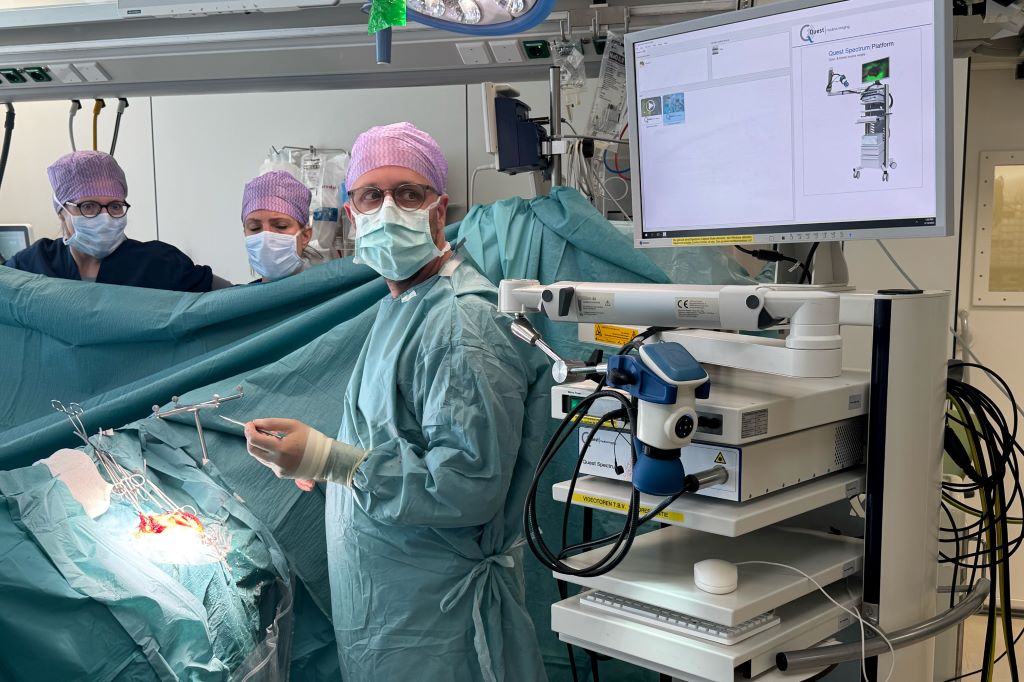
A medical team at Erasmus University Medical Center in the Netherlands uses the new imaging probe with a Quest camera to get a better view of cancerous tumors during non-brain cancer surgery. Photo courtesy of Erasmus University Medical Center
In a significant leap forward for successful cancer surgery, researchers at the University of Missouri and collaborators have developed a new imaging probe to help surgeons more accurately identify and remove aggressive tumours during operations.
The tool is expected to be a critical advancement in the fight against glioblastoma, one of the most difficult-to-treat brain cancers. In the future, it is intended to be expanded for image-guided surgery of various other solid tumours.
Described in a new study in Nature Publishing Group Imaging, the innovation works by pairing a fluorescent dye with a fatty acid molecule that cancer cells readily absorb. When introduced into the body, the compound is taken up by tumour cells, causing them to glow under near-infrared light, revealing cancer that might otherwise remain hidden.
Glioblastoma is considered surgically incurable because the tumour doesn’t stay in one place – it spreads and invades healthy brain tissue in a diffuse, microscopic way. This makes it impossible to remove completely without risking serious damage to brain function.
“Surgery remains one of the primary treatments for many cancers,” Elena Goun, associate professor of chemistry in the College of Arts and Science and one of the lead authors of the study, said. “In breast or prostate cancer, surgeons can often remove the tumour along with surrounding tissue. In brain cancer, that’s simply not possible. You must preserve healthy brain tissue. But if even a few cancer cells are left behind, the disease will return.”
That dilemma is especially acute with glioblastoma, which doesn’t form a neatly contained mass. Instead, it sends out microscopic extensions — finger-like projections that blend into healthy brain tissue and are invisible to the naked eye.
Because of this, surgeons must walk a fine line: removing as much tumour as possible while avoiding harm to vital brain areas. The more thoroughly the tumour is removed, the more effective follow-up treatments like radiation and chemotherapy tend to be.
The new small-molecule probe, known as FA-ICG, is engineered to solve that problem. It links a natural long-chain fatty acid (FA) to indocyanine green (ICG), an FDA-approved near-infrared dye widely used in surgical imaging. This fatty acid-based approach means the probe is highly selective: glioblastoma cells, which thrive on fatty acids, absorb it more than normal brain cells. That makes the cancer stand out more clearly.
The result is a tool that takes advantage of cancer’s altered metabolism to highlight tumour cells from within.
“Surgeons would view a monitor during surgery showing where the probe is lighting up,” Goun explained. “If they still see fluorescent signals, it means cancer is still present and more tissue needs to be removed. When the light disappears, they would know they’ve cleared the area.”
In the operating room, surgeons already use a variety of tools to guide tumour removal – including microscopes, ultrasound and fluorescent dyes. Of those, fluorescent dyes are particularly useful because they make otherwise invisible tumour cells light up under special lighting.
Right now, the only approved imaging dye for glioblastoma surgery is 5-ALA, which fluoresces under blue light. But 5-ALA comes with major limitations: The operating room must be darkened in order to see it, tissue penetration is shallow and the fluorescent signal is often weak and non-specific.
It also comes with side effects, including photosensitivity, meaning patients must avoid bright light exposure after surgery due to the risk of skin and eye damage.
That’s where the FA-ICG probe shines – both literally and functionally.
Compared to 5-ALA, FA-ICG is brighter, works under normal surgical lighting, and offers real-time visualisation under the microscope – no need to turn the lights off mid-surgery. This saves time and makes procedures more efficient. The signal-to-background ratio is also higher, meaning it’s easier to distinguish tumour tissue from healthy brain.
The FA-ICG probe is not only easier to see, it’s also easier to use. Its longer half-life allows more flexibility in scheduling surgeries, and the logistics of administration are simpler than with current probes.
“The upside of fluorescence-guided surgery is that you can make little remnants much more visible using the light emitting properties of these tumour cells when you give them a dye,” said Rutger Balvers, a neurosurgeon at Erasmus University Medical Center in the Netherlands, who is expected to lead human clinical trials of the probe. “And we think that the upside of FA-ICG compared to what we have now is that it’s more select in targeting tumour cells. The visual properties of the probe are better than what we’ve used before.”
Michael Chicoine is a neurosurgeon at MU Health Care and chair of Mizzou’s School of Medicine’s Department of Neurosurgery. While he’s not directly involved in the research, Chicoine understands the potential benefits firsthand.
Currently, he said, MRIs are the gold standard for imaging tumours; however, they’re expensive and time-consuming, especially when required during an operation.
“This fluorescent metabolically linked tool gives you real-time imaging,” he said. “We could merge techniques, using the probe during surgery and saving the MRI for a sort of final exam. It’s definitely an exciting advancement.”
Researchers are also excited about other uses for the probe, including for other types of cancers and for use during follow-up treatments.
“After radiation or chemotherapy, it becomes very difficult to distinguish between scar tissue and active tumor,” Chicoine said. “This probe could give us a definitive answer – helping doctors know whether to continue treatment or adjust it, or consider another surgery. Eliminating the current uncertainty would be really helpful.”
Another promising use of the probe could be in photodynamic therapy either during or after surgery. Since the dye also has light-activated properties that can kill cancer cells, researchers are exploring whether it could double as a treatment tool, not just a diagnostic one.
Clinical trials for use in glioblastoma cases are expected to start in Europe, with strong interest already growing among neurosurgical teams.
The upcoming Phase 1 trial will focus on how patients tolerate the probe, whether there are any side effects at an effective dose and how its performance compares to existing tools. Ultimately, the goal is to make brain tumour surgery safer, helping surgeons remove all cancerous tissues while preserving as much healthy brain tissue as possible.
If results are positive, future studies could expand the use of FA-ICG beyond brain tumours to other cancers with high fatty acid metabolism, such as pancreatic cancer,according to fellow corresponding author Laura Mezzanotte from the Erasmus’ Department of Radiology and Nuclear Medicine.
A new study led by Keck Medicine of USC researchers may have uncovered an effective combination therapy for glioblastoma, a brain tumour diagnosis with few available effective treatments. According to the National Brain Tumor Society, the average survival for patients diagnosed with glioblastoma is eight months.
The study, which was published in the journal Med, finds that using Tumour Treating Fields therapy (TTFields), which delivers targeted waves of electric fields directly into tumours to stop their growth and signal the body’s immune system to attack cancerous tumour cells, may extend survival among patients with glioblastoma, when combined with immunotherapy (pembrolizumab) and chemotherapy (temozolomide).
TTFields disrupt tumour growth using low-intensity, alternating electric fields that push and pull key structures inside tumour cells in continually shifting directions, making it difficult for the cells to multiply. Preventing tumour growth gives patients a better chance of successfully fighting the cancer. When used to treat glioblastoma, TTFields are delivered through a set of mesh electrodes that are strategically positioned on the scalp, generating fields at a precise frequency and intensity focused on the tumour. Patients wear the electrodes for approximately 18 hours a day.
Researchers observed that TTFields attract more tumour-fighting T cells, which are white blood cells that identify and attack cancer cells, into and around the glioblastoma. When followed by immunotherapy, these T cells stay active longer and are replaced by even stronger, more effective tumour-fighting T cells.
“By using TTFields with immunotherapy, we prime the body to mount an attack on the cancer, which enables the immunotherapy to have a meaningful effect in ways that it could not before,” said David Tran, MD, PhD, chief of neuro-oncology with Keck Medicine, co-director of the USC Brain Tumor Center and corresponding author of the study. “Our findings suggest that TTFields may be the key to unlocking the value of immunotherapy in treating glioblastoma.”
TTFields are often combined with chemotherapy in cancer treatment. However, even with aggressive treatment, the prognosis for glioblastoma remains poor. Immunotherapy, while successful in many other cancer types, has also not proved effective for glioblastoma when used on its own.
However, in this study, adding immunotherapy to TTFields and chemotherapy was associated with a 70% increase in overall survival. Notably, patients with larger, unresected (not surgically removed) tumours showed an even stronger immune response to TTFields and lived even longer. This suggests that, when it comes to kick-starting the body’s immune response against the cancer, having a larger tumour may provide more targets for the therapy to work against.
Using alternating electric fields to unlock immunotherapy
Pembrolizumab, the immunotherapy used in this study, is an immune checkpoint inhibitor (ICI), which enhances the body’s natural ability to fight cancers by improving T cells’ ability to identify and attack cancer cells.
However, there are typically few T cells in and around glioblastomas because these tumours originate in the brain and are shielded from the body’s natural immune response by the blood-brain barrier. This barrier safeguards the brain by tightly regulating which cells and substances enter from the bloodstream. Sometimes, this barrier even blocks T cells and other therapies that could help kill brain tumours.
This immunosuppressive environment inside and around the glioblastoma is what makes common cancer therapies like pembrolizumab and chemotherapy significantly less effective in treating it. Tran theorised the best way to get around this issue was to start an immune reaction directly inside the tumour itself, an approach known as in situ immunisation, using TTFields.
This study demonstrates that combining TTFields with immunotherapy triggers a potent immune response within the tumour – one that ICIs can then amplify to bolster the body’s own defence against cancer.
“Think of it like a team sport – immunotherapy sends players in to attack the tumour (the offence), while TTFields weaken the tumour’s ability to fight back (the defence). And just like in team sports, the best defence is a good offence,” said Tran, who is also a member of the USC Norris Comprehensive Cancer Center.
Study methodology and results
The study analysed data from 2-THE-TOP, a Phase 2 clinical trial, which enrolled 31 newly diagnosed glioblastoma patients who had completed chemoradiation therapy. Of those, 26 received TTFields combined with both chemotherapy and immunotherapy. Seven of these 26 patients had inoperable tumours due to their locations – an especially high-risk subgroup with the worst prognosis and few treatment options.
Patients in the trial were given six to 12 monthly treatments of chemotherapy alongside TTFields for up to 24 months. The number and duration of treatments were determined by patients’ response to treatment. The immunotherapy was given every three weeks, starting with the second dose of chemotherapy, for up to 24 months.
Patients who used the device alongside chemotherapy and immunotherapy lived approximately 10 months longer than patients who had used the device with chemotherapy alone in the past. Moreover, those with large, inoperable tumours lived approximately 13 months longer and showed much stronger immune activation compared to patients who underwent surgical removal of their tumours.
“Further studies are needed to determine the optimal role of surgery in this setting, but these findings may offer hope, particularly for glioblastoma patients who do not have surgery as an option,” said Tran.
The researchers are now moving ahead to a Phase 3 trial.
Results of a trial led by the university and reported in Nature Communications revealed that a unique investigational drug formulation called Rhenium Obisbemeda (186RNL) more than doubled median survival and progression-free time, compared with standard median survival and progression rates, and with no dose-limiting toxic effects.
“As a disease with a pattern of recurrence, resistance to chemotherapies and difficulty to treat, glioblastoma has needed durable treatments that can directly target the tumour while sparing healthy tissue,” said lead author Andrew J. Brenner, MD, PhD, professor and chair of neuro-oncology research with Mays Cancer Center at UT Health San Antonio. “This trial provides hope, with a second trial under way and planned for completion by the end of this year.”
Brenner said that the median overall survival time for patients with glioblastoma after standard treatment fails with surgery, radiation and chemotherapy is only about 8 months. More than 90% of patients have a recurrence of the disease at its original location.
Rhenium Obisbemeda enables very high levels of a specific activity of rhenium-186 (186Re), a beta-emitting radioisotope, to be delivered by tiny liposomes, referring to artificial vesicles or sacs having at least one lipid bilayer. The researchers used a custom molecule known as BMEDA to chelate or attach 186Re and transport it into the interior of a liposome where it is irreversibly trapped.
In this trial, known as the phase 1 ReSPECT-GBM trial, scientists set out to determine the maximum tolerated dose of the drug, as well as safety, overall response rate, disease progression-free survival and overall survival.
After failing one to three therapies, 21 patients who were enrolled in the study between March 5, 2015, and April 22, 2021, were treated with the drug administered directly to the tumours using neuronavigation and convection catheters.
The researchers observed a significant improvement in survival compared with historical controls, especially in patients with the highest absorbed doses, with a median survival and progression-free time of 17 months and 6 months, respectively, for doses greater than 100Gy.
Importantly, they did not observe any dose-limiting toxic effects, with most adverse effects deemed unrelated to the study treatment.
“The combination of a novel nanoliposome radiotherapeutic delivered by convection-enhanced delivery, facilitated by neuronavigational tools, catheter design and imaging solutions, can successfully and safely provide high absorbed radiation doses to tumours with minimal toxicity and potential survival benefit,” Brenner concluded.
Patients with glioblastoma typically survive less than two years after diagnosis, even with cutting-edge therapies. The latest immunotherapies have been unsuccessful, likely because glioblastoma cells have few, if any, natural targets for the immune system to attack.
In a cell-based study, scientists at Washington University School of Medicine have forced glioblastoma cells to display immune system targets, potentially making them visible to immune cells and newly vulnerable to immunotherapies. The strategy involves a combination of two drugs, each already FDA-approved to treat different cancers.
“For patients whose tumours do not naturally produce targets for immunotherapy, we showed there is a way to induce their generation,” said co-senior author Ting Wang, PhD, professor of medicine and Department of Genetics head at WashU Medicine. “In other words, when there is no target, we can create one. This is a very new way of designing targeted and precision therapies for cancer. We are hopeful that in the near future we will be able to move into clinical trials, where immunotherapy can be combined with this strategy to provide new therapeutic approaches for patients with very hard-to-treat cancers.”
To create immune targets on cancer cells, Wang has focused on stretches of DNA in the genome known as transposable elements. In recent years, transposable elements have emerged as a double-edged sword in cancer, according to Wang. His work has shown that transposable elements play a role in causing tumours to develop even as they present vulnerabilities that could be exploited to create new cancer treatment strategies.
For this study, Wang’s team took advantage of the fact that transposable elements naturally can cause a tumour to churn out random proteins that are unique to the tumour and not present in normal cells. Called tumour antigens or neoantigens, these unusual proteins could be the targets for immunotherapies, such as checkpoint inhibitors, antibodies, vaccines and genetically engineered T cell therapies.
Even so, some tumours, including glioblastoma, have few immune targets produced naturally by transposable elements. To address this, Wang and his colleagues, including co-senior author Albert H. Kim, MD, PhD, neurosurgery professor, have demonstrated how to purposely force transposable elements to produce immune system targets on glioblastoma cells that normally lack them.
The researchers used a combination of two drugs that influence the epigenome, which controls gene activation. When treated with the two epigenetic therapy drugs, the tightly packed DNA molecules of the glioblastoma cells unfurled, triggering transposable elements to begin making the unusual proteins that could be used to target the cancer cells. The two drugs were decitabine, which is approved to treat myelodysplastic syndromes, a group of blood cancers; and panobinostat, which is approved for multiple myeloma, a cancer of white blood cells.
Before investigating this strategy in people, the researchers are seeking ways to target the epigenetic therapy so that only the tumour cells are induced to make neoantigens. In the new study, the researchers cautioned that normal cells also produced targets when exposed to the two drugs. Even though normal cells didn’t produce as many neoantigens as the glioblastoma cells did, Wang and Kim said there is a risk of unwanted side effects if normal cells create these targets as well.
In ongoing work, Wang and Kim are investigating how to use CRISPR molecular editing technology to induce specific parts of the genome in cancer cells to produce the same neoantigens from transposable elements that are common across the human population. Such a strategy could give many patients’ tumours – even different cancer types – the same targets that could respond to the same immunotherapy, while sparing healthy cells. There are then multiple possible ways to go after such a shared target, including checkpoint inhibitors, vaccines, engineered antibodies and engineered T cells.
Researchers from Germany have made a surprising discovery about the aggressive, lethal glioblastomas: in the vicinity of glioblastomas, they found islands of highly potent immune cells in the neighbouring bone marrow of the skull, which play a central role in defending against cancer. The new data may open up prospects for innovative therapies. On the other hand, they cast a shadow over conventional strategies.
“What we have found is surprising and fundamentally new,” says Björn Scheffler, German Cancer Consortium (DKTK) researcher at the Essen site. Until now, the body’s own defences have always been thought of as a holistic system that sends its troops to different parts of the body as required. “However,” says Scheffler, “our data show that highly potent immune cells gather in regional bone marrow niches close to the tumour and organise the defence from there. At least this is the case with glioblastomas.”
Immune system on site
Based on new findings from animal experiments, the Essen team took tissue samples from the bone marrow near the tumor in the skull from untreated patients with glioblastoma. “However, the methods for this first had to be established,” reports first author Celia Dobersalske, emphasizing the fact that the new research results were obtained from human tissue samples.
The researchers hit the bull’s eye in their search: Bone marrow niches in close proximity to the glioblastoma appear to be the reservoir from which the anti-tumour defence is recruited. Apart from active lymphoid stem cells that develop into immune cells, the researchers also found mature cytotoxic T lymphocytes (CD8 cells) in the bone marrow close to the tumour. “These are highly effective immune cells that play a central role in the defence against cancer,” adds Celia Dobersalske. They can recognize and destroy malignant cells.
The CD8 cells in the bone marrow near the tumour had an increased number of receptors on their surface, which control the proliferation of mature T lymphocytes. In line with this, descendants of the same cell clones – one clone originates from one and the same cell – were detected both in the bone marrow and in the tumour tissue. This is clear evidence that the immune cells gathered on site are fighting the glioblastoma. “And they are successful – at least for a while,” says Björn Scheffler. “We were able to show that the course of the disease correlates with the activity of the local CD8 cells.”
Valuable immune cells destroyed?
This finding not only turns conventional ideas about how the immune system works on their head. The treatment concepts for glioblastoma must also be reconsidered in light of the new data. “Until now, we hadn’t even considered the skullcap in our considerations. How could we, since there was no evidence that highly potent immune cells could be hiding there,” says senior author Scheffler.
“When we opened the skull, we may have destroyed important immune cells in the process,” confirms Ulrich Sure, Director of the Department of Neurosurgery and member of the Essen research team. “In view of the new findings, we find ourselves in a dilemma: we have to gain access to the tumour in order to remove it and also to be able to confirm the diagnosis. There is currently no other way than through the skull. But we are thinking about how we can minimise damage to the local bone marrow in the future.”
On the other hand, the discovery of the local immune system opens up opportunities for innovative therapies. In particular, so-called checkpoint inhibitors are coming back into play. These are immunotherapeutic agents that aim to boost the body’s own cancer defenses. However, checkpoint inhibitors tested to date have shown little effect on glioblastomas.
“Various explanations have been suggested as an explanation, but perhaps we also need to rethink things in this respect,” says Björn Scheffler. “We now know that highly potent immune cells are indeed present on site. We were able to prove that they are fit to fight tumours, but they are not capable of destroying the tumour on their own. This is where we can start. One challenge will be to deliver drugs in sufficient concentration to the regional bone marrow niches at the right time. If we succeed, we may have a chance of controlling the growth of glioblastomas and improving our patients’ chances of survival.”
In a first-ever human clinical trial of four adult patients, an mRNA cancer vaccine developed at the University of Florida quickly reprogrammed the immune system to attack glioblastoma, the most aggressive and lethal brain tumour.
The results mirror those in 10 pet dog patients suffering from naturally occurring brain tumours whose owners approved of their participation, as they had no other treatment options, as well as results from preclinical mouse models. Next, the researchers will test the treatment in a Phase 1 paediatric clinical trial.
This breakthrough, published in Cell, represents a potential new way to recruit the immune system to fight notoriously treatment-resistant cancers using an iteration of mRNA technology and lipid nanoparticles, similar to COVID vaccines, but with two key differences: use of a patient’s own tumour cells to create a personalised vaccine, and a newly engineered complex delivery mechanism within the vaccine.
“Instead of us injecting single particles, we’re injecting clusters of particles that are wrapping around each other like onions, like a bag full of onions,” said senior author Elias Sayour, MD, PhD, a UF Health paediatric oncologist who pioneered the new vaccine, which like other immunotherapies attempts to “educate” the immune system that a tumour is foreign. “And the reason we’ve done that in the context of cancer is these clusters alert the immune system in a much more profound way than single particles would.”
Among the most impressive findings was how quickly the new method, delivered intravenously, spurred a vigorous immune-system response to reject the tumour, said Sayour, principal investigator of the RNA Engineering Laboratory within UF’s Preston A. Wells Jr. Center for Brain Tumor Therapy and a UF Health Cancer Center and McKnight Brain Institute investigator who led the multi-institution research team.
“In less than 48 hours, we could see these tumours shifting from what we refer to as ‘cold’ – immune cold, very few immune cells, very silenced immune response – to ‘hot,’ very active immune response,” he said. “That was very surprising given how quick this happened, and what that told us is we were able to activate the early part of the immune system very rapidly against these cancers, and that’s critical to unlock the later effects of the immune response.”
Glioblastoma is among the most devastating diagnoses, with median survival around 15 months. Current standard of care involves surgery, radiation and some combination of chemotherapy.
The new publication is the culmination of promising translational results over seven years of studies, starting in preclinical mouse models and then in a clinical trial of 10 pet dogs that had spontaneously developed terminal brain cancer and had no other treatment options. That trial was conducted with owners’ consent in collaboration with the UF College of Veterinary Medicine. Dogs offer a naturally occurring model for malignant glioma because they are the only other species that develops spontaneous brain tumors with some frequency, said Sheila Carrera-Justiz, DVM., a veterinary neurologist at the UF College of Veterinary Medicine who is partnering with Sayour on the clinical trials. Gliomas in dogs are universally terminal, she said.
After treating pet dogs that had spontaneously developed brain cancer with personalised mRNA vaccines, Sayour’s team advanced the research to a small Food and Drug Administration-approved clinical trial designed to ensure safety and test feasibility before expanding to a larger trial.
In a cohort of four patients, RNA was extracted from each patient’s own surgically removed tumour, and then messenger RNA, or mRNA was amplified and wrapped in the newly designed high-tech packaging of biocompatible lipid nanoparticles, to make tumour cells “look” like a dangerous virus when reinjected into the bloodstream and prompt an immune-system response. The vaccine was personalised to each patient with a goal of getting the most out of their unique immune system.
“The demonstration that making an mRNA cancer vaccine in this fashion generates similar and strong responses across mice, pet dogs that have developed cancer spontaneously and human patients with brain cancer is a really important finding, because oftentimes we don’t know how well the preclinical studies in animals are going to translate into similar responses in patients,” said Duane Mitchell, M.D., PhD, director of the UF Clinical and Translational Science Institute and the UF Brain Tumor Immunotherapy Program and a co-author of the paper. “And while mRNA vaccines and therapeutics are certainly a hot topic since the COVID pandemic, this is a novel and unique way of delivering the mRNA to generate these really significant and rapid immune responses that we’re seeing across animals and humans.”
While too early in the trial to assess the clinical effects of the vaccine, the patients either lived disease-free longer than expected or survived longer than expected. The 10 pet dogs lived a median of 139 days, compared with a median survival of 30 to 60 days typical for dogs with the condition.
The next step will be an expanded Phase I clinical trial to include up to 24 adult and paediatric patients to validate the findings. Once an optimal and safe dose is confirmed, an estimated 25 children would participate in Phase 2, said Sayour.
Despite the promising results, the authors said one limitation is continued uncertainty about how best to harness the immune system while minimising the potential for adverse side effects.
“I am hopeful that this could be a new paradigm for how we treat patients, a new platform technology for how we can modulate the immune system,” Sayour said. “I am hopeful for how this could now synergise with other immunotherapies and perhaps unlock those immunotherapies. We showed in this paper that you actually can have synergy with other types of immunotherapies, so maybe now we can have a combination approach of immunotherapy.”
New research by the University of Sussex could help to increase life expectancy and improve treatment for glioblastoma. In the study, published in the Journal of Advanced Science, researchers have discovered that an understudied protein, called PANK4, is able to block cancer cells from responding to chemotherapeutic treatment for the highly intrusive brain cancer, glioblastoma.
Scientists at Sussex have demonstrated that if the protein is removed, cancer cells respond better to temozolomide, the main chemotherapy drug for the treatment of glioblastoma.
Prof Georgios Giamas, Professor of Cancer Cell Signalling at the University of Sussex explains: “Glioblastoma is a devastating brain cancer, and researchers are working hard to identify ways to delay progression of the disease, and tackle cell resistance to treatment. As this is the first time that PANK4 has been linked to glioblastoma, the next step is to develop a drug targeting this protein to try to reverse chemo-resistance and restore sensitivity, ensuring that patients receive the best treatment and have better outcomes.”
Glioblastoma is one of the most aggressive forms of brain cancer. Approximately 250 000 – 300 000 globally are diagnosed with it annually, with a best-case survival rate of just one to 18 months after diagnosis.
Following surgery to remove the tumour, glioblastoma patients are typically treated with radiation and the chemotherapeutic drug, temozolomide. Although patients initially respond well to the drug, the cancer cells quickly develop resistance to this treatment.
The University of Sussex scientists led an international research team to understand the possible reasons for this resistance, helping to guide future therapies to improve quality of life and increase life expectancy for those with glioblastoma.
The team identified a protein called PANK4 which, when removed from the cancer cells, can lead to the cell’s death, and saw patients better responding to temozolomide. Linked to this, the researchers found that patients expressing high levels of the PANK4 protein had lower survival rates.
Dr Viviana Vella, research fellow at the University of Sussex explains: “There are a multitude of under-investigated proteins that may hold great potential for therapeutic intervention. Our study sheds light on this understudied protein, PANK4, unveiling a protective role in temozolomide-resistant cancer cells. Ultimately, PANK4 depletion represents a vulnerability that can now be exploited to restore sensitivity to the drug and improve treatment.”
Glioblastoma is one of the most treatment-resistant cancers, with those diagnosed surviving for less than two years. In a new study in NPJ Genomic Medicine, researchers at the University of Notre Dame have found that a largely understudied cell could offer new insight into how the aggressive, primary brain cancer is able to resist immunotherapy.
“A decade ago, we didn’t even know perivascular fibroblasts existed within the brain, and not just in the lining of the skull,” said senior author Meenal Datta, assistant professor of aerospace and mechanical engineering at Notre Dame.
“My lab’s expertise is examining tumours from an engineering and systems-based approach and looking at the novel mechanical features in rare cancers that may have been understudied or overlooked.”
Using standard bioinformatics and newer AI-based approaches, Datta’s TIME Lab began analysing different genes expressed in the tumour microenvironment related to the extracellular matrix – or the scaffolding cells create to support future cell adhesion, migration, proliferation and differentiation – and other various cell types.
What they found was a surprising, fairly new cell type: perivascular fibroblasts.
These fibroblasts are typically found in the blood vessels of a healthy brain and deposit collagen to maintain the structural integrity and functionality of brain vessels.
“It was a serendipitous discovery,” said first author Maksym Zarodniuk, graduate student in the TIME Lab and the bioengineering doctorate programme.
“We started in a completely different direction and stumbled upon this population of cells by using a combination of both bulk and single-cell RNA sequencing analyses of patient tumours.”
In their data, researchers were able to identify two groups of patients: those with a higher proportion of perivascular fibroblasts and those with significantly less.
They found that brain cancer patients with more perivascular fibroblasts in their tumours were more likely to respond poorly to immunotherapies and have poor survival outcomes.
Further study revealed that perivascular fibroblasts support the creation of an immunosuppressive tumour microenvironment, allowing the cancer to better evade the immune system.
The fibroblasts may also help the cancer resist therapies such as chemotherapy that targets cell division by promoting stem-like cancer cells that rarely divide, which are believed to be a major source of tumour relapse and metastasis.
“Moving forward, we want to do new experiments to confirm what we found in this paper and provide some good ground to start thinking about how to improve response to immunotherapy,” Zarodniuk said.
Because perivascular fibroblasts are a part of a healthy brain’s vasculature, Datta believes that these cells are breaking off and getting close to or infiltrating the glioblastoma tumour.
However, instead of supporting healthy brain function, these fibroblasts are getting reprogrammed and helping the tumour instead.
“Most people think about the brain as being very soft, with soft cells and a soft matrix. But by putting down these fibroblasts and making these very fibrous proteins, it gives us an entirely different perspective on the structure of the brain and how it can be taken advantage of by cancer cells originating in the same organ,” Datta said.