Tag: focused ultrasound

Managing Stiffness in Deep Calf Muscles Using Ultrasound Stimulation

Research shows ultrasound stimulation can reduce passive stiffness in the soleus, suggesting potential for non-invasive management of deep muscle stiffness
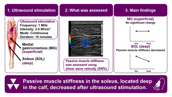
Ultrasound stimulation was applied to the calf for 10 minutes, and passive muscle stiffness was assessed using shear wave velocity. The study compared the superficial medial gastrocnemius (MG) and deeper soleus (SOL) muscles. Passive muscle stiffness decreased in the SOL after ultrasound stimulation, while no significant change was observed in the MG. Image credit: Mr Tomohiro Umeda from Doshisha University, Japan

Passive muscle stiffness is a risk factor for muscle strain injuries. Stretching, heat, and vibration have been evaluated on superficial muscles, but approaches for decreasing stiffness in deeper muscles remain unclear. In a recent study, researchers from Japan showed that 10 minutes of ultrasound stimulation can reduce stiffness in a deep calf muscle called the soleus, hinting at a promising non-invasive strategy for managing deep muscle stiffness, with potential future application in sports injury prevention.

Muscle strain injuries are a common problem in sports, and injuries to the triceps surae, the calf muscle group that includes the gastrocnemius and soleus, can significantly affect athletic performance and return to play. One factor that is thought to influence a muscle’s susceptibility to strain is passive muscle stiffness, which refers to the resistance of a relaxed muscle to being stretched. A recent genetic study has strengthened the idea that stiffer muscles are more prone to injury, suggesting that passive muscle stiffness is an important risk factor. Thus, finding safe and feasible ways to reduce this stiffness might help prevent associated injuries.

Several strategies, including stretching, heat, and vibration, can significantly reduce passive muscle stiffness. However, research on these approaches has focused on muscles located close to the skin. The soleus, one of the main muscles of the triceps surae, sits deep underneath the medial and lateral gastrocnemius, which makes it a difficult target. Ultrasound stimulation is used in sports and rehabilitation and produces both heating and mechanical effects in biological tissue. As lower-frequency ultrasound can penetrate relatively deep into tissue, it raises the question: Could ultrasound actually reduce the stiffness of a deep muscle like the soleus?

To this end, a research team including Ph.D. student Tomohiro Umeda, Professor Tatsuya Hojo, and Professor Taku Wakahara from the Graduate School of Health and Sports Science at Doshisha University, Japan, examined the effects of ultrasound stimulation on calf muscle stiffness in 20 healthy adults. Their work was published in the Journal of Biomechanics.

A randomly selected leg of each participant received 10 minutes of continuous ultrasound stimulation at a frequency of 1 megahertz (MHz) and an intensity of 2.0 W/cm², while the other leg served as an untreated control. The researchers then used shear wave elastography, an imaging technique that measures how quickly mechanical waves travel through tissue, to assess muscle stiffness before and immediately after the ultrasound stimulation. Faster shear waves generally indicate stiffer tissue, so a decrease in shear wave velocity (SWV) was interpreted as a decrease in passive muscle stiffness.

The results showed a clear difference between the soleus and the medial gastrocnemius. SWV, an indicator of passive muscle stiffness, decreased significantly in the soleus after the ultrasound stimulation, while no significant change was observed in the untreated leg. In contrast, no significant change in SWV was observed in the more superficial medial gastrocnemius. Interestingly, participants who initially had stiffer soleus muscles tended to show greater reductions in stiffness following ultrasound stimulation. “Our findings suggest that ultrasound stimulation may have the potential to non-invasively decrease passive stiffness in deep muscles such as the soleus,” remarks Mr Umeda, “In the future, this approach may contribute to the development of conditioning and rehabilitation strategies for the prevention of sports-related muscle injuries.”

The different responses of the soleus and medial gastrocnemius may be related partly to their anatomical locations. The soleus is located about 2.44cm below the skin, whereas the gastrocnemius is much closer to the surface at just about 0.53cm. Moreover, differences in the muscles’ tissue composition may also contribute to their different responses, although the underlying mechanisms remain unclear. “Future research could clarify the optimal ultrasound parameters for different muscles and identify which individuals respond most effectively to ultrasound stimulation. Such knowledge could lead to more individualised conditioning and rehabilitation strategies for athletes,” concludes Mr Umeda.

Overall, the results highlight the potential of ultrasound stimulation as a non-invasive approach for managing stiffness in deep muscles that can be difficult to target with other methods. Further research will be needed to determine whether this approach can be incorporated into individualised conditioning and rehabilitation strategies and ultimately contribute to the prevention of sports-related muscle injuries.

Source: Doshisha University
Focused Ultrasound with Chemotherapy Improves Survival for Glioblastoma Patients

Image credit: University of Maryland School of Medicine

Patients with glioblastoma who received MRI-guided focused ultrasound with standard-of-care chemotherapy had a nearly 40% increase in overall survival in a landmark trial of 34 patients led by University of Maryland School of Medicine (UMSOM) researchers. This is the first time researchers have demonstrated a potential survival benefit from using focused ultrasound to open the blood-brain barrier to improve delivery of chemotherapy to the tumour site in brain cancer patients after surgery.

“Our results are very encouraging. Using focused ultrasound to open the blood-brain barrier and deliver chemotherapy could significantly increase patient survival, which other ongoing studies are seeking to confirm and expand,” said study principal investigator Graeme Woodworth, MD, Professor and Chair of Neurosurgery at UMSOM and Neurosurgeon-In-Chief at the University of Maryland Medical Center (UMMC).

The findings of this groundbreaking safety, feasibility, and comparative trial involved glioblastoma patients who were given focused ultrasound to open their blood-brain barrier before getting chemotherapy; they were matched to a rigorously selected control group of 185 glioblastoma patients with similar characteristics who received the standard dose of the chemotherapy drug, temozolomide, without receiving focused ultrasound. Trial participants were initially treated with surgery to remove their brain tumour, followed by six weeks of chemotherapy and radiation, and up to six monthly focused-ultrasound treatments plus temozolomide.

Results were published in the journal Lancet Oncology and show that trial participants had nearly 14 months of median progression-free survival, compared to eight months in the control group. In terms of overall survival, trial participants, on average, lived for more than 30 months compared to 19 months in the control group.

The study builds on more than a decade of intensive research to test the safety and feasibility of opening the blood-brain barrier using focused ultrasound first in animal studies and then in patients. It was led by Dr Woodworth and was conducted at UMMC and four other university-affiliated clinical sites. “We also demonstrated that this could be a useful technique that enables us to better monitor patients to determine if their brain cancer has progressed,” said Dr Woodworth, who also serves as Director of the Brain Tumor Program at the University of Maryland Marlene and Stewart Greenebaum Comprehensive Cancer Center (UMGCCC).

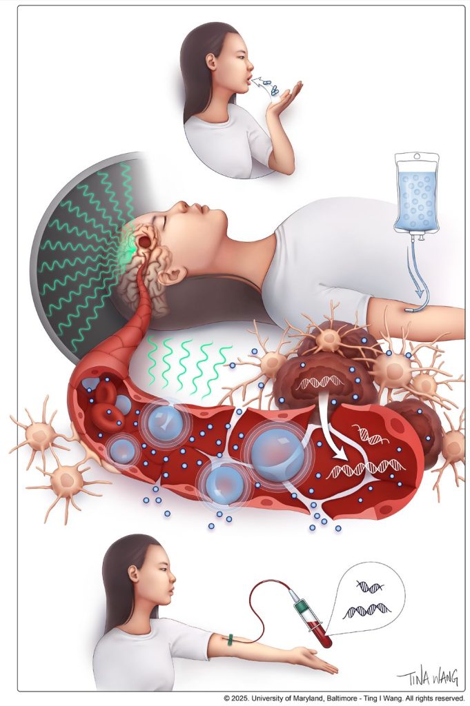
He and his team demonstrated that opening the blood-brain barrier facilitated the use of a “liquid biopsy,” which is a blood test that detects cancer biomarkers, which can include DNA fragments, proteins and other components from the liquid environment surrounding the tumor site.

Such biomarkers have been used in other cancers to determine whether the tumor has remained stable or has the potential to progress or even metastasize. Up until now, however, these tests have not been utilized in brain cancer patients since most components can never pass into the bloodstream from the brain due to the blood-brain barrier.

“These liquid biomarkers were found to be closely concordant with the patient outcomes over time, progression-free survival and overall survival,” said Dr Woodworth.

While temozolomide is the standard treatment for glioblastoma, the drug typically gets blocked by the blood-brain barrier with studies showing that less than 20 percent reaches the brain in patients. This study did not determine the exact amount of temozolomide to reach the brain in each patient, but earlier studies have shown that opening the blood-brain barrier before delivering chemotherapy can dramatically increase the amount that gets to the original tumor site.

Glioblastoma is the most common and deadliest type of malignant brain tumour. The five-year survival rate is only 5.5%, and patients live an average of 14 to 16 months after diagnosis when treated with surgery, radiation, and chemotherapy when appropriate. The malignancy nearly always recurs even after it is removed due to residual infiltrating cancer cells that remain after treatment.

The blood-brain barrier is a specialized network of vascular and brain cells that acts as the brain’s security system to protect against invasion by dangerous toxins and microbes. It can be opened temporarily using a specialised focused ultrasound device. This process starts with injecting microscopic inert gas-filled bubbles into the patient’s bloodstream. Guided by an MRI, precise brain regions are targeted while the injected microbubbles are circulating.

“Upon excitation under low-intensity ultrasound waves, the microbubbles oscillate within the energy field, causing temporary mechanical perturbations in the walls of the brain blood vessels,” said Pavlos Anastasiadis, PhD, an Assistant Professor of Neurosurgery at UMSOM who is an expert in ultrasound biophysics.

Prior studies led by Dr Woodworth and this trial’s co-investigators showed that opening the blood-brain barrier temporarily can be safely and feasibly performed in brain tumour patients. He and his team conducted this procedure in the first brain cancer patient in the US in 2018 at UMMC after the US Food and Drug Administration (FDA) approved the inaugural clinical trial.

Future trials could use focused ultrasound alongside other chemotherapy agents to test the effectiveness of drugs never used in brain cancer due to their ineffectiveness at crossing the blood-brain barrier.

Source: University of Maryland School of Medicine

Prostate Cancer Therapy Improved with Focused Ultrasound

Credit: Darryl Leja National Human Genome Research Institute National Institutes Of Health

Combining an existing small-molecule protein therapy called tumour necrosis factor related apoptosis-inducing ligand (TRAIL) with focused ultrasound (FUS) can significantly reduce tumour size and burden in prostate cancer models, according to a new study published in Advanced Science by researchers at Rice University and Vanderbilt University.

Around the world, about 10 million people die of cancer each year. This collaborative study, led by Michael King, bioengineering professor at Rice, and Charles Caskey, associate professor in radiology and radiological sciences at Vanderbilt, is the first to demonstrate that low-intensity mechanical force in combination with TRAIL can treat cancers.

The study sheds new light on how low-intensity focused ultrasound and soluble TRAIL specifically destroy cancer cells within the compact environment of a primary prostate cancer lesion.

Urgency for safe, effective therapy for prostate cancer

“There is urgent need to improve how we treat advanced and recurrent prostate cancer, which is the second-leading cause of death among men in the United States and is the most frequently diagnosed cancer in more than 100 countries,” said King, who is a Cancer Prevention and Research Institute of Texas Scholar. “We have now found a safe, effective and noninvasive way to enhance the antitumor effects of a specific cancer drug (TRAIL), a promising finding which we are hopeful can soon be translated for clinical care.”

Current standard-of-care prostate cancer treatments are associated with severe adverse effects. In recent years, FUS-based therapies have been gaining attention since they can be localised specifically to tumour tissue, resulting in fewer off-target effects.

Mechanical stimuli amplify anticancer effects of TRAIL via Piezo1

TRAIL protein specifically induces the death of cancer cells without harming nearby healthy cells. However, despite promising results in lab studies, only a few cancer patients have shown improvements with intravenous administration of TRAIL in clinical trials. This is because TRAIL has a very short half-life (~30 minutes) and remains in blood circulation only briefly before it gets destroyed.

Thus, to effectively eliminate cancer cells, TRAIL therapy needs to be administered multiple times per day, which is not only inconvenient but also increases the risk of unwanted side effects.

“Previously, we had found certain mechanical forces like fluid shear stress (FSS) could amplify the anticancer effects of TRAIL with an influx of calcium and activation of a protein called Piezo1 that triggered cell death,” King said.

However, FSS is not clinically applicable for solid tumours because it is only present in the circulatory and lymphatic systems and thus only effective against circulating tumour cells, which are often observed at later stages of malignancy.

“The field is still lacking a straightforward and effective clinical approach that combines the application of mechanical force with soluble TRAIL as a localised therapeutic to treat primary prostate tumours effectively before they metastasise to different locations, which prompted us to undertake this preclinical study to examine if FUS might be a good candidate to be developed into a combination therapy for prostate cancer,” King said.

Low-intensity FUS acts synergistically with TRAIL to reduce prostate tumours in lab

Using prostate cancer cell lines, Abigail Fabiano and Malachy Newman – graduate students mentored by King and Caskey respectively – performed several experiments to refine and optimise several operational parameters of in vitro FUS.

Their initial goal was to ensure that the nearby healthy cells remained unharmed by the mechanical shear forces. Next, they found that combination therapy of FUS and TRAIL was much more effective in reducing the number of cancer cells and size of tumours than FUS or TRAIL alone, supporting the idea that the synergistic action of TRAIL and FUS-mediated Piezo1 activation is key to achieving maximum tumour reduction.

“This foundational study provides crucial preclinical insights that can be used to develop a novel combination therapy for prostate cancer,” King said. “Furthermore, it opens the doors to many new avenues for using mechanotherapy in medicine and has far-reaching implications in how FUS and other mechanical therapies can be combined with small-molecule protein therapy and other drugs to effectively treat various types of cancers with fewer adverse effects in the future.”

Source: Rice University