Tag: cancer screening

Student Designs a Prostate Checking Device to Replace the Digital Exam

Pro check, designed by Loughborough University student Devon Tyso.

A Loughborough University student has developed a new medical device that could transform how prostate health is assessed and monitored.

Devon Tyso, a Product Design and Technology student, has designed ‘PRO check’, an innovative tool designed to replace the traditional digital rectal examination (DRE), which involves a doctor manually assessing the prostate with a finger.

According to Devon, the current approach is heavily reliant on a clinician’s subjective judgement and experience, and many see the method as ‘intrusive’.

“As one in seven men will get prostate cancer, it’s vital to detect abnormalities early and track changes over time,” said Devon, “The current examination method involves a lot of guesswork.

“PRO check provides objective, measurable data and allows prostate health to be visualised – enabling more accurate diagnosis, and improved long-term monitoring.

“Having a device conduct the exam may also feel less invasive, which may encourage more men to get checked, potentially catching issues earlier.”

How the device works

Designed for use by GPs during routine prostate assessments, PRO check allows doctors to evaluate the size and texture of the prostate — two key indicators of potential health issues — in a more objective and consistent way than the traditional digital rectal examination.

The device is a handheld probe, and it is covered with a condom before being inserted into the body. Once in position, the condom inflates to different pressures, pressing against the surface of the prostate, causing it to compress. A laser grid is projected onto the inner surface of the condom so the shape of the underlying prostate can be captured.

Stereoscopic cameras capture images of the laser grid, tracking where the gridlines intersect and how these intersections shift as pressure changes. This information is then fed into mathematical equations to create 3D images — or ‘topographical representations’ — that reveal the prostate’s shape and surface structure under different pressures.

Studying the prostate’s surface details could help clinicians identify areas requiring further investigation. Healthy prostate tissue is typically soft and compressible, so regions that appear stiff or resist pressure could indicate potential abnormalities and warrant further investigation.

The device can also produce data on prostate volume – one of the measurements used to calculate prostate-specific antigen (PSA) density, which helps assess prostate cancer risk. Devon says currently volume estimates are often based on a clinician’s best judgement.

In addition, data from PRO check can be used to generate a compressibility-versus-pressure graph – a novel data type not currently available in clinical practice. This graph shows how the prostate compresses at different pressure levels, which Devon hopes could offer new insights into prostate health and complement existing diagnostic tools.

PRO check is designed to integrate with artificial intelligence, enabling automatic extraction of video data, real-time calculations, and the generation of 3D images for live display on a laptop or tablet during the examination.

The idea is that all examination data from PRO check would be stored on the patient’s records, helping to build a personalised prostate health profile that can be tracked and monitored over time.

Inspiration

Devon’s inspiration for PRO check came from a mix of personal experience – after his grandfather’s prostate cancer diagnosis – and unexpected technical research.

“It really hit home how common prostate issues are after my family member was found to have an enlarged prostate,” said the 22-year-old from Cardiff, “I realised nearly everyone I spoke to about it knew someone affected by it.

“When I started looking into prostate examinations, I kept thinking ‘how can a doctor remember what your prostate felt like four months ago?’ and how horrible it must be just be told whether you’re fine or not without seeing any data or anything visual.”

While researching non-invasive ways to assess tissue structure inside the body, Devon came across a technique used by NASA to map the surface of asteroids — projecting laser grids onto them, capturing images with satellite-mounted cameras, and analysing the gridline intersections to reveal the contours of the surface.

“I saw that NASA were mapping surface heights on a massive scale, and I thought – if they can do that in space, why can’t we use similar principles to examine something here on Earth?” said Devon, “I’ve basically used the exact same technique and scaled it down for PRO check.”

Prototypes

Devon designed PRO check as part of his final year project – which was exhibited at the School of Design and Creative Arts’ 2025 Degree Show – and has prototyped several of its key components.

He has built and tested two working prototypes. The first demonstrates how a laser grid and camera can be setup to map the surface of the prostate.

Devon designed a custom rig that enabled him to capture images of a laser grid projected onto different silicone prostate models — representing a healthy gland, a small tumour, a large tumour, and an enlarged prostate — from an optimal angle using a smartphone camera.

First prototype of pro check
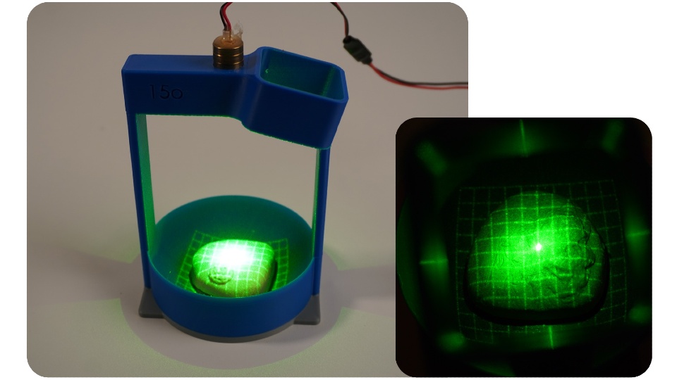
PRO check prototype one demonstrated how laser gridlines and a camera can be used to image the surface of the prostate.

The second prototype features electronics that inflate a small balloon at controlled pressures, regulated by a pressure-sensing chip. Devon consulted three healthcare professionals to measure the pressure typically applied during prostate exams and replicated those levels in his design.

Devon tested the prototype using the silicone prostate models but encased them in a sponge disc to simulate surrounding tissue.

Devon manually extracted data on the gridline intersections from the camera footage and applied mathematical equations to generate 3D images of the prostate surfaces and surrounding tissue under different pressures.

Next steps

Devon hopes to collaborate with medical professionals and product developers to turn PRO check into a fully realised medical device.

When speaking about his ultimate goal, Devon said: “I’d love to see this used in GP surgeries across the UK one day.

“With early detection being so critical, anything that helps men get checked sooner and more comfortably – and provides reliable data and visualisations – has huge potential. I really believe this could make a difference.”

Further information on PRO check can be found on the Degree Show website.

Source: Loughborough University

Celebrate Christmas in July with PinkDrive

Cold Nights, Warm Hearts, Festive Vibes – and Support for Early Detection

Think log fires, festive cheer, a three-course dinner and dancing to a live band – all wrapped up in the joy of giving. On Saturday, 5th July 2025, PinkDrive will host a Christmas in July dinner at the Indaba Hotel in Johannesburg, a night of holiday cheer and hope in action: raising funds for a life-changing cause. And you’re invited.

PinkDrive is a non-profit (NPC) committed to prolonging lives through the early detection of gender-related cancers. It delivers essential health services to thousands of South Africans every year by bringing mobile mammography units directly to communities that need them most, from rural villages to peri-urban areas across the nine provinces. These trucks provide clinical breast exams, mammograms, pap smears, and PSA testing, helping to detect cancer early in areas where healthcare access is often limited or unavailable.

Like many non-profits, PinkDrive depends on the support of corporate partners, sponsors, government, and the public to sustain its vital work, among them, Lee-Chem Laboratories. “This is a cause that is close to our hearts,” says Bhavna Sanker, Marketing Manager at Lee-Chem Laboratories. “Each year, we proudly support PinkDrive through our Mandy’s brand sponsorship, focusing on spreading awareness, sharing survivor stories, and making sure people understand their healthcare options.”

As part of their continued support, every guest at the Christmas in July function will receive a goodie bag from Lee-Chem, filled with products from their Mandy’s brand. “We are truly honoured to have Lee-Chem and the Mandy’s brand as valued sponsors of our Christmas in July Dinner fundraiser,” comments Nelius du Preez, Operations Manager at PinkDrive NPC. “Their continued support, alongside our other generous sponsors, makes events like this possible and helps us not only raise vital funds but also amplify the message of early detection and health education across South Africa.”

The fundraiser will spotlight the resilience of breast cancer survivors, with Deputy Minister of Electricity and Energy, Samantha Graham-Maré, sharing her personal journey with cancer. PinkDrive CEO and Founder, Noelene Kotschan, a passionate advocate for early detection prolonging  lives, will also address guests, and your charming host for the evening? A dashing Mister Global SA finalist will take the mic as MC, steering the evening from heartfelt reflections to lively fundraising with raffles and auctions featuring holiday packages, original artworks, and other exclusive prizes.

“We call the event ‘a night of giving back’ because it is occasions like these that allow us to keep our mobile health units on the road, reaching men and women who might otherwise not have access to screening services,” says du Preez. “Together with compassionate partners like Lee-Chem, we are driving change and prolonging lives, one screening at a time. And we are grateful to every corporate and individual who supports the event by purchasing tickets,” he adds.

All proceeds from the evening will be ringfenced to build and operate a new mobile mammography unit, expanding PinkDrive’s reach to screen more South Africans and detect cancer early, when treatment is most effective. “We encourage the public to join us in this mission by purchasing tickets and supporting the evening’s fundraising efforts,” concludes Sanker.

Tickets for the Christmas in July dinner are R600 per person and available now at pinkdrive.org. Don’t miss this chance to dress up, give back, and help bring hope where it’s needed most.

Men’s Health Warning: Prostate Cancer Usually Has no Early Symptoms

More precise treatment options with robotic technology

Friday, 20 June 2025: Prostate cancer is a major risk to men’s health, with South African men facing a one in eight chance of developing this most common of male cancers.

Urologists Dr Hannes Brummer and Dr Johan Coetzee, who practise at Netcare Greenacres Hospital, are encouraging men to prioritise prostate cancer screening this Men’s Health Month. 

“Usually, men do not feel any symptoms until prostate cancer has progressed significantly, which is why they need to be proactive about booking their routine prostate cancer screenings,” explains Dr Coetzee. 

“With the advanced prostate specific antigen [PSA] screening blood test available from GPs these days, there is so much more opportunity for prostate cancer to be detected earlier when it is still at a highly treatable stage.”

“For men who are diagnosed with prostate cancer following a needle biopsy, the treatment options available have improved to such an extent that there is more hope than ever before. Even where surgery is needed, prostate cancer does not necessarily pose a significant disruption to your life,” Dr Brummer adds. 

“A prostate cancer diagnosis can be daunting. We have walked this path with so many men, and robotic assisted surgery offers some important advantages for the removal of cancerous tissue in the prostate gland, in particular the precision of this minimally invasive option.”

Over 1 000 robotic assisted procedures have been performed at Netcare Greenacres Hospital since the introduction of this technology in August 2017. 

At Netcare Greenacres Hospital, Dr Brummer and Dr Coetzee use the da Vinci X robotic assisted surgical system to operate through tiny punctures in the skin using slender instruments more dexterous than the human hand.

Dr Brummer and Dr Coetzee emphasise that the surgeon remains in control of the robotic system at all times. With magnified 3D imaging capabilities, including a large fixed-focus area at the highest resolution, the nerves, blood vessels and tumour are visible with great clarity for the intricate procedure. 

“This robotic system is especially useful for operating on the prostate, as we can more clearly distinguish the nerves controlling erectile function and urinary continence. In most cases, there is less need for blood transfusion and reduced risk of complications,” Dr Brummer explains. 

“Another of the advantages of this robotic technology for prostate tumours is that there is much less tissue damage in this sensitive area. Compared with traditional surgery, this means men usually experience much less downtime with less discomfort after the procedure. This translates into shorter hospital stays and faster recovery with robotic assisted prostatectomies overall.”

General manager of Netcare Greenacres Hospital, Reon van Rensburg, joined the urologists in reinforcing the importance of prostate cancer awareness. “Let’s talk to our brothers, fathers, sons and grandsons about health issues, and get to know your family risk for both prostate cancer and breast cancer.”

Van Rensburg thanked Dr Brummer and Dr Coetzee for their continued dedication to making the world-class minimally-invasive robotic assisted surgical option available for patients local to Gqeberha and from as far afield as Knysna, George and East London, inland regions of the Eastern Cape, and parts of the southern Free State, the Northern Cape and the north-eastern region of the Western Cape.

“This Men’s Health Month, let’s pledge to be decisive about booking those routine health checks. Making the time now and every year could help to save your life in future,” Dr Brummer and Dr Coetzee concluded. 

Closure of US-funded Cancer Clinic Further Burdens Public Hospitals

The Cervical Cancer Screening and Prevention Clinic at Helen Joseph Hospital in Johannesburg was forced to shut down in mid-May after losing all its funding from the US President’s Emergency Plan for AIDS Relief (PEPFAR). Photos: Elna Schütz

By Elna Schütz

Hundreds of cervical cancer patients will likely be referred to overburdened hospitals following the closure of the Cervical Cancer Screening and Prevention Clinic at Helen Joseph Hospital in Johannesburg.

Following over 20 years of operations, the clinic was forced to shut down in mid-May after losing all its funding from the US President’s Emergency Plan for AIDS Relief (PEPFAR). It relied on some financial reserves to taper its activities over several months. Most clinic staff have been let go.

The clinic served women who were referred from across Johannesburg and as far as Springs. A significant part of that group lives with HIV.

“Many of these women are from underserved communities with limited access to specialist care,” says Dr Mark Faesen, Specialist Gynaecologist with the Clinical HIV Research Unit (CHRU).

The clinic offered critical cervical cancer screening and follow-up services, including Pap smears and colposcopies – a cervical examination for abnormalities. The clinic was managing around 1,400 patients annually. “It served as a clinical and research hub, preventing many cancers,” Faesen says.

We spoke to Zinhle (name changed) who was screened at the clinic after feeling ill for a year and who sought help at four different hospitals.

“When I got [to this clinic], I was received with a warm welcome,” she says, emphasising that every step of the process was explained to her and she was made to feel comfortable. “Where else are we supposed to go?”

Zinhle says she is deeply upset that she can no longer be treated at the clinic if she needs it again.

Faesen says the clinic’s closure will put immense pressure on other public hospitals offering these services, like Rahima Moosa or Chris Hani Baragwanath. This is likely to lead to longer waiting times for screening, diagnosis and treatments. “Early detection is important,” Faesen says. “Without timely diagnosis, outcomes are far poorer.”

Lorraine Govender, the National Manager of Health Programmes at the Cancer Association of South Africa (CANSA) says they are deeply concerned by the closure, as it is a serious setback in the ongoing fight against the disease.

Cervical cancer is the second most common cancer in women in South Africa, and results in the most deaths. It is curable if diagnosed and treated early. A Human Papillomavirus (HPV) vaccination also reduces the risk of cervical cancer. While low screening rates and backlogs in treatment have been long-standing across the country, Johannesburg appears to be particularly burdened. The shutdown of this clinic adds to a larger shortage of screening and treatment in Gauteng.

The Department of Health has previously stated that while it has improved vaccination efforts against cervical cancer, “screening and treatment are lagging behind”. The national health policy calls for women aged 30 to 50 to be screened at least three times in their lives. Women living with HIV should be screened at least every three years.

Cervical cancer screening services are limited and overwhelmed at most public hospitals, Faesen says. “The funding cuts have a knock-on effect: increasing patient loads at the few remaining colposcopy clinics.”

Lorraine Govender, the National Manager of Health Programmes at the Cancer Association of South Africa (CANSA) says they are deeply concerned by the closure, as it is a serious setback in the ongoing fight against the disease.

“Cervical cancer is both preventable and treatable when detected early, making continued access to screening services vital … The closure of this Johannesburg clinic must be a call to action,” Govender says.

Faesen stresses the urgent need for increased funding for decentralised screening services to fill the gaps created by clinics like the one at Helen Joseph Hospital. “Equipping more public sector sites with colposcopy capability and training personnel is also essential.”

Republished from GroundUp under a Creative Commons Attribution-NoDerivatives 4.0 International License.

Read the original article.

Three out of Ten Breast Cancers are Detected Between Screenings

Photo by National Cancer Institute

In a new study, researchers from Karolinska Institutet have shown that so-called interval cancers, which are detected between two screening sessions, account for a significant proportion of breast cancer cases and that certain risk factors may increase the likelihood of developing this type of cancer. The study was published in the journal JAMA Oncology.

Mammography screening has been shown to be effective in reducing breast cancer mortality by detecting cancer at an early stage. Despite this, some cancers are not diagnosed during screening but between screening rounds, known as interval cancers. This type of cancer can be more aggressive and difficult to treat than breast cancer detected during a scheduled screening.

The study, which covers half a million women in Stockholm between 1989 and 2020, shows that interval cancer accounts for a significant proportion of all breast cancer cases.

“We found that interval cancers account for about 30 per cent of all breast cancers detected by screening and this percentage has remained constant over three decades, despite advances in screening technology,” says Yuqi Zhang, postdoctoral fellow at the Department of Medical Epidemiology and Biostatistics, Karolinska Institutet and lead author of the study.

The researchers identified several factors that increase the risk of developing interval cancer. These factors include high breast density, hormone therapy, higher education level and older age at first childbirth.

“Women with high breast density and use of hormone therapy are at increased risk of being missed at screening due to false negative results,” says Yuqi Zhang.

Rapid growth of tumour

In addition, the study showed that women with a family history of breast cancer, especially interval cancer, are at a higher risk of developing interval cancer themselves. Women with a family history of breast cancer were 1.9 times more likely to develop interval cancer, and this risk increased to 2.9 times if they had a family history of interval cancer specifically.

“This is often due to the rapid growth of tumours between screening rounds rather than missed detections. The study therefore emphasises the need for more frequent or improved screening methods specifically designed for women with these particular risk factors,” says Yuqi Zhang. 

“Adapting screening protocols to reflect individual risk profiles – whether through more frequent mammography, supplemental imaging like contrast-enhanced mammography, or incorporating genetic testing – could significantly improve early detection rates,” says last author Professor Kamila Czene at the same department. 

“By identifying cancers earlier, we can offer more effective and less invasive treatments, improve long-term survival outcomes, and reduce the emotional and physical toll on patients.”

Source: Karolinska Institutet

Analysis of Repeat Mammograms Improves Cancer Prediction

Photo by National Cancer Institute on Unsplash

A new study describes an innovative method of analysing mammograms that significantly improves the accuracy of predicting the risk of breast cancer development over the following five years. Using up to three years of previous mammograms, the new method identified individuals at high risk of developing breast cancer 2.3 times more accurately than the standard method, which is based on questionnaires assessing clinical risk factors alone, such as age, race and family history of breast cancer.

The study, from Washington University School of Medicine in St. Louis, appears in JCO Clinical Cancer Informatics.

“We are seeking ways to improve early detection, since that increases the chances of successful treatment,” said senior author Graham A. Colditz, MD, DrPH, associate director, prevention and control, of Siteman Cancer Center, based at Barnes-Jewish Hospital and WashU Medicine. “This improved prediction of risk also may help research surrounding prevention, so that we can find better ways for women who fall into the high-risk category to lower their five-year risk of developing breast cancer.”

This risk-prediction method builds on past research led by Colditz and lead author Shu (Joy) Jiang, PhD, a statistician, data scientist and associate professor at WashU Medicine. The researchers showed that prior mammograms hold a wealth of information on early signs of breast cancer development that can’t be perceived even by a well-trained human eye. This information includes subtle changes over time in breast density, which is a measure of the relative amounts of fibrous versus fatty tissue in the breasts.

For the new study, the team built an algorithm based on artificial intelligence that can discern subtle differences in mammograms and help identify those women at highest risk of developing a new breast tumour over a specific timeframe. In addition to breast density, their machine-learning tool considers changes in other patterns in the images, including in texture, calcification and asymmetry within the breasts.

“Our new method is able to detect subtle changes over time in repeated mammogram images that are not visible to the eye,” said Jiang, yet these changes hold rich information that can help identify high-risk individuals.

At the moment, risk-reduction options are limited and can include drugs such as tamoxifen that lower risk but may have unwanted side effects. Most of the time, women at high risk are offered more frequent screening or the option of adding another imaging method, such as an MRI, to try to identify cancer as early as possible.

“Today, we don’t have a way to know who is likely to develop breast cancer in the future based on their mammogram images,” said co-author Debbie L. Bennett, MD, an associate professor of radiology and chief of breast imaging for the Mallinckrodt Institute of Radiology at WashU Medicine. “What’s so exciting about this research is that it indicates that it is possible to glean this information from current and prior mammograms using this algorithm. The prediction is never going to be perfect, but this study suggests the new algorithm is much better than our current methods.”

AI improves prediction of breast cancer development

The researchers trained their machine-learning algorithm on the mammograms of more than 10 000 women who received breast cancer screenings through Siteman Cancer Center from 2008–2012. These individuals were followed through 2020, and in that time 478 were diagnosed with breast cancer.

The researchers then applied their method to predict breast cancer risk in a separate set of 18 000 women who received mammograms from 2013–2020. Subsequently, 332 women were diagnosed with breast cancer during the follow-up period, which ended in 2020.

According to the new prediction model, women in the high-risk group were 21 times more likely to be diagnosed with breast cancer over the following five years than were those in the lowest-risk group. In the high-risk group, 53 out of every 1000 women screened developed breast cancer over the next five years. In contrast, in the low-risk group, 2.6 women per 1000 screened developed breast cancer over the following five years. Under the old questionnaire-based methods, only 23 women per 1000 screened were correctly classified in the high-risk group, providing evidence that the old method, in this case, missed 30 breast cancer cases that the new method found.

The mammograms were conducted at academic medical centres and community clinics, demonstrating that the accuracy of the method holds up in diverse settings. Importantly, the algorithm was built with robust representation of Black women, who are usually underrepresented in development of breast cancer risk models. The accuracy for predicting risk held up across racial groups. Of the women screened through Siteman, most were white, and 27% were Black. Of those screened through Emory, 42% were Black.

Source: Washington University School of Medicine in St. Louis

Telltale Chemical in the Breath can Warn of Lung Cancer

Credit: Scientific Animations CC4.0

Exhaled breath contains chemical clues to what’s going on inside the body, including diseases like lung cancer. And devising ways to sense these compounds could help doctors provide early diagnoses — and improve patients’ prospects. In a study in ACS Sensors, researchers report the development of ultrasensitive, nanoscale sensors that in small-scale tests distinguished a key change in the chemistry of the breath of people with lung cancer.

Besides carbon dioxide, people also exhale other airborne compounds. Researchers have determined that declines in one exhaled chemical — isoprene — can indicate the presence of lung cancer. However, to detect such small shifts, a sensor would need to be highly sensitive, capable of detecting isoprene levels in the parts-per-billion (ppb) range. It would also need to differentiate isoprene from other volatile chemicals and withstand breath’s natural humidity. Previous attempts to engineer gas sensors with characteristics like these have focused on metal oxides, including one particularly promising compound made with indium oxide. A team led by Pingwei Liu and Qingyue Wangset out to refine indium oxide-based sensors to detect isoprene at the level at which it naturally occurs in breath.

The researchers developed a series of indium(III) oxide (In2O3)-based nanoflake sensors. In experiments, they found one type, which they called Pt@InNiOx for the platinum (Pt), indium (In) and nickel (Ni) it contains, performed best. These Pt@InNiOx sensors:

  • Detected isoprene levels as low as 2ppb, a sensitivity that far surpassed earlier sensors.
  • Responded to isoprene more than other volatile compounds commonly found in breath.
  • Performed consistently during nine simulated uses.

More importantly, the authors’ real-time analysis of the nanoflakes’ structure and electrochemical properties revealed that Pt nanoclusters uniformly anchored on the nanoflakes catalyzed the activation of isoprene sensing, leading to the ultrasensitive performance.

Finally, to showcase the potential medical use of these sensors, the researchers incorporated the Pt@InNiOnanoflakes into a portable sensing device. Into this device they introduced breath collected earlier from 13 people, five of whom had lung cancer. The device detected isoprene levels lower than 40 ppb in samples from participants with cancer and more than 60 ppb from cancer-free participants. This sensing technology could provide a breakthrough in non-invasive lung cancer screening and has the potential to improve outcomes and even save lives, the researchers say.

Source: American Chemical Society

Most Accurate Ultrasound Test Could Detect 96% of Women with Ovarian Cancer

Photo by Mart Production on Pexels

An ultrasound test that detected 96% of ovarian cancers in postmenopausal women should replace current standard of care test in the UK according to a new study.

In a paper published in Lancet Oncology, research led by Professor Sudha Sundar from the University of Birmingham compared all currently available tests to diagnose ovarian cancer in postmenopausal women head-to-head in a high-quality diagnostic test accuracy study.

Of the six diagnostic tests investigated, the IOTA ADNEX model which looks at ultrasound features (how the lump looked like on ultrasound) had the best accuracy of all and could detect up to 96% of women with ovarian cancer.

The ultrasound test outperforms the current standard of care in the UK significantly and so the researchers recommend that the IOTA ultrasound ADNEX model should replace the current standard of care test called risk of malignancy (RMI1) test in the UK which identifies 83% of ovarian cancers.

Sudha Sundar, Professor of Gynaecological Cancer at the University of Birmingham and consultant in gynaecological cancer surgery at Sandwell and West Birmingham NHS Trust said:

“This is the first time that a head-to-head study of all available ovarian cancer tests have been done in the same population. Here we studied their use with symptomatic, postmenopausal women who are most at risk of this cancer. Our trial found that the IOTA ADNEX ultrasound protocol had highest sensitivity for detecting ovarian cancer compared to the standard of care and other test.

“The ultrasound test also performs well when delivered by a trained sonographer who have received specific training and certification and quality assurance, and as the vast majority of ultrasound scans are performed by sonographers it is important that a new standard is able to be delivered by as many clinical professionals as possible.

“We found that the higher sensitivity of the IOTA ADNEX model is likely to lead to some women who don’t have cancer also being flagged up as having a higher risk of cancer. We however did discuss this extensively with patients, cancer charity Target ovarian cancer and NHS experts who all agreed that in postmenopausal women who are at higher risk of ovarian cancer, picking up more women with cancer would benefit women overall.”

The research team note that the IOTA ADNEX model achieved 96% accuracy when delivered by NHS sonographers who were appropriately trained and received quality assurance. As most scans worldwide are carried out by sonographers rather than gynaecologists, introductory free online resources have been created by the researchers for NHS staff to undergo the specialist ultrasound training and get certification and quality assurance.

Source: University of Birmingham

Faster Detection of Pancreatic Cancer

Glycopeptide probes detect tumour-associated antibodies in blood samples

Pancreatic cancer. Credit: Scientific Animations CC BY-SA 4.0

Pancreatic cancer is one of the most lethal forms of cancer, primarily because it is usually diagnosed very late. Current markers are too insensitive and unspecific for early detection screenings. In the journal Angewandte Chemie, a research team has now introduced a new method that could lead to a significantly more precise and reliable diagnosis. It is based on the selective detection of specific antibodies in blood samples.

Tumours produce certain proteins (tumour-associated antigens) that draw the attention of our constantly “patrolling” immune system and trigger an immune response. As a consequence, antibodies directed against the tumours (tumour-associated autoantibodies) are formed, circulating in the blood at very early stages of the disease – which makes them useful for early detection. An international team led by Roberto Fiammengo and Giovanni Malerba at the University of Verona (Italy) as well as Alfredo Martínez at the Center for Biomedical Research of La Rioja (Logroño, Spain) and Francisco Corzana at the Universidad de La Rioja, has now developed an approach to diagnostic testing for pancreatic cancer that is based on the detection of such special tumour-associated autoantibodies.

They chose to use autoantibodies directed against the tumour-associated form of mucin-1 (TA-MUC1). Mucin-1 is a heavily glycosylated protein (a protein with sugar components) that occurs, for example, in glandular tissue. In many types of tumours, including pancreatic cancer, it is found in significantly elevated concentrations. In addition, the pattern of glycosylation is different from the normal form. The team’s goal was to detect autoantibodies that are directed specifically against TA-MUC1 and are a clear indicator of pancreatic cancer.

Based on structural analyses and computer simulations of known antibodies against TA-MUC1 (SM3 and 5E5), the team designed a collection of synthetic glycopeptides that mimic different segments (epitopes) of TA-MUC1. They also made unnatural modifications to increase the chances of identifying autoantibody subgroups indicative of the disease. The team immobilised these model antigens on gold nanoparticles achieving probes suitable for a serological assay (dot-blot assay). The diagnostic assay was validated with real samples from patients with pancreatic cancer and a healthy control group. Some of the nanoparticle probes could differentiate very well between samples from diseased and healthy individuals demonstrating they detected tumour associated autoantibodies. Notably, these specific autoantibodies displayed significantly better correct positive/false positive ratios than current clinical biomarkers for pancreatic cancer.

Probes with smaller glycopeptide antigens that correspond to only a single epitope, gave better results than larger probes that mimic multiple epitopes – an advantage for easier synthetic production. A short glycopeptide with an unnatural modification to its sugar component was found to be particularly effective for the detection of discriminating autoantibodies. This new structure-based approach could help in the selection of autoantibody subgroups with higher tumour specificity.

Source: Wiley

Routine Bloods can Improve Cancer Screening in Patients with Abdominal Symptoms

Risk of cancer by specific site based on blood test abnormalities in symptomatic patients can help guide referral strategies

Photo by National Cancer Institute on Unsplash

Incorporating information from common blood tests can enhance cancer risk assessment in patients with abdominal symptoms, according to a study publishing July 30th in the open-access journal PLOS Medicine by Meena Rafiq from University College London, UK, and colleagues.

Early cancer detection is key to successful treatment. However, many undiagnosed cancer patients present to their primary care provider with non-specific symptoms that can be a result of several other benign causes, making it difficult to determine who warrants additional diagnostic testing or referral. Most guidelines focus on “alarm” symptoms specific to a given type of cancer to guide referrals. There is limited guidance on non-specific symptoms to guide cancer assessment and referral decisions across different cancer types.

In this study, researchers used data from the UK Clinical Practice Research Datalink to identify more than 470 000 patients aged 30 years or older who had visited a general practitioner due to abdominal pain or bloating. Within a year of that visit, approximately 9000 patients with abdominal pain and 1000 patients with bloating were diagnosed with cancer. The researchers looked at 19 abnormal blood test results collected during the initial primary care visit to see if they could predict who was more likely to be diagnosed with cancer.

Several blood abnormalities were predictive of cancer risk across sex and age groups. For example, in patients aged 30–59 years with abdominal symptoms, anaemia, low albumin, raised platelets, abnormal ferritin, and increased inflammatory markers strongly predicted a risk of undiagnosed cancer. Among older patients (aged 60 years and above), the presence of abdominal pain or bloating alone was enough to warrant a cancer referral.

The study also showed which types of cancer were most common based on age, sex, and blood test abnormality. For example, among women aged 50–59 years with anaemia and abdominal bloating, the most common types of cancer were bowel and ovarian cancer. This level of granularity can help guide providers on which diagnostic strategies to prioritise.

The study shows that common, routine blood test results can provide additional context in patients with non-specific abdominal symptoms to improve cancer risk assessment and identify patients who warrant additional testing and/or referral to a specialist.

The authors add, “Using existing blood tests can be an effective and affordable way to improve early diagnosis of cancer in people who see their GP with vague symptoms. Our study identified several commonly used GP blood tests where abnormal results increase a patient’s risk of having cancer and these can be used to diagnose cancer earlier.”

Provided by PLOS