Tag: autism spectrum disorder

Differences in Cortical Development Seen for Autistic Boys and Girls

Photo by Ben Wicks on Unsplash

A new study led by UC Davis researchers finds widespread differences in brain development between autistic boys and girls ages 2-13. The study, published recently in Molecular Psychiatry, found sex-specific changes in the thickness of the brain’s cortex, or outer layer.

The findings are notable because so few studies have addressed cortical development in autistic girls, who are diagnosed with autism less often than males. Nearly four males are diagnosed with autism for every one female.

“It is clear that this sex bias is due, in part, to underdiagnosis of autism in females,” said Christine Wu Nordahl, a professor in the Department of Psychiatry and Behavioral Sciences and the UC Davis MIND Institute and a senior author on the paper.

“But this study suggests that differences in diagnosis are not the full story – biological differences also exist.”

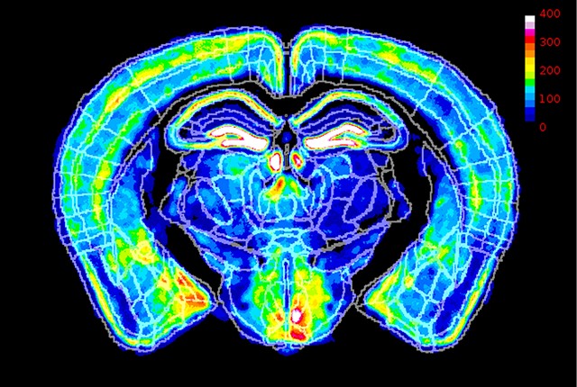
The cortex is made up of distinct layers comprised of millions of neurons. Until about age 2, the cortex rapidly thickens as new neurons are created. After this peak, the outer cortical layer thins. Previous studies have found that this thinning process is different in autistic children than non-autistic children, but whether autistic boys and girls share the same differences had not been examined.

“It’s important to learn more about how sex differences in brain development may interact with autistic development and lead to different developmental outcomes in boys and girls,” explained Derek Andrews, lead author on the study and an assistant project scientist in the Department of Psychiatry and Behavioral Sciences and at the MIND Institute.

A changing cortex in childhood

The research team studied the brain scans of 290 autistic children – 202 males and 88 females, and 139 non-autistic, typically developing individuals – 79 males and 60 females.

All participants were in the MIND Institute’s Autism Phenome Project (APP), one of the largest longitudinal autism studies in the world.

The project includes the Girls with Autism Imaging of Neurodevelopment (GAIN) study, launched to increase the number of females represented in research.

The researchers took MRI scans at up to four time periods between the ages of 2 and 13.

They found that at age 3, autistic girls had a thicker cortex than non-autistic girls of the same age, comprising about 9% of the total cortical surface. Differences in autistic males when compared to non-autistic males of the same age were much less widespread.

In addition, when compared to males, autistic females had faster rates of cortical thinning into middle childhood. The cortical differences were present across multiple neural networks.

“We found differences in the brain associated with autism across nearly all networks in the brain,” Andrews said.

He noted that it was a surprise at first that the differences were greatest at younger ages. Because autistic girls had a more rapid rate of cortical thinning, by middle childhood, the differences between autistic males and females were much less pronounced.

“We typically think of sex differences as being larger after puberty. However, brain development around the ages of 2-4 is highly dynamic, so small changes in timing of development between the sexes could result in large differences that then converge later,” Andrews explained.

The importance of long-term studies of both sexes

These findings make it clear that longitudinal studies that include both sexes are necessary, Nordahl said.

“If we had only looked at boys at age 3, we may have concluded that there were no differences. If we had both boys and girls, but only investigated differences at 11 years of age, we may have concluded that there were very few sex differences in the cortex. We needed to follow both boys and girls across development to see the full picture,” she explained.

This was why Nordahl, who now directs the APP, launched the GAIN study in 2014. “The APP had a wonderfully large sample of about 150 autistic boys, but only about 30 autistic girls. This was too few autistic girls to really examine how they might be similar or different to boys, so we worked to increase the representation of autistic females in our research,” she said.

GAIN is unique, and Andrews said he hopes other researchers will follow suit in including more autistic girls in autism research. “Autistic females represent about 20% of the autistic population. Any successful effort to understand autism will need to include autistic females.”

Source: University of California – Davis Health

Genomic ‘Butterfly Effect’ Explains Risk for Autism spectrum Disorder

Photo by Sangharsh Lohakare on Unsplash

Researchers in Japan discovered that a special kind of genetic mutation works differently from typical mutations in how it contributes to autism spectrum disorder (ASD). In essence, because of the three-dimensional structure of the genome, mutations are able to affect neighbouring genes that are linked to ASD, thus explaining why ASD can occur even without direct mutations to ASD-related genes. This study appeared in the scientific journal Cell Genomics.

ASD is a group of conditions characterised in part by repetitive behaviours and difficulties in social interaction. Although it runs in families, the genetics of its heritability are complex and remain only partially understood. Studies have shown that the high degree of heritability cannot be explained simply by looking at the part of the genome that codes for proteins. Rather, the answer could lie in the non-coding regions of the genome, particularly in promoters, the parts of the genome that ultimately control whether or not the proteins are actually produced. The team led by Atsushi Takata at in the RIKEN Center for Brain Science (CBS) examined de novo gene variants (new, non-inherited mutations) in these parts of the genome.

The researchers analysed an extensive dataset of over 5000 families, making this one of the world’s largest genome-wide studies of ASD to date. They focused on TADs – three-dimensional structures in the genome that allow interactions between different nearby genes and their regulatory elements. They found that de novo mutations in promoters heightened the risk of ASD only when the promoters were located in TADs that contained ASD-related genes. Because they are nearby and in the same TAD, these de novo mutations can affect the expression of ASD-related genes. In this way, the new study explains why mutations can increase the risk of ASD even when they aren’t located in protein-coding regions or in the promotors that directly control the expression of ASD-related genes.

“Our most important discovery was that de novo mutations in promoter regions of TADs containing known ASD genes are associated with ASD risk, and this is likely mediated through interactions in the three-dimensional structure of the genome,” says Takata.

To confirm this, the researchers edited the DNA of stem cells using the CRISPR/Cas9 system, making mutations in specific promoters. As expected, they observed that a single genetic change in a promotor caused alterations in an ASD-associated gene within the same TAD. Because numerous genes linked to ASD and neurodevelopment were also affected in the mutant stem cells, Takata likens the process to a genomic “butterfly effect” in which a single mutation dysregulates disease-associated genes that are scattered in distant regions of the genome.

Takata believes that this finding has implications for the development of new diagnostic and therapeutic strategies. “At the very least, when assessing an individual’s risk for ASD, we now know that we need to look beyond ASD-related genes when doing genetic risk assessment, and focus on whole TADs that contain ASD-related genes,” explains Takata. “Further, an intervention that corrects aberrant promoter-enhancer interactions caused by a promotor mutation may also have therapeutic effects on ASD.”

Further research involving more families and patients is crucial for better understanding ASD’s genetic roots. “By expanding our research, we will gain a better understanding of the genetic architecture and biology of ASD, leading to clinical management that enhances the well-being of affected individuals, their families, and society,” says Takata.

Source: RIKEN

X-chromosome Inactivation may Reduce Females’ Autism Risk

X-chromosome inactivation varies across different areas of brains. Here, fluorescent imaging data from a mouse reveal where the father’s X chromosome is most active (white) and least active (blue). Credit: Eric Szelenyi

A study using mice published in the journal Cell Reports suggests how chromosome inactivation may protect women from autism disorder inherited from their father’s X chromosome.

Because cells do not need two copies of the X chromosome, the cells inactivate one copy early in embryonic development, a well-studied process known as X chromosome inactivation. As a result of this inactivation, every female is made up of a mix of cells, some have an active X chromosome from her father and others from her mother, a phenomenon known as mosaicism. 

For many years, it has been thought that this was random and would result, on average, in a roughly 50/50 mix of cells, with 50% having an active paternal X chromosome and 50% an active maternal X chromosome.

Now a new study finds that, in the mouse brain at least, this is not the case. Instead, there appears to be a bias in the process that results in the paternal X chromosome being inactivated in 60% of the cells rather than the expected 50%.

When the X-linked mutation that is the most common cause of autism spectrum disorder is inherited from the father, the pattern of X-chromosome inactivation in the brain circuitry of females can prevent the effects of that mutation, the study found.

“This bias may be a way to reduce the risk of harmful mutations, which occur more frequently in male chromosomes,” said corresponding author Eric Szelenyi, acting assistant professor of biological structure at the University of Washington School of Medicine in Seattle.

The X-chromosome is of particular interest because it carries more genes involved in brain development than any other chromosome. Mutations in the chromosome are linked to more than 130 neurodevelopmental disorders, including fragile X syndrome and autism.

In the study, the researchers first determined the ratio of X chromosome inactivation in healthy mice by analyzing roughly 40 million brain cells per mouse. The scientists did this by using high-throughput volumetric imaging and automated counting. This analysis revealed a systematic 60:40 ratio across all possible anatomical regions.

They then examined what would happen if they genetically added a mouse model for fragile X syndrome. This syndrome is the most common form of inherited intellectual and developmental disability in humans.

They first tested the mice for behaviors thought to be analogous to those impaired in people with fragile X syndrome. These tests evaluate such things as their sensorimotor function, spatial memory and tendencies towards anxiety and sociability.

They found that the mice who inherited the mutation on their mother’s X chromosome, which are less likely to be inactivated in the 60:40 ratio, were more likely to exhibit behaviour analogous to fragile X syndrome. They exhibited more signs of anxiety, less sociability, poor performance in spatial learning, and deficits in sensorimotor function. 

But mice that inherited the mutation from one their father’s X chromosomes, which were more likely to be inactivated, did not appear impaired. 

“What was most interesting is that using each animal’s behavioural performance was most accurately predicted by X chromosome inactivation in brain circuits, rather than just looking at the brain as a whole, or single brain regions,” said Szelenyi. “This suggests that having more mutant X-active cells due to maternal inheritance increases overall disease risk, but specific mosaic pattern within brain circuitry ultimately decides which behaviors are impacted the most.”

“This suggests that the 20% difference in mutant X-active cells created by the bias can be protective against X mutations from the father, which occur more commonly,” he said.

The findings may also explain why symptoms of X-linked syndromes, like X-linked autism spectrum disorder, vary more in females than males.

Source: University of Washington

Autism and ADHD are Linked to Gut Flora Disturbance in First Year of Life

Photo by Christian Bowen on Unsplash

Disturbed gut flora during the first years of life is associated with diagnoses such as autism and ADHD later in life. One explanation for this disturbance could be from antibiotic treatment. This is according to a study led by researchers at the University of Florida and Linköping University and published in the journal Cell.

The study is the first prospective study to examine gut flora composition and a large variety of other factors in infants, in relation to the development of the children’s nervous system. The researchers have found many biological markers that seem to be associated with future neurological development disorders, such as autism spectrum disorder, ADHD, communication disorder and intellectual disability.

“The remarkable aspect of the work is that these biomarkers are found at birth in cord blood or in the child’s stool at one year of age over a decade prior to the diagnosis,” says Eric W Triplett, professor at the Department of Microbiology and Cell Science at the University of Florida, USA, one of the study leaders.

Antibiotic treatment could be involved

The study is part of the ABIS (All Babies in Southeast Sweden) study led by Johnny Ludvigsson at Linköping University. More than 16 000 children born in 1997–1999, representing the general population, have been followed from birth into their twenties. Of these, 1197 children (7.3%), have been diagnosed with autism spectrum disorder, ADHD, communication disorder or intellectual disability. Many lifestyle and environmental factors have been identified through surveys conducted on several occasions during the children’s upbringing. For some of the children, the researchers have analysed substances in umbilical cord blood and bacteria in their stool at the age of one.

“We can see in the study that there are clear differences in the intestinal flora already during the first year of life between those who develop autism or ADHD and those who don’t. We’ve found associations with some factors that affect gut bacteria, such as antibiotic treatment during the child’s first year, which is linked to an increased risk of these diseases,” says Johnny Ludvigsson, senior professor at the Department of Biomedical and Clinical Sciences at Linköping University, who led the study together with Eric W. Triplett.

Children who had repeated ear infections before one year of age had a higher risk of a developmental neurological disorder diagnosis later in life. It is probably not the infection itself that is the culprit, but the researchers suspect a link to antibiotic treatment. They found that the presence of Citrobacter bacteria or the absence of Coprococcus bacteria increased the risk of future diagnosis. One possible explanation may be that antibiotic treatment has disturbed the composition of the gut flora in a way that contributes to neurodevelopmental disorders. The risk of antibiotic treatment damaging the gut flora and increasing the risk of diseases linked to the immune system, such as type 1 diabetes and childhood rheumatism, has been shown in previous studies.

Coprococcus and Akkermansia muciniphila have potential protective effects. These bacteria were correlated with important substances in the stool, such as vitamin B and precursors to neurotransmitters which play vital roles orchestrating signalling in the brain. Overall, we saw deficits in these bacteria in children who later received a developmental neurological diagnosis,” says study first author Angelica Ahrens, Assistant Scientist in Eric Triplett’s research group at the University of Florida.

The present study also confirms that the risk of developmental neurological diagnosis in the child increases if the parents smoke. Conversely, breastfeeding has a protective effect, according to the study.

Differences at birth

In cord blood taken at the birth of children, the researchers measured substances such as fatty acids and amino acids, as well as exogenous ones such as nicotine and environmental toxins. They compared substances in the umbilical cord blood of 27 children diagnosed with autism with the same number of children without a diagnosis.

It turned out that children who were later diagnosed had low levels of several important fats in the umbilical cord blood. One of these was linolenic acid, which is needed for the formation of omega 3 fatty acids with anti-inflammatory properties and other effects in the brain. The same group also had higher levels than the control group of a PFAS substance, used as flame retardants and shown to negatively affect the immune system in several different ways. PFAS substances can enter the body via drinking water, food and the air we breathe.

Opens up new possibilities

As the relationships found in the Swedish children may not be generalisable to other populations, studies in other populations are needed. Another question is whether gut flora imbalance is a triggering factor or whether it has occurred as a result of underlying factors, such as diet or antibiotics. Yet even accounting for risk factors that might affect the gut flora, they found that the link between future diagnosis remained for many of the bacteria.

The research is at an early stage and more studies are needed, but the discovery that many biomarkers for future developmental neurological disorders can be observed at an early age opens up the possibility of developing screening protocols and preventive measures in the long term.

Source: Linköping University

Wide-ranging Animal Studies Link pH Changes to Cognitive and Psychiatric Disorders

Source: CC0

A global collaborative research group has identified brain energy metabolism dysfunction leading to altered pH and lactate levels as common hallmarks in numerous animal models of neuropsychiatric and neurodegenerative disorders. These include models of intellectual disability, autism spectrum disorders, schizophrenia, bipolar disorder, depressive disorders, and Alzheimer’s disease. The findings were published in eLife.

The research group, comprising 131 researchers from 105 laboratories across seven countries, sheds light on altered energy metabolism as a key factor in various neuropsychiatric and neurodegenerative disorders. While considered controversial, an elevated lactate level and the resulting decrease in pH is now also proposed as a potential primary component of these diseases. Unlike previous assumptions associating these changes with external factors like medicationa, the research group’s previous findings suggest that they may be intrinsic to the disorders. This conclusion was drawn from five animal models of schizophrenia/developmental disorders, bipolar disorder, and autism, which are exempt from such confounding factorsb. However, research on brain pH and lactate levels in animal models of other neuropsychiatric and neurological disorders has been limited. Until now, it was unclear whether such changes in the brain were a common phenomenon. Additionally, the relationship between alterations in brain pH and lactate levels and specific behavioural abnormalities had not been clearly established.

This study, encompassing 109 strains/conditions of mice, rats, and chicks, including animal models related to neuropsychiatric conditions, reveals that changes in brain pH and lactate levels are a common feature in a diverse range of animal models of conditions, including schizophrenia/developmental disorders, bipolar disorder, autism, as well as models of depression, epilepsy, and Alzheimer’s disease. This study’s significant insights include:

I. Common Phenomenon Across Disorders: About 30% of the 109 types of animal models exhibited significant changes in brain pH and lactate levels, emphasising the widespread occurrence of energy metabolism changes in the brain across various neuropsychiatric conditions.

II. Environmental Factors as a Cause: Models simulating depression through psychological stress, and those induced to develop diabetes or colitis, which have a high comorbidity risk for depression, showed decreased brain pH and increased lactate levels. Various acquired environmental factors could contribute to these changes.

III. Cognitive Impairment Link: A comprehensive analysis integrating behavioural test data revealed a predominant association between increased brain lactate levels and impaired working memory, illuminating an aspect of cognitive dysfunction.

IV. Confirmation in Independent Cohort: These associations, particularly between higher brain lactate levels and poor working memory performance, were validated in an independent cohort of animal models, reinforcing the initial findings.

V. Autism Spectrum Complexity: Variable responses were noted in autism models, with some showing increased pH and decreased lactate levels, suggesting subpopulations within the autism spectrum with diverse metabolic patterns.

“This is the first and largest systematic study evaluating brain pH and lactate levels across a range of animal models for neuropsychiatric and neurodegenerative disorders. Our findings may lay the groundwork for new approaches to develop the transdiagnostic characterisation of different disorders involving cognitive impairment,” states Dr Hideo Hagihara, the study’s lead author.

Professor Tsuyoshi Miyakawa, the corresponding author, explains, “This research could be a stepping stone towards identifying shared therapeutic targets in various neuropsychiatric disorders. Future studies will centre on uncovering treatment strategies that are effective across diverse animal models with brain pH changes. This could significantly contribute to developing tailored treatments for patient subgroups characterized by specific alterations in brain energy metabolism.”

The exact mechanism behind the reduction in pH and the increase in lactate levels remains elusive. But the authors suggest that, since lactate production increases in response to neural hyperactivity to meet the energy demand, this might be the underlying reason.

Source: Fujita Health University

Could a Simple Eye Reflex Test Pick up Autism in Children?

Photo by Helena Lopes on Unsplash

Scientists at UC San Francisco that they may have discovered a new way to test for autism by measuring how children’s eyes move when they turn their heads. They found that children with a variant of a gene that is associated with severe autism are hypersensitive to this motion.

The gene, SCN2A, makes an ion channel that is found throughout the brain, including the region that coordinates movement – the cerebellum. Several variants of this gene are also associated with severe epilepsy and intellectual disability.

The researchers found that children with these variants have an unusual form of the reflex that stabilizes the gaze while the head is moving, called the vestibulo-ocular reflex (VOR). In children with autism, it seems to go overboard, and this can be measured with a simple eye-tracking device.

The discovery, published in the journal Neuron, could help to advance research on autism, which affects 1 out of every 36 children in the United States. And it could help to diagnose kids earlier and faster with a method that only requires them to don a helmet and sit in a chair.

“We can measure it in kids with autism who are non-verbal or can’t or don’t want to follow instructions,” said Kevin Bender, PhD, a professor in the UCSF Weill Institute for Neurosciences and co-senior author of the study. “This could be a game-changer in both the clinic and the lab.”

A telltale sign of autism in an eye reflex

Of the hundreds of gene mutations associated with autism, variants of the SCN2A gene are among the most common.

Since autism affects social communication, ion channel experts like Bender had focused on the frontal lobe of the brain, which governs language and social skills in people. But mice with an autism-associated variant of the SCN2A gene did not display marked behavioral differences associated with this brain region.

Chenyu Wang, a UCSF graduate student in Bender’s lab and first author of the study, decided to look at what the SCN2A variant was doing in the mouse cerebellum. Guy Bouvier, PhD, a cerebellum expert at UCSF and co-senior author of the paper, already had the equipment needed to test behaviors influenced by the cerebellum, like the VOR.

The VOR is easy to provoke. Shake your head and your eyes will stay roughly centered. In mice with the SCN2A variant, however, the researchers discovered that this reflex was unusually sensitive. When these mice were rotated in one direction, their eyes compensated perfectly, rotating in the opposite direction.

But this increased sensitivity came at a cost. Normally, neural circuits in the cerebellum can refine the reflex when needed, for example to enable the eyes to focus on a moving object while the head is also moving. In SCN2A mice, however, these circuits got stuck, making the reflex rigid.

A mouse result translates nearly perfectly to kids with autism

Wang and Bender had uncovered something rare: a behaviour that arose from a variant to the SCN2A gene that was easy to measure in mice. But would it work in people?

They decided to test it with an eye-tracking camera mounted on a helmet. It was a “shot in the dark,” Wang said, given that the two scientists had never conducted a study in humans.

Bender asked several families from the FamilieSCN2A Foundation, the major family advocacy group for children with SCN2A variants in the US, to participate. Five children with SCN2A autism and eleven of their neurotypical siblings volunteered.

Wang and Bender took turns rotating the children to the left and right in an office chair to the beat of a metronome. The VOR was hypersensitive in the children with autism, but not in their neurotypical siblings.

The scientists could tell which children had autism just by measuring how much their eyes moved in response to their head rotation.

A CRISPR cure in mice

The scientists also wanted to see if they could restore the normal eye reflex in the mice with a CRISPR-based technology that restored SCN2A gene expression in the cerebellum.

When they treated 30-day-old SCN2A mice – equivalent to late adolescence in humans – their VOR became less rigid but was still unusually sensitive to body motion. But when they treated 3-day-old SCN2A mice – early childhood in humans – their eye reflexes were completely normal.

“These first results, using this reflex as our proxy for autism, point to an early window for future therapies that get the developing brain back on track,” Wang said.

It’s too early to say whether such an approach might someday be used to directly treat autism. But the eye reflex test, on its own, could clear the way to more expedient autism diagnosis for kids today, saving families from long diagnostic odysseys.

“If this sort of assessment works in our hands, with kids with profound, nonverbal autism, there really is hope it could be more widely adopted,” Bender said.

Source: University of California – San Francisco

Study Finds Screen Time for Toddlers is a Bad Idea

Photo by Helena Lopes on Unsplash

Babies and toddlers exposed to television or video viewing may be more likely to exhibit atypical sensory behaviours, such as being disengaged and disinterested in activities, seeking more intense stimulation in an environment, or being overwhelmed by sensations like loud sounds or bright lights, according to data from researchers at Drexel’s College of Medicine published in the journal JAMA Pediatrics.

According to the researchers, children exposed to greater TV viewing by their second birthday were more likely to develop atypical sensory processing behaviours, such as “sensation seeking” and “sensation avoiding,” as well as “low registration” – being less sensitive or slower to respond to stimuli, such as their name being called, by 33 months old.

Sensory processing skills reflect the body’s ability to respond efficiently and appropriately to information and stimuli received by its sensory systems, such as what the toddler hears, sees, touches, and tastes.

The team pulled 2011-2014 data on television or DVD-watching by babies and toddlers at 12- 18- and 24-months from the National Children’s Study of 1471 children (50% male) nationwide.

Sensory processing outcomes were assessed at 33 months using the Infant/Toddler Sensory Profile (ITSP), a questionnaire completed by parents/caregivers, designed to give insights on how children process what they see, hear and smell, etc.

ITSP subscales examine children’s patterns of low registration, sensation seeking, such as excessively touching or smelling objects; sensory sensitivity, such as being overly upset or irritated by lights and noise; and sensation avoiding – actively trying to control their environment to avoid things like having their teeth brushed. Children score in “typical,” “high” or “low” groups based on how often they display various sensory-related behaviours. Scores were considered “typical” if they were within one standard deviation from the average of the ITSP norm.

Measurements of screen exposure at 12-months were based on caregiver responses to the question: “Does your child watch TV and/or DVDs? (yes/no),” and at 18- and 24- months based on the question: “Over the past 30 days, on average, how many hours per day did your child watch TV and/or DVDs?”

The findings suggest:

  • At 12 months, any screen exposure compared to no screen viewing was associated with a 105% greater likelihood of exhibiting “high” sensory behaviours instead of “typical” sensory behaviours related to low registration at 33 months
  • At 18 months, each additional hour of daily screen time was associated with 23% increased odds of exhibiting “high” sensory behaviours related to later sensation avoiding and low registration.
  • At 24 months, each additional hour of daily screen time was associated with a 20% increased odds of “high” sensation seeking, sensory sensitivity, and sensation avoiding at 33 months.

The researchers adjusted for age, whether the child was born prematurely, caregiver education, race/ethnicity and other factors, such as how often the child engages in play or walks with the caregiver.

The findings add to a growing list of concerning health and developmental outcomes linked to screen time in infants and toddlers, including language delay, autism spectrum disorder, behavioural issues, sleep struggles, attention problems and problem-solving delays.

“This association could have important implications for attention deficit hyperactivity disorder and autism, as atypical sensory processing is much more prevalent in these populations,” said lead author Karen Heffler, MD, an associate professor of Psychiatry in Drexel’s College of Medicine. “Repetitive behaviour, such as that seen in autism spectrum disorder, is highly correlated with atypical sensory processing. Future work may determine whether early life screen time could fuel the sensory brain hyperconnectivity seen in autism spectrum disorders, such as heightened brain responses to sensory stimulation.”

Atypical sensory processing in kids with autism spectrum disorder (ASD) and ADHD manifests in a range of detrimental behaviours. In children with ASD, greater sensation seeking or sensation avoiding, heightened sensory sensitivity and low registration have been associated with irritability, hyperactivity, eating and sleeping struggles, as well as social problems. In kids with ADHD, atypical sensory processing is linked to trouble with executive function, anxiety and lower quality of life.

“Considering this link between high screen time and a growing list of developmental and behavioural problems, it may be beneficial for toddlers exhibiting these symptoms to undergo a period of screen time reduction, along with sensory processing practices delivered by occupational therapists,” said Heffler.

The American Academy of Pediatrics (AAP) discourages screen time for babies under 18–24 months. Live video chat is considered by the AAP to be okay, as there may be benefit from the interaction that takes place. AAP recommends time limitations on digital media use for children ages two to five years to typically no more than one hour per day.

“Parent training and education are key to minimising, or hopefully even avoiding, screen time in children younger than two years,” said senior author David Bennett, PhD, a professor of Psychiatry in Drexel’s College of Medicine.

Source: Drexel’s College of Medicine

CRISPR Untangles the Connections between Genome Organisation and Autism

Photo by Peter Burdon on Unsplash

Using CRISPR gene editing, stem cells and human neurons, researchers have isolated the impact of a gene that is commonly mutated in autism. This new study, published today in The American Journal of Human Genetics, ties mutations in the gene CHD8 with a broad spectrum of molecular and cellular defects in human cortical neurons.

Autism is a highly heritable disorder with a recent increase in incidence – approximately 1 in 40 children in the US are diagnosed with autism. Over the past decade, sequencing studies have found many genes associated with autism but it has been challenging to understand how mutations in certain genes drive complex changes in brain activity and function.

The team, led by researchers at the New York Genome Center and New York University (NYU) and the Broad Institute, team developed an integrated approach to understand how mutations in the CHD8 gene alter genome regulation, gene expression, neuron function, and are tied to other key genes that play a role in autism. 

For more than a decade, it has been known that individuals with mutations in the CHD8 gene tend to have many similar ailments, such as autism, an abnormally large head size, digestive issues and difficulty sleeping. The CHD8 gene is a regulator of proteins called chromatin that surround the DNA but it is unclear how this particular gene might relate to major alterations in neural development and, in turn, result in autism. 

The research team identified numerous changes in physical state of DNA, which makes the genome more accessible to regulators of gene expression, and, in turn, drives aberrant expression of hundreds of genes. These molecular defects resulted in clear functional changes in neurons that carry the CHD8 mutation. These neurons are much less talkative: They are activated less often and send fewer messages across their synapses. 

The study authors initially observed these changes using human cortical neurons differentiated from stem cells where CRISPR was used to insert a CHD8 mutation. These findings were further bolstered by similar reductions in neuron and synapse activity when examining neurons from mice with a CHD8 mutation. These substantial defects in neuron function were circumvented when extra CHD8 was added to the cell using a gene therapy approach. In this case, extra copies of a healthy CHD8 gene without any mutation were added using a viral vector. Upon differentiation, the team found that the neurons rescued by the treatment returned to a normal rate of activity and synaptic communication, indicating that this gene therapy approach may be sufficient to restore function.

Lastly, when examining disrupted genes, the authors found that the CHD8 mutation seemed to specifically alter other genes that have been implicated in autism or intellectual disability, but not genes associated with unrelated disorders like cardiovascular disease. This suggest that CHD8 might influence selectively those genes that tend to be involved in neurodevelopmental disorders, providing an explanation for some of the particular characteristics of individuals carrying a CHD8 mutation.

Source: EurekAlert!

Children with Autism Have Memory Impairments, Study Finds

Photo by Ben Wicks on Unsplash

Children with autism have memory challenges that hinder not only their memory for faces but also their ability to remember other kinds of information, according to new research. These impairments are reflected in distinct connection patterns children’s brains, the study found.

Published in Biological Psychiatry: Cognitive Neuroscience and Neuroimaging, the study findings clarify a debate about memory function in children with autism, showing that their memory struggles surpass their ability to form social memories. The finding should prompt broader thinking about autism in children and about treatment of the developmental disorder, according to the scientists who conducted the study.

“Many high-functioning kids with autism go to mainstream schools and receive the same instruction as other kids,” said lead author Jin Liu, PhD at Stanford University. Memory is a key predictor of academic success, said Liu, adding that memory challenges may academically disadvantage children with autism.

The study’s findings also raise a philosophical debate about the neural origins of autism, the researchers said. Social challenges are recognised as a core feature of autism, but it’s possible that memory impairments might significantly contribute to the ability to engage socially.

“Social cognition can not occur without reliable memory,” said senior author Vinod Menon, PhD.

“Social behaviours are complex, and they involve multiple brain processes, including associating faces and voices to particular contexts, which require robust episodic memory,” Menon said. “Impairments in forming these associative memory traces could form one of the foundational elements in autism.”

Comprehensive memory tests

Affecting about one in every 36 children, autism is characterised by social impairments and restricted, repetitive behaviours. The condition exists on a wide spectrum, with those on one end having severe intellectual disability and about a third of people with autism have intellectual impairments. On the other end of the spectrum, many people with high-functioning autism have normal or high IQ, complete higher education and work in a variety of fields.

Children with autism are known to have difficulty remembering faces. Some small studies have also suggested that children with autism have broader memory difficulties. They included children with wide ranges of age and IQ, both of which influence memory.

To clarify the impact of autism on memory, the new study included 25 children with high-functioning autism and normal IQ who were 8 to 12 years old, and a control group of 29 typically developing children with similar ages and IQs.

All participants completed a comprehensive evaluation of their memory skills, including their ability to remember faces; written material; and non-social photographs, or photos without any people. The scientists tested participants’ ability to accurately recognise information (identifying whether they had seen an image or heard a word before) and recall it (describing or reproducing details of information they had seen or heard before). The researchers tested participants’ memory after delays of varying lengths. All participants also received fMRI scans of their brains to evaluate how memory-associated regions are connected to each other.

Distinct brain networks drive memory challenges

In line with prior research, children with autism had more difficulty remembering faces than typically developing children, the study found.

The research showed they also struggled to recall non-social information. On tests about sentences they read and non-social photos they viewed, their scores for immediate and delayed verbal recall, immediate visual recall and delayed verbal recognition were lower.

“We thought that behavioural differences might be weak because the study participants with autism had fairly high IQ, comparable to typically developing participants, but we still observed very obvious general memory impairments in this group,” Liu said.

Among typically developing children, memory skills were consistent: If a child had good memory for faces, he or she was also good at remembering non-social information.

This wasn’t the case in autism. “Among children with autism, some kids seem to have both impairments and some have more severe impairment in one area of memory or the other,” Liu said.  

“It was a surprising finding that these two dimensions of memory are both dysfunctional, in ways that seem to be unrelated – and that maps onto our analysis of the brain circuitry,” Menon said.

The brain scans showed that, among the children with autism, distinct brain networks drive different types of memory difficulty.

For children with autism, the ability to retain non-social memories was predicted by connections in a network centred on the hippocampus. But face memory was predicted by a separate set of connections centred on the posterior cingulate cortex, a key region of the brain’s default mode network, which has roles in social cognition and distinguishing oneself from other people.

“The findings suggest that general and face-memory challenges have two underlying sources in the brain which contribute to a broader profile of memory impairments in autism,” Menon said.

In both networks, the brains of children with autism showed over-connected circuits relative to typically developing children. Over-connectivity, likely from insufficient selective pruning of neural circuits, has been found in other studies of brain networks in children with autism.

New autism therapies should account for the breadth of memory difficulties the research uncovered, as well as how these challenges affect social skills, Menon said. “This is important for functioning in the real world and for academic settings.”

Source: Stanford University Medical Center

New Analysis Strengthens Evidence Linking Autism and the Microbiome

Photo by Peter Burdon on Unsplash

In spite of burgeoning studies, the biological roots of autism remain elusive. Microbial approaches however have shown some promise, and now a study published in Nature Neuroscience has uncovered a microbial signature associated with autism, which clearly overlaps with metabolic pathways.

The study re-analysed of dozens of previously published datasets and found that they align with a recent, long-term study of autistic individuals that used a microbiome-focused intervention. These findings also underscore the importance of longitudinal studies in elucidating the interplay between the microbiome and complex conditions such as autism.

“We were able to harmonise seemingly disparate data from different studies and find a common language with which to unite them. With this, we were able to identify a microbial signature that distinguishes autistic from neurotypical individuals across many studies,” says Jamie Morton, one of the study’s corresponding authors. “But the bigger point is that going forward, we need robust long-term studies that look at as many datasets as possible and understand how they change when there is a [therapeutic] intervention.”

With 43 authors, this study brought together leaders in computational biology, engineering, medicine, autism and the microbiome who hailed from institutions in North America, South America, Europe and Asia. “The sheer number of fields and areas of expertise in this large-scale collaboration is noteworthy and necessary to get a new and consistent picture of autism,” says Rob Knight, the director of the Center for Microbiome Innovation at the University of California San Diego and a study co-author.

Autism is inherently complex, and studies that attempt to pinpoint specific gut microbes involved in the condition have been confounded by this complexity. First, autism presents in heterogeneous ways – autistic individuals differ from each other genetically, physiologically and behaviourally. Second, the microbiome presents unique difficulties. Microbiome studies typically report simply the relative proportions of specific microbes, requiring sophisticated statistics to understand which microbial population changes are relevant to a condition of interest.

This makes it challenging to find the signal amongst the noise. Making matters more complicated, most studies to date have been one-time snapshots of the microbial populations present in autistic individuals. “A single time point is only so powerful; it could be very different tomorrow or next week,” says study co-author Brittany Needham, assistant professor of anatomy, cell biology and physiology at the Indiana University School of Medicine.

“We wanted to address the constantly evolving question of how the microbiome is associated with autism, and thought, ‘let’s go back to existing datasets and see how much information we may be able to get out of them,'” says co-corresponding author Gaspar Taroncher-Oldenburg, director of Therapeutics Alliances at New York University, who initiated the work with Morton while he was a consultant-in-residence for SFARI.

In the new study, the research team developed an algorithm to re-analyse 25 previously published datasets containing microbiome and other “omic” information, such as gene expression, immune system response and diet, from both autistic and neurotypical cohorts. Within each dataset, the algorithm found the best matched pairs of autistic and neurotypical individuals in terms of age and sex, two factors that can typically confound autism studies.

Novel computational methodologies

“Rather than comparing average cohort results within studies, we treated each pair as a single data point, and thus were able to simultaneously analyse over 600 ASD-control pairs corresponding to a de facto cohort of over 1200 children,” says Taroncher-Oldenburg. “From a technical standpoint, this required the development of novel computational methodologies altogether,” he adds. Their new computational approach enabled them to reliably identify microbes that have differing abundances between ASD and neurotypical individuals.

The analysis identified autism-specific metabolic pathways associated with particular human gut microbes. Importantly, these pathways were also seen elsewhere in autistic individuals, from their brain-associated gene expression profiles to their diets. “We hadn’t seen this kind of clear overlap between gut microbial and human metabolic pathways in autism before,” says Morton.

Even more striking was an overlap between microbes associated with autism, and those identified in a recent long-term faecal microbiota transplant study led by James Adams and Rosa Krajmalnik-Brown at Arizona State University. “Another set of eyes looked at this, from a different lens, and they validated our findings,” says Krajmalnik-Brown, who was not involved in this study.

“What’s significant about this work is not only the identification of major signatures, but also the computational analysis that identified the need for future studies to include longitudinal, carefully designed measurements and controls to enable robust interpretation,” says Kelsey Martin, executive vice president of SFARI and the Simons Foundation Neuroscience Collaborations, who was not involved in the study.

“Going forward, we need more long-term studies that involve interventions, so we can get at cause-and-effect,” says Morton. Taroncher-Oldenburg, who cites the compliance issues often faced by traditional long-term studies, suggests that study designs could more effectively take into account the realities of long-term microbiome sampling of autistic individuals. “Practical, clinical restrictions must inform the statistics, and that will inform the study design,” he says. Further, he points out that long-term studies can reveal insights about both the group and the individual, as well as how that individual responds to specific interventions over time.

Importantly, researchers say these findings go beyond autism. The approach set forth here could also be employed across other areas of biomedicine that have long proved challenging. “Before this, we had smoke indicating the microbiome was involved in autism, and now we have fire. We can apply this approach to many other areas, from depression to Parkinson’s to cancer, where we think the microbiome plays a role, but where we don’t yet know exactly what the role is,” says Knight.

Source: EurekAlert!