Tag: antivirals

Surprising Drug Duo Outperforms Oseltamivir in Treating Flu

Photo by Andrea Piacquadio on Pexels

In a potential game-changer for how we treat the flu, scientists at the Hebrew University of Jerusalem have unveiled a new drug pairing that outperforms oseltamivir – the most widely used anti-influenza medication – against even the deadliest flu strains, including bird(avian) and swine flu.

The surprising duo? One of them is theobromine, a compound found in chocolate.

In a study recently published in PNAS, researchers, led by Prof Isaiah (Shy) Arkin, have developed a novel combination therapy that targets a key weakness in the influenza virus: its ion channel, a microscopic gate the virus uses to replicate and spread. By blocking this gate, the team effectively cut off the virus’s ability to survive.

Their study, conducted at Israel’s new Barry Skolnick Biosafety Level 3 facility, tested this combo, consisting of theobromine and a lesser-known compound called arainosine, against a broad range of flu viruses. In both cell cultures and animal trials, the treatment dramatically outperformed oseltamivir (Tamiflu), especially against drug-resistant strains.

“We’re not just offering a better flu drug,” said Prof Arkin. “We’re introducing a new way to target viruses – one that may help us prepare for future pandemics.”

Why It Matters

The stakes are high: Influenza continues to sweep the globe each year, with unpredictable mutations that challenge vaccines and existing drugs. In the U.S. alone, seasonal flu costs an estimated $87 billion annually in healthcare and lost productivity. Past pandemics – like the 2009 swine flu – have inflicted even deeper global costs, and the cost of future pandemics was estimated to rise even further up to $4.4 trillion.

Meanwhile, outbreaks of avian flu have devastated poultry industries and sparked fears of cross-species transmission to humans. Just one recent outbreak in the U.S. led to the loss of 40 million birds and billions in economic damage.

Current flu treatments, like oseltamivir, are losing ground as the virus adapts. Most drugs in use target a viral protein that mutates frequently, rendering treatments less effective over time. That’s where Arkin’s team saw an opening.

A New Strategy for Old Viruses

Instead of fighting the virus head-on with traditional antivirals, the researchers zeroed in on the M2 ion channel – a crucial viral feature that helps the virus replicate. Past efforts to block this channel have largely failed due to drug resistance. But the new theobromine–arainosine combo sidesteps this resistance, even neutralising hard-to-treat strains.

The team discovered the combo by scanning a library of repurposed compounds, many originally developed for other diseases, and testing their effects on both drug-sensitive and drug-resistant versions of the virus.

Broader Implications

The implications extend beyond influenza. Because many viruses, including coronaviruses, also rely on ion channels, this new approach could form the basis of future antiviral strategies.

The next steps include human clinical trials, but the early results offer hope not just for a better flu treatment, but for a smarter way to fight viral disease in general. ViroBlock, a startup company emanating from the Hebrew University, has been entrusted to develop the discoveries to reach the public.

Source: Hebrew University of Jerusalem

Researchers Debunk Concerns over Common Flu Antiviral in Children

Photo by Andrea Piacquadio on Unsplash

For decades, medical professionals debated whether a common antiviral medication used to treat flu in children caused neuropsychiatric events or if the infection itself was the culprit.

Now researchers at Monroe Carell Jr. Children’s Hospital at Vanderbilt have debunked a long-standing theory about oseltamivir, known as Tamiflu.

According to the study, published in JAMA Neurology, oseltamivir treatment during flu episodes was associated with a reduced risk of serious neuropsychiatric events, such as seizures, altered mental status and hallucination.

“Our findings demonstrated what many pediatricians have long suspected, that the flu, not the flu treatment, is associated with neuropsychiatric events,” said principal investigator James Antoon, MD, PhD, MPH, assistant professor of Pediatrics in the Division of Pediatric Hospital Medicine at Monroe Carell. “In fact, oseltamivir treatment seems to prevent neuropsychiatric events rather than cause them.”

Key points:

  • Influenza itself was associated with an increase in neuropsychiatric events compared to children with no influenza, regardless of oseltamivir use.
  • Among children with influenza, those treated with oseltamivir had about 50% reduction in neuropsychiatric events.
  • Among children without influenza, those who were treated with oseltamivir prophylactically had the same rate of events as the baseline group with no influenza.

“Taken together, these three findings do not support the theory that oseltamivir increases the risk of neuropsychiatric events,” said Antoon. “It’s the influenza.”

The team reviewed the de-identified data from a cohort of children and adolescents ages 5-17 who were enrolled in Tennessee Medicaid between July 1, 2016, and June 30, 2020.

During the four-year period, 692 295 children, with a median age of 11 years, were included in the study cohort. During follow-up, study children experienced 1230 serious neuropsychiatric events (898 neurologic and 332 psychiatric).

The clinical outcomes definition included both neurologic (seizures, encephalitis, altered mental status, ataxia/movement disorders, vision changes, dizziness, headache, sleeping disorders) and psychiatric (suicidal or self-harm behaviours, mood disorders, psychosis/hallucination) events.

“The 2024-2025 influenza season highlighted the severity of influenza-associated neurologic complications, with many centres reporting increased frequency and severity of neurologic events during the most recent season,” said Antoon. “It is important for patients and families to know the true risk-benefit profile of flu treatments, such as oseltamivir, that are recommended by the American Academy of Pediatrics.”

“These flu treatments are safe and effective, especially when used early in the course of clinical disease,” added senior author Carlos Grijalva, MD, MPH, professor of Health Policy and Biomedical Informatics at Vanderbilt University Medical Center.

Investigators hope the findings will provide reassurance to both caregivers and medical professionals about the safety of oseltamivir and its role in preventing flu-associated complications.

Source: Vanderbilt University Medical Center

Flawed Data on Key SARS-CoV-2 Enzyme Trips up Research

Detail from Small’s reprocessed cryo-EM data zooming in on an unoccupied area of the SARS-CoV-2 NiRAN domain. (Courtesy of Campbell lab)

The COVID pandemic illustrated how urgently we need antiviral medications capable of treating coronavirus infections. To aid this effort, researchers quickly homed in on part of SARS-Cov-2’s molecular structure known as the NiRAN domain – an enzyme region essential to viral replication that’s common to many coronaviruses. A drug targeting the NiRAN domain would likely work broadly to shut down a range of these pathogens, potentially treating known diseases like COVID as well as helping to head off future pandemics caused by related viruses.

In 2022, scientists (Yan et. al.) published a structural model describing exactly how this domain works. It should have been a tremendous boon for drug developers.

But the model was wrong.

“Their work contains critical errors,” says Gabriel Small, a graduate fellow in the laboratories of Seth A. Darst and Elizabeth Campbell at Rockefeller. “The data does not support their conclusions.”

Now, in a new study published in Cell, Small and colleagues demonstrate exactly why scientists still don’t know how the NiRAN domain works. The findings could have sweeping implications for drug developers already working to design antivirals based on flawed assumptions, and underscore the importance of rigorous validation.

“It is absolutely important that structures be accurate for medicinal chemistry, especially when we’re talking about a critical target for antivirals that is the subject of such intense interest in industry,” says Campbell, head of the Laboratory of Molecular Pathogenesis. “We hope that our work will prevent developers from futilely trying to optimise a drug around an incorrect structure.”

A promising lead

By the time the original paper was published in Cell, the Campbell and Darst labs were already quite familiar with the NiRAN domain and its importance as a therapeutic target. Both laboratories study gene expression in pathogens, and their work on SARS-CoV-2 focuses in part on characterizing the molecular interactions that coordinate viral replication.

The NiRAN domain is essential for helping SARS-CoV-2 and other coronaviruses cap their RNA, a step that allows these viruses to replicate and survive. In one version of this process, the NiRAN domain uses a molecule called GDP to attach a protective cap to the beginning of the virus’s RNA. Small previously described that process in detail, and its structure is considered solved. But the NiRAN domain can also use a related molecule, GTP, to form a protective cap. Determined to develop antivirals that comprehensively shut down the NiRAN domain, scientists were keen to discover the particulars of the latter GTP-related mechanism.

In the 2022 paper, researchers described a chain of chemical steps, beginning with a water molecule breaking a bond to release the RNA’s 5′ phosphate end. That end then attaches to the beta-phosphate end of the GTP molecule, which removes another phosphate and, with the help of a magnesium ion, transfers the remaining portion of the GTP molecule to the RNA, forming a protective cap that allows the virus to replicate and thrive.

The team’s evidence? A cryo-electron microscopy image that showed the process caught in action. To freeze this catalytic intermediate, the team used a GTP mimic called GMPPNP.

Small read the paper with interest. “As soon as they published, I went to download their data,” he says. It wasn’t there. This raised a red flag—data is generally available upon release of a structural biology paper. Months later, however, when Small was finally able to access the data, he began to uncover significant flaws. “I tried to make a figure using their data, and realized that there were serious issues,” he says. Small brought his concerns to Campbell and Darst.

They agreed. “Something was clearly wrong,” Campbell says. “But we decided to give the other team the benefit of the doubt, and reprocess all of their data ourselves.”

An uphill battle

It was painstaking work, with Small leading the charge. Working frame by frame, he compared the published atomic model to the actual cryo-EM map and found something striking: the key molecules that Yan and colleagues claimed to have seen, specifically, the GTP mimic GMPPNP and a magnesium ion in the NiRAN domain’s active site, simply were not there.

Not only was there no supporting image data, but the placement of these molecules in the original model also violated basic rules of chemistry, causing severe atomic clashes and unrealistic charge interactions. Small ran additional tests, but even advanced methods designed to pick out rare particles turned up empty. He could find no evidence to support the model previously produced by Yan and colleagues.

Once the Rockefeller researchers validated their results, they submitted their findings to Cell. “It was very important that we publish our corrective manuscript in the same journal that published the original model,” Campbell says, noting that corrections to high-profile papers are often overlooked when published in lower tier journals.

Otherwise, this confusion in the field could cause problems that reach far beyond the lab bench, Campbell adds – a costly reminder that rigorous basic biomedical research is not just academic, but essential to real-world progress. “Companies keep their cards close to their chests, but we know that several industry groups are studying this,” she says. “Efforts based on a flawed structural model could result in years of wasted time and resources.”

Source: The Rockerfeller University

Bird flu is Mutating, but Antivirals Still Work

Professor Luis Martinez-Sobrido, Ph.D., (left) and Staff Scientist Ahmed Mostafa Elsayed, PhD, (right) review test results for the presence of bird flu while wearing protective equipment required for biosafety level-3 laboratories.

One of the earliest strains of bird flu isolated from a human in Texas shows a unique constellation of mutations that enable it to more easily replicate in human cells and cause more severe disease in mice compared to a strain found in dairy cattle, researchers from Texas Biomedical Research Institute (Texas Biomed) report in Emerging Microbes & Infections.

The finding highlights a key concern about the H5N1 strains of bird flu currently circulating in the U.S.: the speed at which the virus can mutate when introduced to a new host.

Naturally found in wild birds and lethal in chickens, H5N1 has spread to a wide variety of mammals and began infecting dairy cows for the first time in spring 2024. As of early 2025, the outbreak had spread through herds across multiple states in the U.S. and infected dozens of people, mostly farm workers. So far, most people infected experience mild illness and eye inflammation and the virus is not spreading between people. The first H5N1 death in the U.S. was reported in January 2025 following exposure to infected chickens.

“The clock is ticking for the virus to evolve to more easily infect and potentially transmit from human to human, which would be a concern,” said Texas Biomed Professor Luis Martinez-Sobrido, PhD, whose lab specialises in influenza viruses and has been studying H5N1 since the outbreak began last year. The team has developed specialised tools and animal models to test prophylactic vaccines and therapeutic antivirals.

Human vs bovine

In the recent study, they compared H5N1 strains isolated from a human patient and from dairy cattle in Texas.

“There are nine mutations in the human strain that were not present in the bovine strain, which suggests they occurred after human infection,” Dr Martinez-Sobrido said.

In mouse studies, they found that compared to the bovine strain, the human strain replicated more efficiently, caused more severe disease and was found in much higher quantities in brain tissue. They also tested several FDA-approved antiviral medications to see if they were effective against both virus strains in cells.

“Fortunately, the mutations did not affect the susceptibility to FDA-approved antivirals,” said Staff Scientist Ahmed Mostafa Elsayed, PhD, first author of the study.

Antivirals will be a key line of defence should a pandemic occur before vaccines are widely available, Dr Martinez-Sobrido said. This is especially true since humans have no preexisting immunity against H5N1 and seasonal flu vaccines appear to offer very limited protection, according to a separate study conducted in collaboration with Aitor Nogales, PhD, at the Center for Animal Health Research in Spain.

Dr Elsayed shows the host species of the four types of influenza viruses: A, B, C and D. Avian influenza is part of the influenza A group and has infected a wide range of species. Influenza A and B are responsible for seasonal flu in humans.

Next steps and recommendations

Texas Biomed is now exploring the human H5N1 mutations individually to determine which are responsible for increased pathogenicity and virulence. The team wants to figure out what allows H5N1 to infect such a wide range of mammal species; why H5N1 causes mild disease in cows but is lethal in cats; and why infections via cows are less harmful to people than infections from chickens.

In a third paper, Dr Elsayed and collaborators analysed the history of H5N1 in dairy cattle for the journal mBio and called for a One Health approach to protect both animals and people.

“A key priority will be to eradicate bird flu from dairy cows to minimise risk of mutations and transmission to people and other species,” Dr Elsayed said. “Steps that can be taken now include thorough decontamination of milking equipment and more stringent quarantine requirements, which will help eliminate the virus more quickly in cows.”

Source: Texas Biomedical Research Institute

“Two for the Price of One” – New Process that Drives Anti-viral Immunity is Discovered

Scientists at Trinity College Dublin have discovered a new process in the immune system that leads to the production of an important family of anti-viral proteins called interferons. They hope the discovery will now lead to new, effective therapies for people with some autoimmune and infectious diseases.

Reporting in Nature Metabolism, Luke O’Neill, Professor of Biochemistry in the School of Biochemistry and Immunology at Trinity, and his team have found that a natural metabolite called Itaconate can stimulate immune cells to make interferons by blocking an enzyme called SDH. 

Co-lead author, Shane O’Carroll, from Trinity’s School of Biochemistry and Immunology, said: “We have linked the enzyme SDH to the production of interferons in an immune cell type called the macrophage. We hope our work will help the effort to develop better strategies to fight viruses because interferons are major players in how our innate immune system eliminates viruses – including COVID-19.” 

Co-lead author, Christian Peace, from Trinity’s School of Biochemistry and Immunology, added: “Itaconate is a fascinating molecule made by macrophages during infections. It’s already known to suppress damaging inflammation but now we have found how it promotes anti-viral interferons.”

Working with drug companies Eli Lilly and Sitryx Ltd, the next step is to test new therapies  based on Itaconate in various diseases, with some autoimmune diseases and some infectious diseases on the likely list. And the work potentially extends to other disease contexts in which SDH is inhibited, such as cancer, and could reveal a new therapeutic target for SDH-deficient tumours.

Prof O’Neill said: “With Itaconate you get two for the price of one – not only can it block harmful inflammation, but it can also help fight infections. We have discovered important mechanisms for both and the hope now is that patients will benefit from new therapies that exploit Itaconate and its impacts.” 

Clinical trials in patients are set to start next year.  

Source: Trinity College Dublin

Wood May Have Natural Antiviral Properties

Photo by National Cancer Institute on Unsplash

Thinking about getting a new desk for your practice? That might be a good idea. Viruses, including SARS-CoV-2, can get passed from person to person via contaminated surfaces. But can some surfaces reduce the risk of this type of transmission without the help of household disinfectants? As reported in ACS Applied Materials & Interfaces, wood has natural antiviral properties that can reduce the time viruses persist on its surface – and some species of wood are more effective than others at reducing infectivity.

Enveloped viruses, like the coronavirus, can live up to five days on surfaces; nonenveloped viruses, including enteroviruses linked to the common cold, can live for weeks, in some cases even if the surfaces are disinfected. Previous studies have shown that wood has antibacterial and antifungal properties, making it an ideal material for cutting boards. But wood’s ability to inactivate viruses has yet to be explored, which is what Varpu Marjomäki and colleagues set out to study.

The researchers looked at how long enveloped and nonenveloped viruses remained infectious on the surface of six types of wood: Scots pine, silver birch, gray alder, eucalyptus, pedunculate oak and Norway spruce. To determine viral activity, they flushed a wood sample’s surface with a liquid solution at different time points and then placed that solution in a petri dish that contained cultured cells. After incubating the cells with the solution, they measured the number (if any) infected with the virus.

Results from their demonstrations with an enveloped coronavirus showed that pine, spruce, birch and alder need one hour to completely reduce the virus’ ability to infect cells, with eucalyptus and oak needing two hours. Pine had the fastest onset of antiviral activity, beginning after five minutes. Spruce came in second, showing a sharp drop in infectivity after 10 minutes.

For a nonenveloped enterovirus, the researchers found that incubation on oak and spruce surfaces resulted in a loss of infectivity within about an hour, with oak having an onset time of 7.5 minutes and spruce after 60 minutes. Pine, birch and eucalyptus reduced the virus’ infectivity after four hours, and alder showed no antiviral effect.

Based on their study data, the researchers concluded that the chemical composition of a wood’s surface is primarily responsible for its antiviral functionality. While determining the exact chemical mechanisms responsible for viral inactivation will require further study, they say these findings point to wood as a promising potential candidate for sustainable, natural antiviral materials.

Source: American Chemical Society

Scientists Discover Immune Key for Chronic Viral Infections

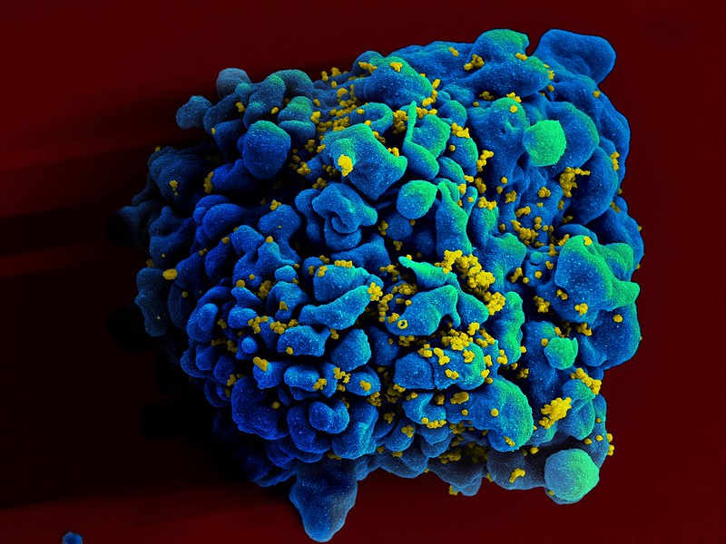
Colourised scanning electron micrograph of HIV (yellow) infecting a human T9 cell (blue). Credit: NIH

Australian researchers have discovered a previously unknown rogue immune cell that can cause poor antibody responses in chronic viral infections. The finding, published in the journal, Immunity, may lead to earlier intervention and possibly prevention of some types of viral infections such as HIV or hepatitis.

One of the remaining mysteries of the human immune system is why ‘memory’ B cells often only have a weak capacity to protect us from persistent infections.

In an answer to this, researchers from the Monash University Biomedicine Discovery Institute have now discovered that chronic viral infection induces a previously unknown immune B memory cell that does not produce high levels of antibodies.

Importantly the research team, led by Professor Kim Good-Jacobson and Dr Lucy Cooper, also determined the most effective time during the immune response for therapeutics such as anti-viral and anti-cancer drugs to better boost immune memory cell development.

“What we discovered was a previously unknown cell that is produced by chronic viral infection. We also determined that early intervention with therapeutics was the most effective to stop this type of memory cell being formed, whereas late intervention could not,” Professor Good-Jacobson said.

According to Dr Cooper, chronic viral infections have been known to alter our ability to form effective long-term protective antibody responses, but how that happens is unknown.

“In the future, this research may result in new therapeutic targets, with the aim to reduce the devastating effect of chronic infectious diseases on global health, specifically those that are not currently preventable by vaccines,” she said.

“Revealing this new immune memory cell type, and what genes it expresses, allows us to determine how we can target it therapeutically and whether that will lead to better antibody responses.”

The research team are also looking to see whether this population is a feature of long COVID, which results in some people having a reduced capacity to fight off the symptoms of COVID infection long after the virus has dissipated.

Source: Monash University

Scientists Reveal how Drug Locks Hepatitis D Virus out of Liver Cells

Colourised transmission electron micrograph of hepatitis B virus particles (colourised red and yellow). Credit: NIAID and CDC (Transmission electron micrograph image courtesy of CDC; colourisation by NIAID).

Over 12 million people worldwide suffer from a chronic infection with the hepatitis D virus. This most severe viral liver disease is associated with a high risk of dying from liver cirrhosis and liver cancer. It is caused by the hepatitis D virus (HDV), which uses the surface proteins of the hepatitis B virus (HBV) as a vehicle to specifically enter liver cells via a protein in the cell membrane – the bile salt transporter protein NTCP. This cell entry can be prevented by the active agent bulevirtide.

An international research team has now succeeded in deciphering the molecular structure of bulevirtide in complex with the HBV/HDV receptor NTCP at the molecular level. The research results published in the journal Nature Communications pave the way for more targeted and effective treatments for millions of people chronically infected with HBV/HDV.

The entry inhibitor bulevirtide is the first and currently only approved drug (under the drug name Hepcludex) for the treatment of chronic infections with the hepatitis D virus. The active agent effectively inhibits the replication of hepatitis D viruses and leads to a significant improvement in liver function. But the exact mechanism by which bulevirtide interacts with the virus entry receptor on the surface of the liver cells – the bile salt transporter protein NTCP (sodium taurocholate cotransporting polypeptide) – and thereby inhibits the entry of the viruses into the cells was previously unknown.

In order to understand the molecular interaction of bulevirtide and NTCP at the molecular level, the researchers first generated an antibody fragment that specifically recognises the NTCP-bulevirtide complex and makes it accessible for analysis when bound to nanoparticles. This complex was then analysed using cryo-electron microscopy, which allowed to visualise structural details with atomic resolution. The research results represent a milestone in understanding both the interaction of HBV and HDV with their cellular entry receptor NTCP and the mechanism of cell receptor blockade by bulevirtide.

How bulevirtide blocks the cell entry receptor NTCP

The analysis showed that bulevirtide forms three functional domains in the interaction with the HBV/HDV receptor NTCP: a myristoyl group that interacts with the cell membrane on the outside of the cell; an essential core sequence (‘plug’) that fits precisely into the bile salt transport tunnel of the NTCP like the bit of a key into a lock; and an amino acid chain that stretches across the extracellular surface of the receptor, enclosing it like a brace.

“The formation of a ‘plug’ in the transport tunnel and the associated inactivation of the bile salt transporter is so far unique among all known virus-receptor complexes. This structure explains why the physiological function of the NTCP is inhibited when patients are treated with bulevirtide,” says Prof Stephan Urban, DZIF Professor of Translational Virology and Deputy Coordinator of the DZIF research area Hepatitis, in whose laboratory at Heidelberg University the active agent bulevirtide was developed.

“Thanks to the structural details of the interaction with bulevirtide, we have also gained insights that enable the development of smaller active agents – so-called peptidomimetics – with improved pharmacological properties. Our structural analysis also lays the foundation for the development of drugs that are not only based on peptides and possibly enable oral administration,” adds the co-author of the study, Prof Joachim Geyer from the Institute of Pharmacology and Toxicology at Justus Liebig University Giessen.

Evolutionary adaptation of hepatitis B viruses to host species

The structural analysis also helped to decode an important factor in the species specificity of hepatitis B and D viruses. According to the findings of the analysis, the amino acid at position 158 of the NTCP amino acid chain plays an essential role in virus-receptor interaction. A change in the amino acid at this position prevents the binding of HBV/HDV. This explains why certain Old World monkeys, such as macaques, cannot be infected by HBV/HDV.

“Our findings enable a deeper understanding of the evolutionary adaptation of human and animal hepatitis B viruses to their hosts and also provide an important molecular basis for the development of new and targeted drugs,” adds co-author Prof Dieter Glebe, DZIF scientist at the Institute of Medical Virology at Justus Liebig University Giessen.

“Thanks to the structural details of the interaction with bulevirtide, we have also gained insights that enable the development of smaller active agents — so-called peptidomimetics — with improved pharmacological properties. Our structural analysis also lays the foundation for the development of drugs that are not only based on peptides and possibly enable oral administration,” adds the co-author of the study, Prof Joachim Geyer from the Institute of Pharmacology and Toxicology at Justus Liebig University Giessen.

Evolutionary adaptation of hepatitis B viruses to host species

The structural analysis also helped to decode an important factor in the species specificity of hepatitis B and D viruses. According to the findings of the analysis, the amino acid at position 158 of the NTCP amino acid chain plays an essential role in virus-receptor interaction. A change in the amino acid at this position prevents the binding of HBV/HDV. This explains why certain Old World monkeys, such as macaques, cannot be infected by HBV/HDV.

“Our findings enable a deeper understanding of the evolutionary adaptation of human and animal hepatitis B viruses to their hosts and also provide an important molecular basis for the development of new and targeted drugs,” adds co-author Prof Dieter Glebe, DZIF scientist at the Institute of Medical Virology at Justus Liebig University Giessen.

Source: German Center for Infection Research

Time is Running out to Develop a Paxlovid Alternative

Photo by CDC on Unsplash

Researchers from Rutgers University in the U.S. believe that they are ahead in a race to find an oral COVID-19 treatment to supplement or replace the antiviral Paxlovid. Their report, published in Science, shows that an alternative medication, a viral papain-like protease inhibitor, inhibits disease progression in animals while also possessing an important advantage over Paxlovid – fewer prescription drug contraindications.

“COVID-19 remains the nation’s third leading cause of death, so there’s already a massive need for additional treatment options,” said Jun Wang, senior author of the study and associate professor at Rutgers. “That need will grow more urgent when, inevitably, COVID-19 mutates in ways that prevent Paxlovid from working.”

The Rutgers team hoped to make a drug that interfered with viral papain-like protease (PLpro), a protein that performs important functions in all known strains of COVID-19.

Creating such a drug required detailed information about PLpro’s structure, which Wang’s team got from the Arnold Lab at Rutgers’ Center for Advanced Biotechnology and Medicine (CABM).

Precise knowledge of PLpro’s structure enabled Wang’s team to design and synthesise 85 drug candidates that would bond to – and interfere with – this vital protein.

“The PLpro crystal structures showed an unexpected arrangement of how the drug candidate molecules bind to its protein target, leading to innovative design ideas implemented by professor Wang’s medicinal chemistry team,” said Eddy Arnold, who is a professor at CABM.

Laboratory testing established that the most effective of those drug candidates, a compound dubbed Jun12682, inhibited several strains of the SARS-CoV-2 virus, including strains that resist treatment with Paxlovid.

Oral treatment with Jun12682 on SARS-CoV-2-infected mice was shown to reduce viral lung loads and lesions while improving survival rates.

“Our treatment was about as effective in mice as Paxlovid was in its initial animal tests,” said Wang, who added the experimental drug appears to have at least one major advantage over the older drug.

“Paxlovid interferes with many prescription medications, and most people who face the highest risk of severe COVID-19 take other prescription medicines, so it’s a real problem,” Wang said.

“We tested our candidate Jun12682 against major drug-metabolising enzymes and saw no evidence that it would interfere with other medications.”

Source: Rutgers University

Searching for Broad-spectrum Antiviral Agents for the Next Pandemic

Photo by National Cancer Institute on Unsplash

A new study has identified potential broad-spectrum antiviral agents that can target multiple families of RNA viruses with pandemic potential. The study, published in Cell Reports Medicine, tested an array of innate immune agonists that work by targeting pathogen recognition receptors, and found several agents that showed promise, including one that exhibited potent antiviral activity against members of RNA viral families.

The authors say recent epidemics as well as global climate change and the continuously evolving nature of the RNA genome indicate that arboviruses, viruses spread by arthropods such as mosquitoes, are prime candidates for the next pandemic after COVID. These include Chikungunya virus (CHIKV), Dengue virus, West Nile virus and Zika virus. The researchers write: “Given their already-demonstrated epidemic potential, finding effective broad-spectrum treatments against these viruses is of the utmost importance as they become potential agents for pandemics.”

Led by Gustavo Garcia Jr. in the UCLA Department of Molecular and Medical Pharmacology, researchers found that several antivirals inhibited these arboviruses to varying degrees. “The most potent and broad-spectrum antiviral agents identified in the study were cyclic dinucleotide (CDN) STING agonists, which also hold promise in triggering an immune defence against cancer,” said senior author Vaithi Arumugaswami, Associate Professor in the UCLA Department of Molecular and Medical Pharmacology.

“A robust host antiviral response induced by a single dose treatment of STING agonist cAIMP is effective in preventing and mitigating the debilitating viral arthritis caused by Chikungunya virus in a mouse model. This is a very promising treatment modality as Chikungunya virus-affected individuals suffer from viral arthritis years and decades from the initial infection,” Arumugaswami added.

“At molecular level, CHIKV contributes to robust transcriptional (and chemical) imbalances in infected skin cells (fibroblasts) compared to West Nile Virus and ZIKA Virus, reflecting a possible difference in the viral-mediated injury (disease pathogenesis) mechanisms by viruses belonging to different families despite all being mosquito-borne viruses,” said senior author Arunachalam Ramaiah, Senior Scientist in the City of Milwaukee Health Department.

“The study of transcriptional changes in host cells reveals that cAIMP treatment rescues (reverses) cells from the harmful effect of CHIKV-induced dysregulation of cell repair, immune, and metabolic pathways,” Ramaiah added.

The study concludes that the STING agonists exhibited broad-spectrum antiviral activity against both arthropod-borne- and respiratory viruses, including treaded SARS-CoV-2 and Enterovirus D68 in cell culture models.

Garcia notes, “The next step is to develop these broad-spectrum antivirals in combination with other existing antivirals and be made readily available in the event of future respiratory and arboviral disease outbreaks.”

Source: University of California – Los Angeles Health Sciences