Scientists have discovered that an important brain protein that quiets overactive brain cells and is abnormally low in children with autism, which may explain why so many children with autism also have epilepsy. The findings were published in Neuron.
This protein can be detected in the cerebrospinal fluid, making it a promising marker to diagnose autism and potentially treat the epilepsy that accompanies the disorder.
Mutated versions of this gene were known to cause autism combined with epilepsy, and epilepsy appears in 30% to 50% of children with autism. Autism, which is 90% genetic, affects 1/58 children in the US.
Appropriately nicknamed ‘catnap2’, the protein, CNTNAP2, is produced by the brain cells when they become overactive. Because the brains of children with autism and epilepsy lack sufficient CNTNAP2, scientists found, their brains become overactive, leading to seizures.
For the study, the researchers analysed the cerebrospinal fluid in individuals with autism and epilepsy, and in mouse models. Though, cerebrospinal fluid has been used in researching disorders such as Parkinson’s, this is the first study showing it is an important biomarker in autism.
The new finding about CNTNAP2’s role in calming the brain in autism and epilepsy may lead to new treatments.
“We can replace CNTNAP2,” said lead study author Peter Penzes, the director of the Center for Autism and Neurodevelopment at Northwestern University Feinberg School of Medicine. “We can make it in a test tube and should be able to inject it into children’s spinal fluid, which will go back into their brain.”
Penzes’ lab is currently working on this technique in preclinical research.
The level in the spinal cord is proxy for the level in the brain, explained Penzes. When brain cells are too active because of overstimulation, they produce more CNTNAP2, which floats away and binds to other brain cells to calm them. The protein also leaks into the cerebrospinal fluid, where scientists were able to measure it, giving them a clue for how much is produced in the brain.
The findings of a study published in Science Translational Medicine paint a new picture of how current antidepressant drugs work and suggest a new drug target in depression. As with most drugs, antidepressants were developed through trial and observation. Some 40% of patients with the disorder don’t respond adequately to the drugs, and when they do work, antidepressants take weeks to provide relief. Why this is has remained largely a mystery.
To figure out why these drugs have a delayed onset, the team examined a mouse model of chronic stress that leads to changes in behaviours controlled by the hippocampus. The hippocampus is vulnerable to stress and atrophies in people with major depression or schizophrenia. Mice exposed to chronic stress show cognitive deficits, a hallmark of impaired hippocampal function.
“Cognitive impairment is a key feature of major depressive disorder, and patients often report that difficulties at school and work are some of the most challenging parts of living with depression. Our ability to model cognitive impairment in lab mice gives us the chance to try and understand how to treat these kinds of symptoms,” said Professor Dane Chetkovich, MD, PhD, who led the study.
The study focussed on an ion transporter channel in nerve cell membranes known as the HCN channel. Previous work has shown HCN channels have a role in depression and separately to have a role in regulation of cognition. According to the authors, this was the first study to explicitly link the two observations.
Examination of postmortem hippocampal samples led the team to establish that HCN channels are more highly expressed in people with depression. HCN channel activity is modulated by a small signaling molecule called cAMP, which is increased by antidepressants. The team used protein receptor engineering to increase cAMP signaling in mice and establish in detail the effects this has on hippocampal HCN channel activity and, through that connection, on cognition.
Turning up cAMP was found to initially increase HCN channel activity, limit the intended effects of antidepressants and negatively impact cognition (as measured in standard lab tests).
However, a total reversal took place over a period of some weeks. Previous work by the researchers had established that an auxiliary subunit of the HCN channel, TRIP8b, is essential for the channel’s role in regulating animal behaviour. The new study shows that, over weeks, a sustained increase in cAMP starts to interfere with TRIP8b’s ability to bind to the HCN channel, thereby quieting the channel and restoring cognitive abilities.
“This leaves us with acute and chronic changes in cAMP, of the sort seen in antidepressant drug therapy, seen here for the first time to be regulating the HCN channel in the hippocampus in two distinct ways, with opposing effects on behaviour,” Prof Chetkovich said. “This appears to carry promising implications for new drug development, and targeting TRIP8b’s role in the hippocampus more directly could help to more quickly address cognitive deficits related to chronic stress and depression.”
During childhood, about one in 20 people go through a period of stuttering. Until the latter half of the 20th century, stuttering was believed to be a psychological problem stemming from lack of effort or from trauma.
Nowadays, neuroimaging techniques are leading to a much better understanding of brain function during speech and how stuttering arises. Frank Guenther, from Boston University, reported findings from a new study at the 181st Meeting of the Acoustical Society of America.
Guenther gives the example of speech being a jukebox that plays CDs. The jukebox has two circuits: one that chooses a CD and one that plays the CD.
In the brain, this corresponds to one circuit initiating the desired speech in the basal ganglia, while another circuit coordinates the muscles needed to generate the speech. Stuttering stems from the initiation of speech, so only the first of the two circuits is impaired.
“In stuttering, the CDs themselves are fine, but the mechanism for choosing them is impaired,” said Guenther.
This theory is in agreement with behavioural observations of stuttering: people will often speak words fluently later in a sentence, even if those same words cause stuttering at the start of a sentence.
Guenther and his team created computational models of how the speech initiation circuit performs in a non-stuttering individual. Since Parkinson’s disease also affects the initiation circuit, they can compare these models directly to data taken from the basal ganglia during deep brain stimulation surgery in patients with the disease.
“This gives us a fighting chance of finding the specific problems underlying stuttering and addressing them with highly targeted drugs or technological treatments that have minimal unwanted side effects,” said Guenther.
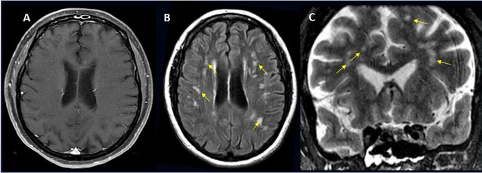
49-year-old female with past medical history of mitral valve disease and tricuspid valve regurgitation who developed headache followed by cough and fever presented to the ER with right upper eyelid ptosis (drooping). Credit: Radiological Society of North America and Scott H. Faro, M.D.
Approximately one in 100 patients hospitalised with COVID will likely develop complications of the central nervous system, according to a large international study. These can include stroke, haemorrhage, and other potentially fatal complications. The study was presented at the annual meeting of the Radiological Society of North America (RSNA).
“Much has been written about the overall pulmonary problems related to COVID, but we do not often talk about the other organs that can be affected,” said study lead author Scott H. Faro, MD, FASFNR, professor of radiology and neurology at Thomas Jefferson University. “Our study shows that central nervous system complications represent a significant cause of morbidity and mortality in this devastating pandemic.”
Dr Faro initiated the study after finding that only a small number of cases informed existing literature on central nervous system complications in hospitalised COVID patients.
To build a more complete picture, he and his colleagues analysed nearly 40 000 cases of hospitalised COVID patients, admitted between September 2019 and June 2020. Their average age was 66 years old, and two thirds were men.
Confusion and altered mental status were the most common causes of admission followed by fever. Comorbidities such as hypertension, cardiac disease and diabetes were common.
There were 442 acute neuroimaging findings most likely associated with the viral infection, with central nervous system complications in 1.2% of this large patient group.
“Of all the inpatients who had imaging such as MRI or a CT scan of the brain, the exam was positive approximately 10% of the time,” Dr Faro said. “The incidence of 1.2% means that a little more than one in 100 patients admitted to the hospital with COVID are going to have a brain problem of some sort.”
Ischaemic stroke, with an incidence of 6.2%, was the most common complication, followed by intracranial haemorrhage (3.72%) and encephalitis (0.47%).
A small percentage of unusual findings was uncovered, such as acute disseminating encephalomyelitis, an inflammation of the brain and spinal cord, and posterior reversible encephalopathy syndrome, a syndrome that mimics many of the symptoms of a stroke.
“It is important to know an accurate incidence of all the major central nervous system complications,” Dr Faro said. “There should probably be a low threshold to order brain imaging for patients with COVID.”
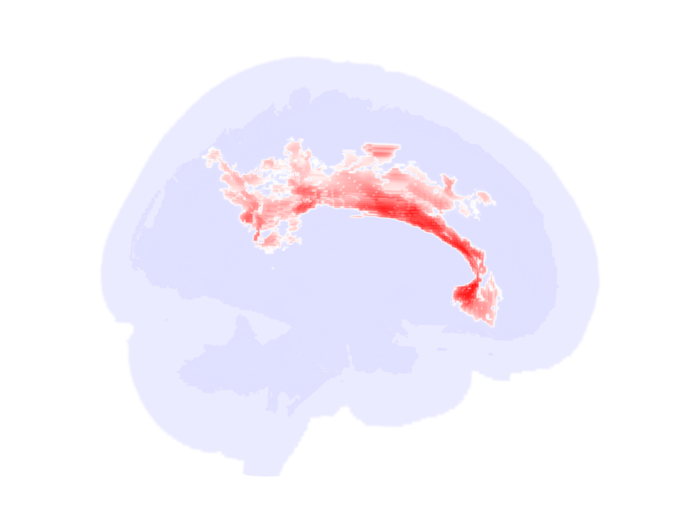
Significant alterations in the brain’s white matter in adolescents with autism spectrum disorder (ASD). Credit: RSNA and researcher, Clara Weber
Using specialised MRI, researchers found significant changes in the microstructure of the brain’s white matter, especially in thecorpus callosum in adolescents and young adults with autism spectrum disorder (ASD) compared to controls. This research will be presented next week at the annual meeting of the Radiological Society of North America (RSNA).
“One in 68 children in the U.S. is affected by ASD, but high variety in symptom manifestation and severity make it hard to recognise the condition early and monitor treatment response,” explained Clara Weber, postgraduate research fellow at Yale University School of Medicine. “We aim to find neuroimaging biomarkers that can potentially facilitate diagnosis and therapy planning.”
Researchers reviewed diffusion tensor imaging (DTI) brain scans from a large dataset of patients between the age of six months and 50 years. DTI is an MRI technique that measures connectivity in the brain by detecting how water moves along its white matter tracts. Water molecules diffuse differently through the brain, depending on the integrity, architecture and presence of barriers in tissue.
“If you think of gray matter as the computer, white matter is like the cables,” Weber said. “DTI helps us assess how connected and intact those cables are.”
For the study, clinical and DTI data from 583 patients from four existing studies of distinct patient populations were analysed: infants (median age 7 months), toddlers (median age 32 months), adolescents, and young adults.
“One of the strengths of our study is that we looked at a wide range of age groups, not just school-aged children,” Weber said.
To assess the influences of age and ASD diagnosis on white matter microstructure, the research team created fractional anisotropy, mean diffusivity and radial diffusivity maps using data from the four studies.
Fractional anisotropy is the extent water diffusion is restricted to just one direction. A value of zero means that diffusion is unrestricted in all directions, while one means that diffusion is unidirectional. Mean diffusivity is the overall mobility of water molecules, indicating how densely cells are packed together. Radial diffusivity is the extent water diffuses perpendicular to a white matter tract.
“When white matter integrity is disrupted, we see more water diffusing perpendicularly, which translates to a higher radial diffusivity,” Weber said.
The key finding of the analysis was reduced fractional anisotropy within the anterior/middle tracts of the corpus callosum in adolescent and young adult ASD patients compared to individuals in the control group. The corpus callosum is a thick bundle of nerve fibers that connects and allows the two sides of the brain to communicate. Corresponding increases in ASD-related mean diffusivity and radial diffusivity were found in young adults.
“In adolescents, we saw a significant influence of autism,” Weber said. “In adults, the effect was even more pronounced. Our results support the idea of impaired brain connectivity in autism, especially in tracts that connect both hemispheres.”
Compared to controls, no reduction in fractional anisotropy was seen in the same tracts in toddlers and infants with ASD.
The researchers hope the findings can help improve early diagnosis of ASD and provide potential objective biomarkers to monitor treatment response.
“We need to find more objective biomarkers for the disorder that can be applied in clinical practice,” Weber said.
A new study has found that hypertension may double an adult’s risk of developing epilepsy, according to a new study published in Epilepsia.
The study recruited 2986 US participants with an average age of 58 years, 55 new cases of epilepsy were identified during an average follow-up of 19 years. Hypertension, defined as presence of elevated blood pressure or use of antihypertensive medications, was linked to a nearly 2-fold higher risk of epilepsy. After excluding participants with normal blood pressure who were taking antihypertensive medications, hypertension was linked to a 2.44-times higher risk of epilepsy.
“Our study shows that hypertension, a common, modifiable, vascular risk factor, is an independent predictor of epilepsy in older age,” said co–lead author Maria Stefanidou, MD, MSc, of Boston University School of Medicine. “Even though epidemiological studies can only show association and not causation, this observation may help identify subgroups of patients who will benefit from targeted, aggressive hypertension management and encourage performance of dedicated clinical studies that will focus on early interventions to reduce the burden of epilepsy in older age.”
Patients with this unusual combination of conditions were referred to Mehul Dattani (UCL), and affected individuals were found to carry the same homozygous mutation in the PRDM13 gene, which encodes a chromatin modifying factor that contributes to regulating cell fate. Intriguingly, an unaffected heterozygous carrier of this mutation was identified by screening 42 unaffected individuals in the Maltese population, suggesting that this mutation is present at low levels in the population.
The researchers set out to model this condition and identify the underlying causes using a PRDM13-deficient mouse model. The researchers found evidence that both the cerebellar hypoplasia and reproductive phenotypes resulted from defects in the specification of specific populations of GABAergic neuronal progenitors in the developing cerebellum and hypothalamus, respectively.
The results indicate that this condition results from abnormal cell fate specification during development. Consequently, the hypoplastic cerebellum is deficient in molecular layer interneurons, which play critical roles in regulating cerebellar circuits. In the hypothalamus, fewer Kisspeptin neurons, which are important regulators of gonadotropin releasing hormone and puberty, were present in PRDM13 mutant mice.
Together, these findings identify PRDM13 as a critical regulator of neuronal cell fate in the cerebellum and hypothalamus, providing a mechanistic explanation for the co-occurrence of hypogonadism and cerebellar hypoplasia in this syndrome.
A healthy neuron. Credit: National Institutes of Health
Human Neurons Differ From Animal Ones in a Surprising WayIn a surprising new finding published in Nature, neuroscientists have shown that human neurons have a much smaller number of ion channels than expected, compared to the neurons of other mammals.
Ion channels are integral membrane proteins that contain pathways through which ions can flow. By shifting between closed and open conformational states (‘gating’ process), they control passive ion flow through the plasma membrane.
The researchers hypothesise that lower channel density may have helped the human brain evolve energy efficiency, letting it divert resources elsewhere.
“If the brain can save energy by reducing the density of ion channels, it can spend that energy on other neuronal or circuit processes,” said senior author Mark Harnett, an associate professor of brain and cognitive sciences.
Analysing neurons from 10 different mammals, the researchers identified a “building plan” that holds true for every examined species — save humans. They found that as the size of neurons increases, the density of channels found in the neurons also increases.
However, human neurons proved to be a striking exception to this rule.
“Previous comparative studies established that the human brain is built like other mammalian brains, so we were surprised to find strong evidence that human neurons are special,” said lead author and former MIT graduate student Lou Beaulieu-Laroche.
Neurons in the mammalian brain can receive electrical signals from thousands of other cells, and that input determines whether or not they will fire an electrical impulse called an action potential. In 2018, Prof Harnett and Beaulieu-Laroche discovered that human and rat neurons differ in some of their electrical properties, primarily in dendrites.
One of the findings from that study was that human neurons had a lower density of ion channels than neurons in the rat brain. The researchers were surprised by this observation, as ion channel density was generally assumed to be constant across species. In their new study, Harnett and Beaulieu-Laroche decided to compare neurons from several different mammalian species to see if they could find any patterns that governed the expression of ion channels. They studied two types of voltage-gated potassium channels and the HCN channel, which conducts both potassium and sodium, in layer 5 pyramidal neurons, a type of excitatory neurons found in the brain’s cortex.
They were able to obtain brain tissue from a range of 10 mammalian species, including human tissue removed from patients with epilepsy during brain surgery. This variety allowed the researchers to cover a range of cortical thicknesses and neuron sizes across the mammalian kingdom.
In nearly every mammalian species the researchers examined, the density of ion channels increased as the size of the neurons went up. Human neurons bucked this trend, having a much lower density of ion channels than expected.
The increase in channel density across species was a surprise, Prof Harnett explained, because the more channels there are, the more energy is required to pump ions in and out of the cell. However, it started to make sense once the researchers began thinking about the number of channels in the overall volume of the cortex, he said.
In the tiny brain of the Etruscan shrew, which is packed with very small neurons, there are more neurons in a given volume of tissue than in the same volume of tissue from the rabbit brain, which has much larger neurons. But because the rabbit neurons have a higher density of ion channels, the density of channels in a given volume of tissue is the same in both species, or any of the nonhuman species the researchers analysed.
“This building plan is consistent across nine different mammalian species,” Prof Harnett said. “What it looks like the cortex is trying to do is keep the numbers of ion channels per unit volume the same across all the species. This means that for a given volume of cortex, the energetic cost is the same, at least for ion channels.”
The human brain represents a striking deviation from this building plan, however. Instead of increased density of ion channels, the researchers found a dramatic decrease in the expected density of ion channels for a given volume of brain tissue.
The researchers believe this lower density may have evolved as a way to expend less energy on pumping ions, which allows the brain to use that energy for something else, like creating more complicated synaptic connections between neurons or firing action potentials at a higher rate.
“We think that humans have evolved out of this building plan that was previously restricting the size of cortex, and they figured out a way to become more energetically efficient, so you spend less ATP per volume compared to other species,” Prof Harnett said.
He now hopes to study where that extra energy might be going, and whether there are specific gene mutations that help neurons of the human cortex achieve this high efficiency. The researchers are also interested in exploring whether primate species that are more closely related to humans show similar decreases in ion channel density.
A pioneering new study from Taiwan showed that focused ultrasound, which can be used to non-invasively target circuits in the brain, may benefit some patients with epilepsy who experience seizures which remain unresponsive to standard anti-seizure medications.
The results showed that of six patients with drug-resistant seizures, two patients had fewer seizures within three days of receiving focused ultrasound; however, one patient showed signs of more frequent subclinical seizures (which are not felt by the individual). The findings from the study were published in the journal Epilepsia.
Imaging tests performed after the treatment show that there were no negative effects on the brain. One patient reported a sensation of heat on the scalp during the treatment, and another patient experienced temporary memory impairment that resolved within three weeks.
“Neuromodulation is an alternative treatment for drug-resistant epilepsy. Compared with the present modalities used in neuromodulation for epilepsy, focused ultrasound can access deeper brain regions and focus on the main target of the epileptic network in a relatively less invasive approach,” explained senior author Hsiang-Yu Yu, MD, of Taipei Veterans General Hospital, in Taiwan. “It gives new hope and sheds new light for patients with drug-resistant epilepsy.”
University of Utah scientists have discovered a new type of neuron in the retina, which will help fill in our understanding of how sensory information is relayed.
In the central nervous system a complex network of neurons communicate with each other to relay sensory and motor information. In this chain of communication, a type of neuron called interneurons serve as intermediaries . A research team led by Ning Tian, PhD, identified a previously unknown type of interneuron in the mammalian retina. Their findings were published in the journal PNAS.
This discovery is a major step forward for the field as scientists strive to build a better understanding of the central nervous system by identifying all classes of neurons and their connections.
“Based on its morphology, physiology, and genetic properties, this cell doesn’t fit into the five classes of retinal neurons first identified more than 100 years ago,” said Dr Tian. “We propose they might belong to a new retinal neuron class by themselves.”
The research team called their discovery the Campana cell after its shape, which resembles a hand bell. Campana cells relay visual signals from both types of light-sensing rod and cone photoreceptors in the retina, however their exact purpose is the subject of ongoing research. Experiments revealed that Campana cells remain activated for an unusually long time – as long as 30 seconds – in response to a 10 millisecond light flash stimulation.
“In the brain, persistent firing cells are believed to be involved in memory and learning,” said Dr Tian. “Since Campana cells have a similar behaviour, we theorise they could play a role in prompting a temporal ‘memory’ of a recent stimulation.”