Category: Neurology

Scientists Record Powerful Signals in the Brain’s White Matter

Scientists have concentrated on the grey matter of the cortex, composed of nerve cell bodies , while ignoring white matter, composed of axons, even though it makes up half the brain. Now, in the Proceedings of the National Academy of Sciences, Vanderbilt University researchers report strong signs of brain activity when performing certain tasks.

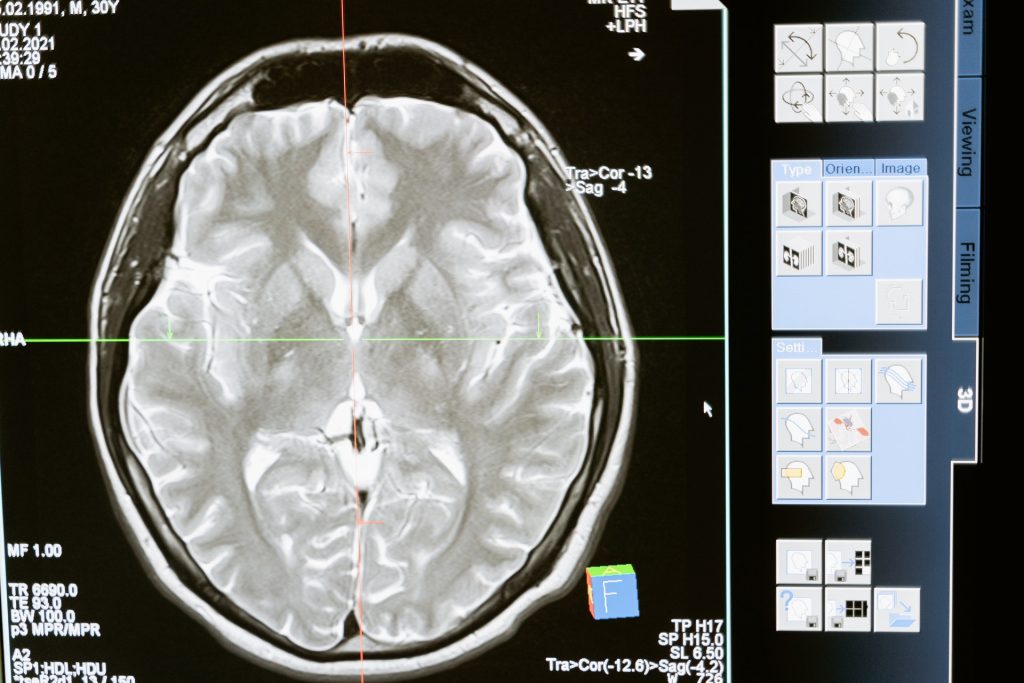
For several years, John Gore, PhD, director of the Vanderbilt University Institute of Imaging Science, and his colleagues have used functional magnetic resonance imaging (fMRI) to detect blood oxygenation-level dependent (BOLD) signals, a key marker of brain activity, in white matter.

In this latest paper, the researchers report that when people who are having their brains scanned by fMRI perform a task, like wiggling their fingers, BOLD signals increase in white matter throughout the brain.

“We don’t know what this means,” said the paper’s first author, Kurt Schilling, PhD, research assistant professor of Radiology and Radiological Sciences at VUMC. “We just know that something is happening. There truly is a powerful signal in the white matter.”

It is important to pursue this because disorders as diverse as epilepsy and multiple sclerosis disrupt the “connectivity” of the brain, Schilling said. This suggests that something is going on in white matter.

To find out, the researchers will continue to study changes in white matter signals they’ve previously detected in schizophrenia, Alzheimer’s disease and other brain disorders. Through animal studies and tissue analysis, they also hope to determine the biological basis for these changes.

In grey matter, BOLD signals reflect a rise in blood flow (and oxygen) in response to increased nerve cell activity.

Perhaps the axons, or the glial cells that maintain the protective myelin sheath around them, also use more oxygen when the brain is ‘working’. Or perhaps these signals are somehow related to what’s going on in the grey matter.

But even if nothing biological is going on in white matter, “there’s still something happening here,” Schilling said. “The signal is changing. It’s changing differently in different white matter pathways and it’s in all white matter pathways, which is a unique finding.”

One reason that white matter signals have been understudied is that they have lower energy than grey matter signals, and thus are more difficult to distinguish from the brain’s background “noise.”

The VUMC researchers boosted the signal-to-noise ratio by having the person whose brain was being scanned repeat a visual, verbal or motor task many times to establish a trend and by averaging the signal over many different white matter fibre pathways.

“For 25 or 30 years, we’ve neglected the other half of the brain,” Schilling said. Some researchers not only have ignored white matter signals but have removed them from their reports of brain function.

The Vanderbilt findings suggest that many fMRI studies thus “may not only underestimate the true extent of brain activation, but also … may miss crucial information from the MRI signal,” the researchers concluded.

Source: Vanderbilt University

The Eyes may Hold the Secret to the Greatest Benefits from TMS Therapy

Photo by Victor Freitas on Pexels

A pair of recently published studies from researchers at UCLA Health suggest that measuring changes in how pupils react to light could help predict recovery from depression and personalise transcranial magnetic stimulation (TMS) treatment of major depressive disorder.

TMS is a safe, non-invasive therapy that uses magnetic fields to stimulate parts of the brain involved in mood regulation. While TMS is proven effective, not all patients respond equally well to the therapy. The ability to predict who will benefit most could allow doctors to better customise and target treatments.

In two recent studies, UCLA scientists found that the pupil’s response to light before treatment correlated with improvements in depression symptoms over the course of therapy. Pupil size reflects activation of the autonomic nervous system, which controls involuntary functions and is negatively impacted in people with depression.

The first study, appearing in the Journal of Affective Disorders, reports on outcomes for 51 patients who underwent daily TMS sessions. Before receiving treatment, researchers measured the patients’ baseline pupillary constriction amplitude, or CA: how much the pupil shrinks when exposed to light. The pupil’s constriction is an indicator of parasympathetic nervous system function. The researchers found a significant association between baseline pupil constriction amplitude and symptom improvement, indicating that a greater constriction amplitude at baseline was associated with a better outcome. In other words, those with larger pupil constriction in response to light at baseline showed greater symptom improvement over their full treatment.

The second study, published in Brain Stimulation, went further and compared patients who were treated for depression with one of two common TMS protocols: 10Hz stimulation and intermittent theta burst stimulation (iTBS). In 10Hz stimulation, magnetic pulses are delivered in a continuous and relatively high-frequency stimulation. iTBS is a faster form of stimulation with bursts of three pulses at 50Hz, repeated with short breaks between bursts. This pattern is thought to mimic the natural rhythm of certain brain activities.

The researchers found that people with slower pupillary constriction had significantly greater improvement in depression after 10 sessions if they received iTBS rather than 10Hz treatment.

“These results suggest we may be able to use a simple test of the pupil to identify who is most likely to respond to electromagnetic stimulation of the brain to treat their depression,” said researcher Cole Citrenbaum, lead author of both studies.

Tailored TMS treatments

The researchers propose that measuring pupillary reactivity before starting TMS could guide treatment selection. “Additionally, we may be able to tailor the frequency of stimulation to the individual patient to maximise their benefit from treatment,” Citrenbaum said.

“At the present time, about 65% of patients treated with TMS have a substantial improvement in their depression,” said Dr Andrew F. Leuchter, senior author of both studies. “Our goal is to have more than 85% of patients fully recover from depression. As we better understand the complex brain activity underlying depression, we move closer to matching patients with the treatments that ensure their full recovery. Pupil testing may be one useful tool in reaching this goal.”

The studies add to growing evidence on the benefits of biologically-based personalization in treating major depression. UCLA researchers plan further trials to confirm the value of pupillometry in optimizing transcranial magnetic stimulation.

Source: University of California – Los Angeles Health Sciences

Converting Brain Immune Cells into Neurons Boosts Stroke Recovery in Mice

Source: CC0

Japanese researchers have turned microglia (brain immune cells) into neurons, successfully restoring brain function after stroke-like injury in mice. These findings, published in PNAS, suggest that replenishing neurons from immune cells could be a promising avenue for treating stroke in humans.

Recovery from stroke, where blood supply to neurons is disrupted by blockage or bleeding, is often poor, with patients suffering from severe physical disabilities and cognitive problems.

“When we get a cut or break a bone, our skin and bone cells can replicate to heal our body. But the neurons in our brain cannot easily regenerate, so the damage is often permanent,” says Professor Kinichi Nakashima, from Kyushu University’s Graduate School of Medical Sciences. “We therefore need to find new ways to replace lost neurons.”

One possible strategy is to convert other cells in the brain into neurons. Here, the researchers focused on microglia, the main immune cells in the central nervous system. Microglia are tasked with removing damaged or dead cells in the brain, so after a stroke, they move towards the site of injury and replicate quickly.

“Microglia are abundant and exactly in the place we need them, so they are an ideal target for conversion,” says first author Dr Takashi Irie, from Kyushu University Hospital.

In prior research, the team demonstrated that they could coax microglia to develop into neurons in the brains of healthy mice. Dr Irie and Professor Nakashima and colleagues, now showed that this strategy of replacing neurons also works in injured brains and contributes to brain recovery.

To conduct the study, the researchers caused a stroke-like injury in mice by temporarily blocking the right middle cerebral artery — a major blood vessel in the brain that is commonly associated with stroke in humans. A week later, the researchers examined the mice and found that they had difficulties in motor function and had a marked loss of neurons in a brain region known as the striatum. This part of the brain is involved in decision making, action planning and motor coordination.

The researchers then used a lentivirus to insert DNA into microglial cells at the site of the injury. The DNA held instructions for producing NeuroD1, a protein that induces neuronal conversion. Over the subsequent weeks, the infected cells began developing into neurons and the areas of the brain with neuron loss decreased. By eight weeks, the new induced neurons had successfully integrated into the brain’s circuits.

At only three weeks post-infection, the mice showed improved motor function in behavioural tests. These improvements were lost when the researchers removed the new induced neurons, providing strong evidence that the newly converted neurons directly contributed to recovery.

“These results are very promising. The next step is to test whether NeuroD1 is also effective at converting human microglia into neurons and confirm that our method of inserting genes into the microglial cells is safe,” says Professor Nakashima.

Furthermore, the treatment was conducted in mice in the acute phase after stroke, when microglia were migrating to and replicating at the site of injury. Therefore, the researchers also plan to see if recovery is also possible in mice at a later, chronic phase.

Source: Kyushu University

AI-based CT Scans of the Brain can Nearly Match MRI

Photo by Mart Production on Pexels

A new artificial intelligence (AI)-based method can provide as much information on subtle neurodegenerative changes in the brain captured by computed tomography (CT) as compared to magnetic resonance imaging (MRI). The method, reported in the journal Alzheimer’s & Dementia, could enhance diagnostic support, particularly in primary care, for conditions such as dementia and other brain disorders.

Compared to MRI, which requires powerful superconducting magnetics and their associated cryogenic cooling, computed tomography (CT) is a relatively inexpensive and widely available imaging technology. CT is considered inferior to MRI when it comes to reproducing subtle structural changes in the brain or flow changes in the ventricular system. Certain imaging must therefore currently be carried out by specialist departments at larger hospitals equipped with MRI.

AI trained on MRI images

Created with deep learning, a form of AI, the software has been trained to transfer interpretations from MRI images to CT images of the same brains. The new software can provide diagnostic support for radiologists and other professionals who interpret CT images.

“Our method generates diagnostically useful data from routine CT scans that, in some cases, is as good as an MRI scan performed in specialist healthcare,” says Michael Schöll, a professor at Sahlgrenska Academy who led the work involved in the study, carried out in collaboration with researchers at Karolinska Institutet, the National University of Singapore, and Lund University

“The point is that this simple, quick method can provide much more information from examinations that are already carried out on a routine basis within primary care, but also in certain specialist healthcare investigations. In its initial stage, the method can support dementia diagnosis, however, it is also likely to have other applications within neuroradiology.”

Reliable decision-making support

This is a well-validated clinical application of AI-based algorithms, and has the potential to become a fast and reliable form of decision-making support that effectively reduces the number of false negatives. The researchers believe that this solution can improve diagnostics in primary care, optimising patient flow to specialist care.

“This is a major step forward for imaging diagnosis,” says Meera Srikrishna, a postdoctor at the University of Gothenburg and lead author of the study.

“It is now possible to measure the size of different structures or regions of the brain in a similar way to advanced analysis of MRI images. The software makes it possible to segment the brain’s constituent parts in the image and to measure its volume, even though the image quality is not as high with CT.”

Applications for other brain diseases

The software was trained on images of 1117 people, all of whom underwent both CT and MRI imaging. The current study mainly involved healthy older individuals and patients with various forms of dementia. Another application that the team is now investigating is for normal pressure hydrocephalus (NPH).

With NPH, the team has obtained new results indicating that the method can be used both during diagnosis and to monitor the effects of treatment. NPH is a condition that occurs particularly in older people, whereby fluid builds up in the cerebral ventricular system and results in neurological symptoms. About two percent of all people over the age of 65 are affected. Because diagnosis can be complicated and the condition risks being confused with other diseases, many cases are likely to be missed.

“NPH is difficult to diagnose, and it can also be hard to safely evaluate the effect of shunt surgery to drain the fluid in the brain,” continues Michael. “We therefore believe that our method can make a big difference when caring for these patients.”

The software has been developed over the course of several years, and development is now continuing in cooperation with clinics in Sweden, the UK, and the US together with a company, which is a requirement for the innovation to be approved and transferred to healthcare.

Source: University of Gothenburg

The Vascular System also Plays a Role in Forming Memories

Diagram of a capillary. Source: Wikimedia Commons

Research on long-term memories has largely focused on the role of neurons but in recent years, research is revealing that other cell types are also vital in memory formation and storage. A new study reveals the crucial role of vascular system cells (pericytes) in the formation of long-term memories of life events – memories that are lost in diseases such as Alzheimer’s. The research, published in the journal Neuron, shows that pericytes, which wrap around the capillaries work in concert with neurons to help ensure that long-term memories are formed.

Pericytes help maintain the structural integrity of the capillaries. Specifically, they control the amount of blood flowing in the brain and play a key role in maintaining the barrier that stops pathogens and toxic substances from leaking out of the capillaries and into brain tissue.

“We now have a firmer understanding of the cellular mechanisms that allow memories to be both formed and stored,” says Cristina Alberini, a professor in New York University’s Center for Neural Science and the paper’s senior author. “It’s important because understanding the cooperation among different cell types will help us advance therapeutics aimed at addressing memory-related afflictions.”

“This work connects important dots between the newly discovered function of pericytes in memory and previous studies showing that pericytes are either lost or malfunction in several neurodegenerative diseases, including Alzheimer’s disease and other dementia,” explains author Benjamin Bessières, a postdoctoral researcher in NYU’s Center for Neural Science.

The discovery, reported in the new Neuron article, of the pericytes’ significance in long-term memory emerged because Alberini, Bessières, Kiran Pandey, and their colleagues examined the role of insulin-like growth factor 2 (IGF2) – a protein that was known to increase following learning in brain regions, such as the hippocampus, and to play a critical role in the formation and storage of memories.

They found that IGF2’s highest levels in the brain cells of the hippocampus do not come from neurons or glial cells, or other vascular cells, but, rather, from pericytes.

Source: New York University

3D-Printed Structures Hold Promise for Repair of Traumatic Brain Injuries

Researchers at the University of Oxford have produced an engineered tissue representing a simplified cerebral cortex by 3D printing human stem cells. The results, published in the journal Nature Communications, showed that, when implanted into mouse brain slices, the structures became integrated with the host tissue.

The breakthrough technique could lead to tailored repairs for brain injuries. The researchers demonstrated for the first time that neural cells can be 3D-printed to mimic the architecture of the cerebral cortex.

Brain injuries, including those caused by trauma, stroke and surgery for brain tumours, typically result in significant damage to the cerebral cortex. For example, each year, around 70 million people globally suffer from traumatic brain injury (TBI), with 5 million of these cases being severe or fatal. Currently, there are no effective treatments for severe brain injuries, leading to serious impacts on quality of life.

Tissue regenerative therapies, especially those in which patients are given implants derived from their own stem cells, could be a promising route to treat brain injuries in the future. Up to now, however, there has been no method to ensure that implanted stem cells mimic the architecture of the brain.

In this new study, the University of Oxford researchers fabricated a two-layered brain tissue by 3D printing human neural stem cells. When implanted into mouse brain slices, the cells showed convincing structural and functional integration with the host tissue.

Lead author Dr Yongcheng Jin (Department of Chemistry, University of Oxford) said: ‘This advance marks a significant step towards the fabrication of materials with the full structure and function of natural brain tissues. The work will provide a unique opportunity to explore the workings of the human cortex and, in the long term, it will offer hope to individuals who sustain brain injuries.’

The cortical structure was made from human induced pluripotent stem cells (hiPSCs), which have the potential to produce the cell types found in most human tissues. A key advantage of using hiPSCs for tissue repair is that they can be easily derived from cells harvested from patients themselves, and therefore would not trigger an immune response.

The hiPSCs were differentiated into neural progenitor cells for two different layers of the cerebral cortex, by using specific combinations of growth factors and chemicals. The cells were then suspended in solution to generate two ‘bioinks’, which were then printed to produce a two-layered structure. In culture, the printed tissues maintained their layered cellular architecture for weeks, as indicated by the expression of layer-specific biomarkers.

When the printed tissues were implanted into mouse brain slices, they showed strong integration, as demonstrated by the projection of neural processes and the migration of neurons across the implant-host boundary. The implanted cells also showed signalling activity, which correlated with that of the host cells. This indicates that the human and mouse cells were communicating with each other, demonstrating functional as well as structural integration.

The researchers now intend to further refine the droplet printing technique to create complex multi-layered cerebral cortex tissues that more realistically mimic the human brain’s architecture. Besides their potential for repairing brain injuries, these engineered tissues might be used in drug evaluation, studies of brain development, and to improve our understanding of the basis of cognition.

The new advance builds on the team’s decade-long track record in inventing and patenting 3D printing technologies for synthetic tissues and cultured cells.

Senior author Dr Linna Zhou (Department of Chemistry, University of Oxford) said: “Our droplet printing technique provides a means to engineer living 3D tissues with desired architectures, which brings us closer to the creation of personalised implantation treatments for brain injury.”

Senior author Associate Professor Francis Szele (Department of Physiology, Anatomy and Genetics, University of Oxford) added: “The use of living brain slices creates a powerful platform for interrogating the utility of 3D printing in brain repair. It is a natural bridge between studying 3D printed cortical column development in vitro and their integration into brains in animal models of injury.”

Senior author Professor Zoltán Molnár (Department of Physiology, Anatomy and Genetics, University of Oxford) said: “Human brain development is a delicate and elaborate process with a complex choreography. It would be naïve to think that we can recreate the entire cellular progression in the laboratory. Nonetheless, our 3D printing project demonstrates substantial progress in controlling the fates and arrangements of human iPSCs to form the basic functional units of the cerebral cortex.”

Source: University of Oxford

Evidence Points to Consciousness Emerging Shortly after Birth or in Late Pregnancy

Figure I Neural measurement tools for studying the emergence of consciousness. Examples of techniques for recording brain activity and/or neuroimaging in infants and foetuses. (A) Infant electroencephalography (EEG) with a geodesic electrode net. (B) Foetal magnetoencephalography (MEG) recorded from a pregnant woman. (C) Infant functional near infrared spectroscopy (fNIRS) recording with multichannel optode cap. (D) An infant is prepared for functional magnetic resonance imaging (fMRI). Source: Bayne et al., 2023

There is evidence that some form of conscious experience is present by birth, and perhaps even in late pregnancy, an international team of researchers has found. The findings, published today in Trends in Cognitive Science, have important clinical, ethical and potentially legal implications, according to the authors. 

Converging evidence from studies of functional network connectivity, attention, multimodal integration, and cortical responses to global oddballs suggests that consciousness is likely to be in place in early infancy and may even occur before birth. Over the decades, theorists have argued that consciousness emerges from anywhere from 30 to 35 weeks of pregnancy (based on EEG of the foetus’s brain) to 12 to 15 months of age (based on higher-order representational theory).

In the study, the researchers argue that by birth the infant’s developing brain is capable of conscious experiences that can leave a lasting imprint on their developing sense of self and understanding of their environment.

The team comprised neuroscientists and philosophers from Monash University, in Australia, University of Tübingen, in Germany, University of Minnesota, in the USA, and Trinity College Dublin.

Although each of us was once a baby, infant consciousness remains mysterious, because infants cannot tell us what they think or feel, explains one of the two lead authors of the paper Dr Tim Bayne, Professor of Philosophy at Monash University. 

“Nearly everyone who has held a newborn infant has wondered what, if anything, it is like to be a baby. But of course we cannot remember our infancy, and consciousness researchers have disagreed on whether consciousness arises ‘early’ (at birth or shortly after) or ‘late’ ­– by one year of age, or even much later.”

To provide a new perspective on when consciousness first emerges, the team built upon recent advances in consciousness science. In adults, some markers from brain imaging have been found to reliably differentiate consciousness from its absence, and are increasingly applied in science and medicine. This is the first time that a review of these markers in infants has been used to assess their consciousness.

Co-author of the study, Lorina Naci, Associate Professor in the School of Psychology, who leads Trinity’s ‘Consciousness and Cognition Group, explained: “Our findings suggest that newborns can integrate sensory and developing cognitive responses into coherent conscious experiences to understand the actions of others and plan their own responses.”

The paper also sheds light into ‘what it is like’ to be a baby. We know that seeing is much more immature in babies than hearing, for example. Furthermore, this work suggests that, at any point in time, infants are aware of fewer items than adults, and can take longer to grasp what’s in front of them, but it is easier for them to process more diverse information, such as sounds from other languages.

Source: Trinity College Dublin

New Clinical Guidelines for the Determination of Brain Death

Photo by Rodnae Productions on Pexels

New guidance has been issued for clinicians on the determination of brain death, also known as death by neurologic criteria. A new consensus practice guideline, developed through a collaboration between the American Academy of Neurology (AAN), the American Academy of Pediatrics (AAP), the Child Neurology Society (CNS), and the Society of Critical Care Medicine (SCCM) is published in Neurology, the medical journal of the American Academy of Neurology.

This guideline updates the 2010 AAN adult practice guidelines and the 2011 AAP/CNS/SCCM paediatric practice guidelines on the determination of brain death. Because of a lack of high-quality evidence on the subject, the experts used an evidence-informed consensus process to develop the guideline.

“Until now, there have been two separate guidelines for determining brain death, one for adults and one for children,” said author Matthew P. Kirschen, MD, PhD, FAAN, of the Children’s Hospital of Philadelphia, and a member of the Child Neurology Society and the Society of Critical Care Medicine. “This update integrates guidance for adults and children into a single guideline, providing clinicians with a comprehensive and practical way to evaluate someone who has sustained a catastrophic brain injury to determine if they meet the criteria for brain death.”

Brain death is a state in which there is complete and permanent cessation of function of the brain in a person who has suffered catastrophic brain injury.

“Brain death means that clinicians cannot observe or elicit any clinical signs of brain function,” said author David M. Greer, MD, FAAN, FCCM, of Boston University in Massachusetts. “Brain death is different from comatose and vegetative states. People do not recover from brain death. Brain death is legal death.”

The consensus practice guideline outlines the standardised procedure for trained clinicians to evaluate people for brain death. As part of this procedure, clinicians perform an evaluation to determine whether there is any clinical functioning of the brain and brainstem, including whether the person breathes on their own. Brain death is declared if a person has a catastrophic brain injury, has no possibility of recovering any brain function, is completely unresponsive, does not demonstrate any brain or brainstem function, and does not breathe on their own.

This guideline includes updates on the prerequisites for brain death determination, the examination and the examiners, apnoea testing and ancillary testing.

Source: American Academy of Neurology

Goalies Really are Wired Differently to Other Soccer Players

Photo by Vidar Nordli-Mathisen on Unsplash

In soccer, goalkeepers have a unique role: they must be ready to make split-second decisions based on incomplete information to stop their opponents from scoring a goal. Now researchers reporting in Current Biology on have some of the first solid scientific evidence that goalkeepers show fundamental differences in the way they perceive the world and process multi-sensory information.

“Unlike other football players, goalkeepers are required to make thousands of very fast decisions based on limited or incomplete sensory information,” says Michael Quinn, the study’s first author at Dublin City University who is also a retired professional goalkeeper and son of former Irish international Niall Quinn. “This led us to predict that goalkeepers would possess an enhanced capacity to combine information from the different senses, and this hypothesis was confirmed by our results.”

“While many football players and fans worldwide will be familiar with the idea that goalkeepers are just ‘different’ from the rest of us, this study may actually be the first time that we have proven scientific evidence to back up this claim,” says David McGovern, the study’s lead investigator also from Dublin City University.

Based on his own history as a professional goalkeeper, Quinn already had a feeling that goalkeepers experience the world in a distinctive way. In his final year working on a psychology degree, he wanted to put this notion to the test.

To do it, the researchers enlisted 60 volunteers, including professional goalkeepers, professional outfield players, and age-matched controls who don’t play soccer. They decided to look for differences among the three groups in what’s known as temporal binding windows – that is, the time window within which signals from the different senses are likely to be perceptually fused or integrated.

In each trial, participants were presented with one or two images (visual stimuli) on a screen. Those images could be presented along with one, two, or no beeps (auditory stimuli). Those stimuli were presented with different amounts of time in between.

In these tests, trials with one flash and two beeps generally led to the mistaken perception of two flashes, providing evidence that the auditory and visual stimuli have been integrated. This mistaken perception declines as the amount of time between stimuli increases, allowing researchers to measure the width of a person’s temporal binding window, with a narrower temporal binding window indicating more efficient multisensory processing.

their tests showed that goalkeepers had marked differences in their multisensory processing ability. More specifically, goalkeepers had a narrower temporal binding window relative to outfielders and non-soccer players, indicating a more precise and speedy estimation of the timing of audiovisual cues.

The test results revealed another difference too. Goalkeepers didn’t show as much interaction between the visual and auditory information. The finding suggests that the goalies had a greater tendency to separate sensory signals. In other words, they integrated the flashes and beeps to a lesser degree.

“We propose that these differences stem from the idiosyncratic nature of the goalkeeping position that puts a premium on the ability of goalkeepers to make quick decisions, often based on partial or incomplete sensory information,” the researchers write.

They speculate that the tendency to segregate sensory information stems from goalies need to make quick decisions based on visual and auditory information coming in at different times. For example, goalkeepers watch how a ball is moving in the air and also make use of the sound of the ball being kicked. But the relationship between those cues in time will depend on where the outfielder making the shot is on the field. After repeated exposure to those scenarios, goalkeepers may start to process sensory cues separately rather than combining them.

The researchers say they hope to explore other questions in future studies, including whether players with other highly specialised positions, such as strikers and centre-backs, may also show perceptual differences. They’re also curious to know which comes first. “Could the narrower temporal binding window observed in goalkeepers stem from the rigorous training regimens that goalkeepers engage in from an early age?” McGovern asks. “Or could it be that these differences in multisensory processing reflect an inherent, natural ability that draws young players to the goalkeeping position? Further research that tracks the developmental trajectory of aspiring goalkeepers will be required to tease between these possibilities.”

Source: Cell Press via MedicalXpress

A New Drug Could Provide Hope in Treatment-resistant Epilepsy

Photo by Towfiqu Barbhuiya on Unsplash

In cases where standard therapies fail, an in-development drug called XEN1101 reduces seizure frequency by more than 50% in some patients and in some cases eliminates them, according to a new study published in JAMA Neurology. Unlike several treatments that must be started at low doses and slowly ramped up, the new drug can safety be taken at its most effective dose from the start, the authors say.

Focal seizures, the most common type seen in epilepsy, occur when nerve cells in a particular brain region send out a sudden, excessive burst of electrical signals. Along with seizures, this uncontrolled activity can lead to abnormal behaviour, periods of lost awareness, and mood changes. While many available therapies control or reduce seizures, they fail to stop seizures in about one-third of patients and may cause harsh side effects, experts say.

Led by researchers at NYU Grossman School of Medicine, a new clinical trial found that patients who added XEN1101 to their current antiseizure treatments saw a 33% to 53% drop in monthly seizures, depending on their dose. By contrast, those given a placebo had on average 18% fewer seizures during the treatment phase of the trial, which lasted eight weeks. Most patients then volunteered to extend the trial, with about 18% of those treated with the new drug remaining entirely seizure free after six months, and about 11% having no seizures after a year or longer.

“Our findings show that XEN1101 may offer a swift, safe, and effective way to treat focal epilepsy,” said study lead author, neurologist Jacqueline French, MD. “These promising results offer hope for those who have struggled for decades to get their symptoms under control.”

French, a professor in the Department of Neurology at NYU Langone Health, notes that XEN1101 was well tolerated by the study participants, who reported side effects similar to other antiseizure treatments, including dizziness, nausea, and fatigue, and the majority felt well enough to continue the regimen. Another benefit of the drug, she adds, is that it takes more than a week to break down, so levels in the brain remain consistent over time. This steadiness allows the treatment to be started at full strength and helps to avoid dramatic spikes that worsen side effects, and dips that allow seizures to return. This lengthy breakdown time also allows for a “grace period” if a dose is accidently skipped or taken late.

XEN1101 is part of a class of chemicals called potassium-channel openers, which avert seizures by boosting the flow of potassium out of nerves, stopping them from firing. French notes that while other drugs of this kind have been explored for epilepsy patients in the past, such treatments were taken out of use because the compounds were later found to gradually build up in the skin and eyes, prompting safety concerns, the researchers say.

Meanwhile, XEN1101 combines the effectiveness of potassium-channel openers with the safety of more traditional drugs, says French, who is also a member of NYU Langone’s Comprehensive Epilepsy Center.

For the study, which included 285 men and women with epilepsy and ran from January 2019 to September 2021, the research team recruited adults with epilepsy who had already tried and stopped taking an average of six drugs that failed to treat their focal seizures. Patients in the trial had to have experienced at least four episodes a month despite ongoing treatment to qualify. The patients were randomly provided either a daily oral capsule of XEN1101 (in 10mg, 20mg, or 25mg doses) or placebo.

Among the results, the trial revealed no signs of dangerous side effects such as heart problems, allergic reactions, or concerning skin discolourations. However, French says that the research team plans to expand the number of patients exposed to the drug and monitor for potential issues that could arise in the long term, or include specific groups of people, such as pregnant women. In addition, the team also intends to explore XEN1101 for other types of seizures, including those that broadly affect the brain at the same time (generalised seizures).

“Our study highlights the importance of finding as many therapeutic options as possible for those who suffer from seizures,” says French. “Since everyone responds differently, treating epilepsy cannot be a one-size-fits-all approach.”

Source: NYU Langone Health / NYU Grossman School of Medicine