Category: Neurology

1 in 3 Children with Bacterial Meningitis will Suffer Lasting Neurological Disabilities

Photo by Ben Wicks on Unsplash

One in three children who fall ill from bacterial meningitis go on to live with permanent neurological disabilities due to the infection. This is according to a new epidemiological study led by Karolinska Institutet and published in JAMA Network Open. This marks the first time that researchers have identified the long-term health burden of bacterial meningitis.

The bacterial infection can currently be cured with antibiotics, but it often leads to permanent neurological impairment. And since children are often affected, the consequences are significant.

“When children are affected, the whole family is affected. If a three-year-old child has impaired cognition, a motor disability, impaired or lost vision or hearing, it has a major impact. These are lifelong disabilities that become a major burden for both the individual and society, as those affected need health care support for the rest of their lives,” says Federico Iovino, associate professor in Medical Microbiology at the Department of Neuroscience, Karolinska Institutet, and one of the authors of the current study.

By analyzing data from the Swedish quality register on bacterial meningitis between 1987 and 2021, the researchers have been able to compare just over 3500 people who contracted bacterial meningitis as children with just over 32 000 matched controls from the general population, with an average follow-up time of over 23 years.

The results show that those diagnosed with bacterial meningitis consistently have a higher prevalence of neurological disabilities such as cognitive impairment, seizures, visual or hearing impairment, motor impairment, behavioural disorders, or structural damage to the head.

The risk was highest for structural head injuries – 26 times greater, hearing impairment – almost eight times greater, and motor impairment almost five times greater.

About one in three people affected by bacterial meningitis had at least one neurological impairment compared to one in ten among controls.

“This shows that even if the bacterial infection is cured, many people suffer from neurological impairment afterwards,” says Federico Iovino.

With the long-term effects of bacterial meningitis identified, Federico Iovino and his colleagues will now move forward with their research.

“We are trying to develop treatments that can protect neurons in the brain during the window of a few days it takes for antibiotics to take full effect. We now have very promising data from human neurons and are just entering a preclinical phase with animal models. Eventually, we hope to present this in the clinic within the next few years,” says Federico Iovino.

Source: Karolinska Institutet

Amnesia from Head Injury Reversed in Early Mouse Study

Photo by Olga Guryanova on Unsplash

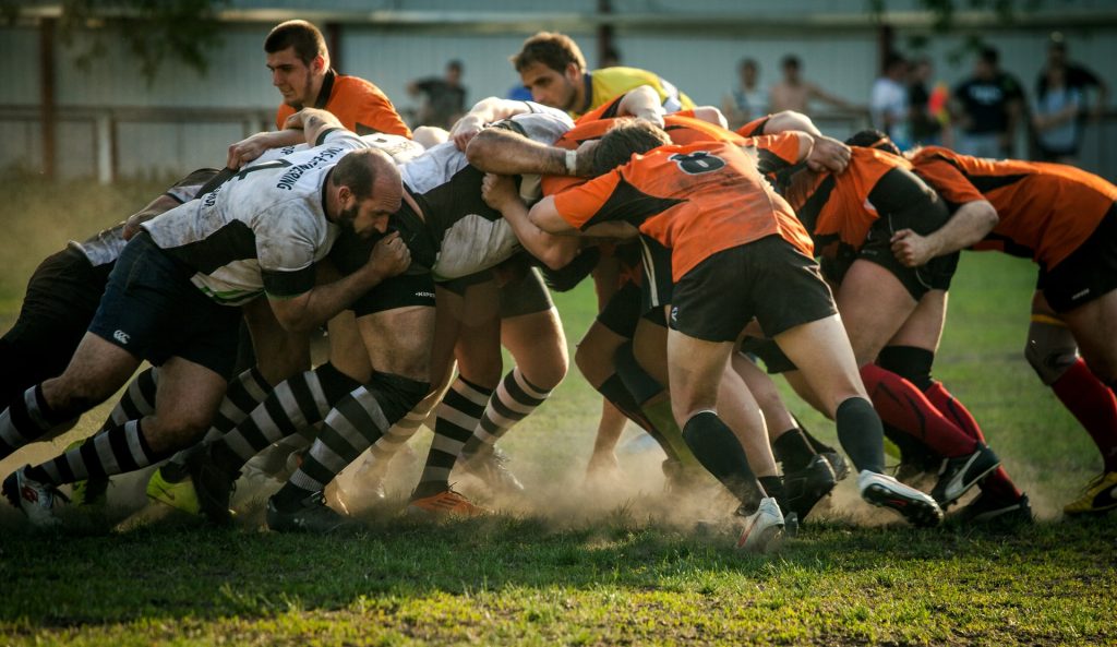
A mouse-based study to investigate memory loss in people who experience repeated head impacts, such as athletes, suggests the condition could potentially be reversed. The research in mice finds that amnesia and poor memory following head injury is due to inadequate reactivation of neurons involved in forming memories.

The study, conducted by researchers at Georgetown University Medical Center in collaboration with Trinity College Dublin, Ireland, is reported in the Journal of Neuroscience.

Importantly for diagnostic and treatment purposes, the researchers found that the memory loss attributed to head injury was not a permanent pathological event driven by a neurodegenerative disease.

Indeed, the researchers could reverse the amnesia to allow the mice to recall the lost memory, potentially allowing cognitive impairment caused by head impact to be clinically reversed.

The Georgetown investigators had previously found that the brain adapts to repeated head impacts by changing the way the synapses in the brain operate, which can cause trouble in memory storage and retrieval.

In their new study, investigators were able to trigger mice to remember memories that had been forgotten due to head impacts.

“Our research gives us hope that we can design treatments to return the head-impact brain to its normal condition and recover cognitive function in humans that have poor memory caused by repeated head impacts,” says the study’s senior investigator, Mark Burns, PhD, a professor and Vice-Chair in Georgetown’s Department of Neuroscience and director of the Laboratory for Brain Injury and Dementia.

In the new study, the scientists gave two groups of mice a new memory by training them in a test they had never seen before. One group was exposed to a high frequency of mild head impacts for one week (similar to contact sport exposure in people) and one group were controls that didn’t receive the impacts. The impacted mice were unable to recall the new memory a week later.

“Most research in this area has been in human brains with chronic traumatic encephalopathy (CTE), which is a degenerative brain disease found in people with a history of repetitive head impact,” said Burns.

“By contrast, our goal was to understand how the brain changes in response to the low-level head impacts that many young football players regularly experience.”

Researchers have found that, on average, college football players receive 21 head impacts per week with defensive ends receiving 41 head impacts per week.

The number of head impacts to mice in this study were designed to mimic a week of exposure for a college football player, and each single head impact by itself was extraordinarily mild.

Using genetically modified mice allowed the researchers to see the neurons involved in learning new memories, and they found that these memory neurons (the “memory engram”) were equally present in both the control mice and the experimental mice.

To understand the physiology underlying these memory changes, study first author Daniel P. Chapman, PhD, said, “We are good at associating memories with places, and that’s because being in a place, or seeing a photo of a place, causes a reactivation of our memory engrams. This is why we examined the engram neurons to look for the specific signature of an activated neuron. When the mice see the room where they first learned the memory, the control mice are able to activate their memory engram, but the head impact mice were not. This is what was causing the amnesia.”

The researchers were able to reverse the amnesia to allow the mice to remember the lost memory using lasers to activate the engram cells.

“We used an invasive technique to reverse memory loss in our mice, and unfortunately this is not translatable to humans,” Burns adds.

“We are currently studying a number of non-invasive techniques to try to communicate to the brain that it is no longer in danger, and to open a window of plasticity that can reset the brain to its former state.”

Source: Georgetown University Medical Center

  1. Daniel P. Chapman, Sarah D. Power, Stefano Vicini, Tomás J. Ryan, Mark P. Burns. Amnesia after repeated head impact is caused by impaired synaptic plasticity in the memory engramThe Journal of Neuroscience, 2024; e1560232024 DOI: 10.1523/JNEUROSCI.1560-23.2024

Scientists may have Found out How Rapid-acting Antidepressants Work

Photo by Marek Piwnicki

Rapid-acting antidepressants, including ketamine, scopolamine and psilocybin, have been found to have immediate and lasting positive effects on mood in patients with major depressive disorder but how these effects arise is unknown. New research led by the University of Bristol and published in Science Translational Medicine explored their neuropsychological effects and found that all three of these drugs can modulate affective biases associated with learning and memory.

Negative affective biases are a core feature of major depressive disorder. Affective biases occur when emotions alter how the brain processes information and negative affective biases are thought to contribute to the development and continuation of depressed mood.

The research team used an affective bias test, based on an associative learning task, to investigate the effects of rapid-acting antidepressants (RAADs) in rats.

They found that all the treatments were able to reduce negative affective biases associated with past experiences but there were additional characteristics of the dissociative anaesthetic, ketamine, and the serotonergic psychedelic, investigational COMP360 psilocybin (Compass Pathways’ proprietary formulation of synthetic psilocybin), which could explain why the effects of a single treatment can be long-lasting.

The findings suggest that these sustained effects are due to adaptive changes in the brain circuits which control affective biases, and these can influence how past experiences are remembered.

The effects at low doses were very specific to affective bias modulation and were localised to the prefrontal cortex of the brain, a region known to play an important role in mood.

Emma Robinson, Professor of Psychopharmacology in the School of Physiology, Pharmacology & Neuroscience at Bristol, and lead author, said: “Using a behavioural task we showed that drugs that are believed to have rapid and sustained benefits in depressed patients, specifically modulate affective biases associated with past experiences, something which we think is really important for understanding why they can improve a patient’s mood so quickly.

“We also found differences in how ketamine, scopolamine and COMP360 psilocybin interact with these neuropsychological mechanisms which may explain why the effects of a single treatment in human patients can be long-lasting, days (ketamine) to months (psilocybin).

“By using an animal model, we have been able to investigate these important interactions with learning and memory processes and neural plasticity and propose a two-stage model that may explain the effects we observe.”

In the task, each animal learnt to associate a specific digging material with a food reward under either treatment or control conditions.

The treatment condition is designed to generate a change in the animal’s affective state and a choice test is used to quantify the affective bias this generates.

Acute treatment with the RAADs ketamine, scopolamine, or psilocybin prevented the retrieval of the negative affective bias induced in this model.

However, the most exciting finding was at 24 hours after treatment when low, but not high, doses of ketamine and psilocybin led to a re-learning effect where the negatively biased memory was retrieved with a more positive affective valence.

Only psilocybin, but not ketamine or scopolamine treatment also positively biased new experiences.

Exploring in more detail the re-learning effects of ketamine in the studies, the researchers found they were protein synthesis-dependent, localised to the medial prefrontal cortex and could be modulated by cue-reactivation, consistent with their predictions of experience-dependent neural plasticity.

The study’s findings propose a neuropsychological mechanism that may explain both the immediate and sustained effects of RAADs, potentially linking their effects on neural plasticity with mood.

Source: University of Bristol

Scientists Give Macrophages First-aid ‘Backpacks’ to Calm TBI Inflammation

Colourised electron micrograph image of a macrophage. Credit: NIH

Scientists have created a new treatment for traumatic brain injury (TBI). The new approach leverages macrophages, which can increase or decrease inflammation in response to infection and injury. The team attached “backpacks” containing anti-inflammatory molecules directly to the macrophages. These molecules kept the cells in an anti-inflammatory state when they arrived at the injury site in the brain, enabling them to reduce local inflammation and mitigate the damage caused. The research is reported in PNAS Nexus.

“Every year, millions of people suffer from a TBI, but there is currently no treatment beyond managing symptoms. We have applied our cellular backpack technology – which we previously used to improve macrophages’ inflammatory response to cancerous tumours – to deliver localised anti-inflammatory treatment in the brain, which helps mitigate the cascade of runaway inflammation that causes tissue damage and death in a human-relevant model,” said senior author Samir Mitragotri, PhD, in whose lab the research was performed.

Stopping a runaway inflammation train

There is currently no treatment for the damage caused to brain tissue during a traumatic brain injury (TBI), beyond managing a patient’s symptoms. One of the main drivers of TBI-caused damage is a runaway inflammatory cascade in the brain.

As cells die from the impact, they release a cocktail of pro-inflammatory cytokine molecules that attract immune cells to clean up the damage. But the same cytokine molecules can also disrupt the blood-brain barrier, which causes blood to leak into the brain. Blood accumulation in the brain causes swelling, impaired oxygen delivery, and increased inflammation, and creates a vicious cycle of bleeding and damage that drives even more cell death.

The Mitragotri lab saw an opportunity in this problem.

“It’s generally believed anti-inflammatory therapies can be effective for treating TBI, but so far, none of them have proven effective clinically. Our previous work with macrophages has shown us that we can use our backpack technology to effectively steer their behaviour when they arrive at the injury site. Since these cells are already active players in the body’s natural immune response to a TBI, we had a hunch we could augment that pre-existing biology to reduce the initial damage,” said co-first author Rick Liao, Ph.D., a Postdoctoral Fellow at the Wyss Institute and SEAS.

“Body, heal thyself”…with backpacks

Macrophages are very malleable cells and can “switch” between pro-inflammatory and anti-inflammatory states. While the team’s previous work in cancer had been focused on keeping macrophages in a pro-inflammatory state when they arrive at the inflammation-reducing microenvironment of a tumour, this new project would be trying to do the opposite: keep the macrophages “calm” in the inflammation-riddled setting of a brain injury.

To do so, they used a disc-shaped “backpack” they had previously designed to treat multiple sclerosis that contained layers of two anti-inflammatory molecules: dexamethasone, a steroid, and interleukin-4, a cytokine that encourages macrophages to adopt an anti-inflammatory state. They then incubated these microparticles with both human and pig macrophages in vitro and saw that the backpacks stably stuck to the cells without causing any negative effect. They also observed that application of their backpacks decreased the expression of pro-inflammatory biomarkers and increased the expression of anti-inflammatory biomarkers, retaining the pig macrophages in a healing state.

But to prove that this shift would work in the body, they had to test the backpack-bearing macrophages in vivo. They chose pigs as their model organism because their brains’ structures and responses to injury more closely mimic those of humans than mice.

“Probably our biggest challenge in this project was scaling up production to match what we needed to run the experiments. Our previous studies were done in rodents, which required about two million macrophages and four million backpacks administered per subject. For the porcine study, we needed 100 million macrophages and 200 million backpacks per subject – on the scale of what would be administered in humans – and lots of helping hands,” said co-first author Neha Kapate, PhD, a Postdoctoral Fellow at the Wyss Institute and SEAS.

Once they had generated enough backpack-wearing porcine macrophages, they infused them into the pigs’ bloodstreams four hours after a TBI. Seven days later, they analysed the animals’ brains. Pigs that had received the macrophage treatment showed a high concentration of the cells in the area immediately surrounding the injury site, their lesions were 56% smaller, and there was significantly less haemorrhaging than in untreated animals.

Local immune cells also displayed a lower amount of a pro-inflammatory activation marker called CD80, indicating that the macrophages had accomplished their damage control by reducing inflammation in the brain. Corroborating that data, the levels of two soluble biomarkers for inflammation in the blood and cerebrospinal fluid were lower in treated animals than in untreated animals. The macrophage treatment also did not cause any negative effects.

The team plans to conduct future studies that focus on elucidating exactly how their anti-inflammatory macrophage therapy affects the blood-brain barrier’s integrity to prevent bleeding, which could also hold promise for treating other conditions like hemorrhagic strokes.

“Macrophages’ susceptibility to their local environment has historically prevented scientists from taking full advantage of their immune-modulating capabilities. This impressive study describes a truly novel and potentially powerful macrophage-based therapy for treating the inflammation that is the root cause of so many human afflictions in an effective and non-invasive way that works with biology rather than against it,” said Wyss Founding Director Donald Ingber, MD, PhD.

Source: Wyss Institute for Biologically Inspired Engineering at Harvard

Foundations Laid for Standardised PET Examination of Diffuse Gliomas

Photo by Mart Production on Pexels

Diffuse gliomas are malignant brain tumours that cannot be optimally examined by means of conventional MRI imaging. So-called amino acid PET (positron emission tomography) scans are better able to image the activity and spread of gliomas. An international team of researchers from the RANO Working Group have drawn up the first ever international criteria for the standardised imaging of gliomas using amino acid PET. It has published its results in the journal The Lancet Oncology.

PET uses a radioactive tracer to measure metabolic processes in the body. Amino acid PET is used in the diagnosis of diffuse gliomas, with tracers that work on a protein basis (amino acids) and accumulate in brain tumours.

The Response Assessment in Neuro-Oncology (RANO) Working Group is an international, multidisciplinary consortium founded to develop standardised new response criteria for clinical studies relating to brain tumours.

Under the joint leadership of nuclear physician Nathalie Albert from LMU and oncologist Professor Matthias Preusser from the Medical University of Vienna, the RANO group has developed new criteria for assessing the success of therapies for diffuse gliomas.

Nathalie Albert explains: “PET imaging with radioactively labelled amino acids has proven extremely valuable in neuro-oncology and permits reliable representation of the activity and extension of gliomas. Although amino acid PET has been used for years, it had not been evaluated in a structured manner before now. In contrast to MRI-based diagnostics, there have been no criteria for interpreting these PET images.” According to the researchers, the new criteria allow PET to be used in clinical studies and everyday clinical practice and create a foundation for future research and the comparison of treatments for improved therapies.

New criteria for PET examinations of brain tumours

Diffuse gliomas are malignant brain tumorus that cannot be optimally examined by means of conventional MRI imaging. So-called amino acid PET scans are better able to image the activity and spread of gliomas.

These malignant brain tumours develop out of glial cells and are generally aggressive and difficult to treat.

The RANO group has developed criteria that permit evaluation of the success of treatment using PET. Called PET RANO 1.0, these PET-based criteria open up new possibilities for the standardised assessment of diffuse gliomas.

Source: Ludwig-Maximilians-Universität München

Seizures Identified as Potential Cause of Sudden Unexplained Death in Children

Photo by Caleb Woods on Unsplash

In a study designed to better understand sudden, unexpected deaths in young children, which usually occur during sleep, researchers have identified brief seizures, accompanied by muscle convulsions, as a potential cause.

Experts estimate in excess of 3000 families each year in the US lose a baby or young child unexpectedly and without explanation. Most are infants in what is referred to as sudden infant death syndrome, or SIDS, but 400 or more cases involve children aged 1 and older, and in what is called sudden unexplained death in children (SUDC). Over half of these children are toddlers.

The study, published in the journal Neurology, used a registry of more than 300 SUDC cases, set up a decade ago by researchers at NYU Grossman School of Medicine. Researchers used extensive medical record analysis and video evidence donated by families to document the inexplicable deaths of seven toddlers between the ages of 1 and 3 that were potentially attributable to seizures. These seizures lasted less than 60 seconds and occurred within 30 minutes immediately prior to each child’s death, say the study authors.

For decades, researchers have sought an explanation to sudden death events in children, noticing a link between those with a history of febrile seizures (seizures accompanied by fever). Earlier research had reported that children who died suddenly and unexpectedly were 10 times more likely to have had febrile seizures than children who did not die suddenly and unexpectedly. Febrile seizures are also noted in one-third of SUDC cases registered at NYU Langone Health.

The new study involved an analysis by a team of eight physicians of the rare SUDC cases for which there were also home video recordings, from either security systems or commercial crib cameras, made while each child was sleeping on the night or afternoon of their death.

Five of seven recordings were running nonstop at the time and showed direct sound and visible motion indicative of a seizure happening. The remaining two recordings were triggered by sound or motion, but only one suggested that a muscle convulsion, a sign of seizure, had occurred. As well, only one toddler had a documented previous history of febrile seizures. All children in the study had previously undergone an autopsy that revealed no definitive cause of death.

“Our study, although small, offers the first direct evidence that seizures may be responsible for some sudden deaths in children, which are usually unwitnessed during sleep,” said study lead investigator Laura Gould, a research assistant professor at NYU Langone. Gould lost her daughter, Maria, to SUDC at the age of 15 months in 1997, a tragedy that prompted her successful lobby for establishment of the NYU SUDC Registry and Research Collaborative. Gould points out that if not for the video evidence, the death investigations would not have implicated a seizure.

“These study findings show that seizures are much more common than patients’ medical histories suggest, and that further research is needed to determine if seizures are frequent occurrences in sleep-related deaths in toddlers, and potentially in infants, older children, and adults,” said study senior investigator and neurologist Orrin Devinsky, MD.

Devinsky, a professor in the Departments of Neurology, Neurosurgery, and Psychiatry at NYU Langone, as well as chief of its epilepsy service, adds that “convulsive seizures may be the ‘smoking gun’ that medical science has been looking for to understand why these children die.

“Studying this phenomenon may also provide critical insight into many other deaths, including those from SIDS and epilepsy,” said Devinsky, who cofounded the SUDC Registry and Research Collaborative at NYU Langone with Gould.

Further research, Devinsky notes, is also needed to determine precisely how seizures with or without fever may induce sudden death. Previous research in epilepsy patients, he says, points to difficulty breathing that is known to occur immediately after a seizure and that can lead to death. This has been found to happen more frequently in epilepsy patients, as it does in the children involved in the study, while they are sleeping face down on the stomach and without anyone witnessing the death.

Continuous monitoring of child deaths and improvements in health records to track how often these convulsive seizures precede death, he explains, will be needed for this to be confirmed. Seizure-related deaths are underreported in people with and without epilepsy.

For the study, experts in forensic pathology, neurology, and sleep medicine analysed each recording for video quality, sound, and motion. From this, they were able to determine which toddlers showed signs of muscle convulsions as a sign of seizures prior to their death and when. Access to the videos was and remains strictly limited to the researchers involved in the study.

Source: NYU Langone Health / NYU Grossman School of Medicine

RSV Shown to Infect Nerve Cells, Causing Inflammation and Damage

Photo by Andrea Piacquadio on Unsplash

Respiratory syncytial virus (RSV), a common infection in children and older adults, can also infect nerve cells and trigger inflammation leading to nerve damage, according to a new Tulane University study.

RSV can cause mild symptoms such as coughing, sneezing and fever or lead to more severe conditions such as pneumonia or bronchiolitis. But since the disease was first discovered in 1956, it has been thought to only infect the respiratory tract.

This study, published in The Journal of Infectious Diseases, is the first to prove that RSV can penetrate nerve cells and may provide the clearest link between RSV and reported neurological symptoms in children.

RSV has been previously detected in the spinal fluid of children with seizures. Additionally, 40% of RSV-positive children under the age of 2 have shown acute encephalopathy, brain damage that can result in confusion, memory loss or cognitive difficulties.

The findings underscore the potential long-term impacts of the disease, as well as the importance of preventative measures such as the two RSV vaccines approved by the FDA in 2023.

“This is the most common respiratory virus in the first years of life as well as an impactful virus among the elderly,” said Dr Giovanni Piedimonte, Tulane University vice president for research and professor of pediatrics, biochemistry and molecular biology.

“This adds a new dimension to the importance of RSV vaccines for both the elderly and mothers to protect their babies.”

Researchers studied the virus using 3D peripheral nerve cultures grown from stem cells and rat embryos.

After finding they can be infected by RSV, researchers found RSV induced the release of chemokines – proteins that fight infections by controlling immune cells – and caused significant inflammation.

With low levels of RSV infection, the nerves became hyperreactive to stimulation. At higher levels, they observed a progressive degeneration of the nerve and increased neurotoxicity due to excess inflammation.

“Until this study, the theory was that the inflammatory response was indirectly activating the nerves,” Piedimonte said.

“This study shows that not only does that happen, but the virus can penetrate directly into the nerves.”

The nerve hyperreactivity could explain why children who get RSV are later more likely to have asthmatic symptoms, Piedimonte said.

The study also found that RSV could enter the spinal cord via peripheral nerves despite not having the ability to enter the spinal neurons directly.

More research is needed to explore that mechanism, but Piedimonte theorises that by using the peripheral nerves to enter the spinal cord, RSV can bypass the blood-brain barrier, enter the central nervous system and infect the brain.

If confirmed, it could signal a connection between RSV and other neurological or developmental disorders, Piedimonte said.

“If indeed it’s confirmed in future studies that viruses like this are able to access the central nervous system, that opens a huge Pandora’s box,” Piedimonte said.

Source: Tulane University

Targeted Neurostimulation Makes People More Hypnotisable

Photo by Bruce Christianson on Unsplash

Hypnotisability appears to be a stable trait that changes little throughout adulthood, much like personality and IQ. But now, for the first time, Stanford Medicine researchers have demonstrated a way to temporarily heighten hypnotisablity, potentially allowing more people to access the benefits of hypnosis-based therapy.

In the new study, published in Nature Mental Health, the researchers found that less than two minutes of electrical stimulation targeting a precise area of the brain could boost participants’ hypnotisability for about one hour.

“We know hypnosis is an effective treatment for many different symptoms and disorders, in particular pain,” said lead author Afik Faerman, PhD, a postdoctoral scholar in psychiatry. “But we also know that not everyone benefits equally from hypnosis.”

Focused attention

Approximately two-thirds of adults are at least somewhat hypnotisable, and 15% are considered highly hypnotisable, meaning they score 9 or 10 on a standard 10-point measure of hypnotisability.

“Hypnosis is a state of highly focused attention, and higher hypnotisability improves the odds of your doing better with techniques using hypnosis,” said David Spiegel, MD, a professor of psychiatry and behavioural sciences and a senior author of the study.

Spiegel has devoted decades to studying hypnotherapy and using it to help patients control pain, lower stress, stop smoking and more. Several years ago, Spiegel led a team that used brain imaging to uncover the neurobiological basis of the practice. They found that highly hypnotisable people had stronger functional connectivity between the left dorsolateral prefrontal cortex, which is involved in information processing and decision making; and the dorsal anterior cingulate cortex, involved in detecting stimuli.

“It made sense that people who naturally coordinate activity between these two regions would be able to concentrate more intently,” Spiegel said. “It’s because you’re coordinating what you are focusing on with the system that distracts you.”

Shifting a stable trait

With these insights, Spiegel teamed up with Nolan Williams, MD, associate professor of psychiatry and behavioural sciences, who has pioneered non-invasive neurostimulation techniques to treat conditions such as depression, obsessive-compulsive disorder and suicidal ideation.

The hope was that neurostimulation could alter even a stable trait like hypnotisability.

In the new study, the researchers recruited 80 participants with fibromyalgia, a chronic pain condition that can be treated with hypnotherapy. They excluded those who were already highly hypnotisable.

Half of the participants received transcranial magnetic stimulation, in which paddles applied to the scalp deliver electrical pulses to the brain. Specifically, they received two 46-second applications that delivered 800 pulses of electricity to a precise location in the left dorsolateral prefrontal cortex. The exact locations depended on the unique structure and activity of each person’s brain.

“A novel aspect of this trial is that we used the person’s own brain networks, based on brain imaging, to target the right spot,” said Williams, also a senior author of the study.

The other half of participants received a sham treatment with the same look and feel, but without electrical stimulation. Hypnotisability was assessed by clinicians immediately before and after the treatments, with neither patients nor clinicians knowing who was in which group.

The researchers found that participants who received the neurostimulation showed a statistically significant increase in hypnotisability, scoring roughly one point higher. The sham group experienced no effect.

When the participants were assessed again one hour later, the effect had worn off and there was no longer a statistically significant difference between the two groups.

“We were pleasantly surprised that we were able to, with 92 seconds of stimulation, change a stable brain trait that people have been trying to change for 100 years,” Williams said. “We finally cracked the code on how to do it.”

The researchers plan to test whether different dosages of neurostimulation could enhance hypnotisability even more.

“It’s unusual to be able to change hypnotisability,” Spiegel said. A study of Stanford University students that began in the 1950s, for example, found that the trait remained relatively consistent when the students were tested 25 years later, as consistent as IQ over that time period. Recent research by Spiegel’s lab also suggests that hypnotisability may have a genetic basis.

Bigger implications

Clinically, a transient bump in hypnotisability may be enough to allow more people living with chronic pain to choose hypnosis as an alternative to long-term opioid use. Spiegel will follow up with the study participants to see how they fare in hypnotherapy.

The new results could have implications beyond hypnosis. Faerman noted that neurostimulation may be able to temporarily shift other stable traits or enhance people’s response to other forms of psychotherapy.

“As a clinical psychologist, my personal vision is that, in the future, patients come in, they go into a quick, non-invasive brain stimulation session, then they go in to see their psychologist,” he said. “Their benefit from treatment could be much higher.”

Story Source: Stanford Medicine

Oestrogen Receptor Involved in Social Anxiety Suppression in Male Mice

Source: CC0

Researchers at the University of Tsukuba in Japan have discovered that oestrogen receptor (ER) β, expressed in the lateral septum of the limbic system, plays a crucial role in suppressing anxiety-like behaviour by male mice in social situations. Publishing their findings in Neuroscience, they also reported that the distribution and expression region of ERβ differs from that of ERα.

Oestradiol, a sex steroid hormone, plays an essential role in social behaviour, including regulating social anxiety, which is anxiety experienced when unknown individuals are encountered.

In males, testosterone secreted by the testes is converted to oestradiol in the brain, and the oestradiol binds to two types of oestrogen receptors (ERs), ERα and ERβ, to regulate social behaviour. However, its neuroendocrine basis has not been understood. In this study, the role of ERα and ERβ expressed in the lateral septum (LS), which regulates social anxiety, was investigated using male mice.

The researchers first investigated the expression of ERα and ERβ in the LS using genetically modified male mice. ERβ-expressing cells in the mice were labelled with red fluorescent protein, which revealed that the distributions of ERα and ERβ are different.

Furthermore, the researchers investigated the knockdown effects of ERα or ERβ gene expression in the LS of male mice during situations of social and nonsocial anxiety. The results show that social anxiety increases with the inhibition of ERβ expression.

Additionally, ERα- and ERβ-positive cells in the LS projected into different regions of the hypothalamus.

Thus, the researchers concluded that ERα- and ERβ-expressing cells in LS are distinct cell populations with different localisations and neuronal projections, and the ERβ population plays a crucial role in neural circuitry that regulates anxiety-like behaviour in social situations.

Source: University of Tsukuba

Trial Finds Argatroban Promising in Acute Ischaemic Stroke with Early Neurological Deterioration

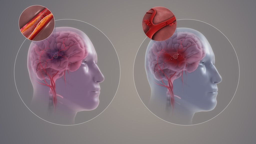
Ischaemic and haemorrhagic stroke. Credit: Scientific Animations CC4.0

Early neurological deterioration (END) within the first 48 hours after acute ischaemic stroke (AIS) onset is relatively common, and is a predictor of poor outcomes. Treatment options are limited and unproven, but but a clinical trial has shown that the anticoagulant argatroban was safe and effective in improving outcomes. The results were published in JAMA Neurology.

Apart from straightforward causes, such as intracerebral haemorrhage and malignant oedema, the mechanism of END remains mostly unclear. Interventions for unexplained END can include plasma volume expansion, induced hypertension, and intensified antithrombotic therapy, but none has been formally proved so far.

The direct thrombin inhibitor argatroban is rapid acting, short acting, and has low bleeding rates, which could help prevent thrombus propagation and provide additional benefit after stroke/TIA. Argatroban has been associated with a reduction in ischaemic stroke damage but the safety and efficacy of argatroban is not well established for AIS treatment, and evidence is lacking for the effect of argatroban in patients with AIS and END.

Researchers conducted a randomised clinical trial that initially included 628 patients, average age 65 and 400 (63.7%) male. Eligible patients were adults with AIS who experienced END, which was defined as an increase of 2 or more points on the National Institutes of Health Stroke Scale within 48 hours from symptom onset.

Patients were randomly assigned to the argatroban group and control group within 48 hours of symptom onset. Both groups received standard therapy based on guidelines, including oral mono or dual antiplatelet therapy. The argatroban group received intravenous argatroban for 7 days (continuous infusion at a dose of 60mg per day for 2 days, followed by 20mg per day for 5 days) in addition to standard therapy.

The results showed that good neurological function at 90 days in those randomised to receive argatroban plus antiplatelet compared with antiplatelet alone was observed in 80.5% vs 73.7%)of participants, a statistically significant difference.

The authors concluded that the trial “shows that the combination of argatroban and antiplatelet therapy resulted in a significantly greater likelihood of good functional outcome at 90 days in patients with END after AIS, with no additional risk of major intracranial or extracranial haemorrhage.”