Maintaining prediabetic status after diagnosis reduces risk of death decades later
Individuals diagnosed with prediabetes can reduce their long-term risk of death and diabetes-related health complications if they delay the onset of diabetes for just four years through diet and exercise. Guangwei Li of the China-Japan Friendship Hospital and colleagues report these findings in a new study published July 9thin the open-access journal PLOS Medicine.
Type 2 diabetes is associated with an increased risk of death and disability, and imposes a significant economic burden on individuals and societies worldwide. Lifestyle changes, such as eating a healthy diet and getting more exercise, can delay or reduce the risk of developing diabetes in people diagnosed with impaired glucose tolerance – commonly called prediabetes. However, it is unknown how long a person must delay diabetes to ensure better long-term health.
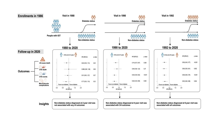
In the new study, researchers looked at health outcomes from 540 prediabetic individuals who participated in the original Da Qing Diabetes Prevention Study, a six-year trial conducted in Da Qing City in China, starting in 1986. Participants belonged to either a control group or one of three lifestyle intervention groups, which involved following a healthy diet, getting more exercise, or both. The trial followed up with participants for more than 30 years.
Li’s team determined the long-term risk of death, cardiovascular events – like heart attack, stroke or heart failure – and other diabetes-related complications for trial participants. They found that individuals who remained non-diabetic for at least four years after their initial diagnosis had a significantly lower risk of dying and a significantly lower risk of experiencing a cardiovascular event compared to those who developed diabetes sooner. This protective effect was not observed in individuals who remained non-diabetic for less than the “four-year threshold.”
Overall, the analysis suggests that the longer a prediabetic person can delay developing diabetes, the better their long-term health outcomes will be. However, even just a few years of maintaining prediabetic status can yield benefits for years to come.
The authors add, “This study suggests that a longer duration of non-diabetes status in those with IGT has beneficial health outcomes and reduces mortality. The implementation of effective interventions targeting those with IGT should be considered as part of preventative management for diabetes and diabetes related vascular complications.”
People with prediabetes are advised to reduce their weight in order to prevent the development of diabetes. For the first time, new research shows that people achieve the best diabetes protection when they reduce their weight and at the same time normalise blood sugar regulation with lifestyle changes and medication. In an article published in Diabetologia, the authors argue that the normalisation of blood sugar levels in prediabetes should be included as a therapeutic goal in the guidelines in order to improve the prevention of type 2 diabetes.
Diabetes is widespread and is associated with an increased risk of a number of life-threatening complications such as stroke, heart attack and kidney failure. “In order to prevent the development of the disease, early therapies are already important in the prediabetes stage, a preliminary stage of type 2 diabetes. Our results can be used to change the goals of these early lifestyle interventions in order to reduce the overall development rates of diabetes,” explains first author Reiner Jumpertz-von Schwartzenberg.
Prediabetes drastically increases the risk of diabetes
Prediabetes is diagnosed when there is no manifest type 2 diabetes yet, but the fasting blood sugar is already elevated and glucose tolerance is impaired. To prevent prediabetes from becoming diabetes, affected patients are advised to reduce their weight. US guidelines from the American Diabetes Association (ADA), for example, recommend reducing body weight by at least 7%. This recommendation is based on the DPP study.
The research team from the University Tübingen and the National Institute of Diabetes and Digestive and Kidney Diseases in the US, investigated whether this weight loss is sufficient, or whether it is not better to prevent diabetes by also reducing blood sugar levels such that prediabetes goes into remission.
Prevention through one-year lifestyle intervention
They analysed data from 480 people with prediabetes who participated in the US Diabetes Prevention Program (DPP) and had lost at least 7% of their body weight through a one-year lifestyle intervention. In 114 of them, prediabetes also went into remission during the intervention, meaning that their fasting blood sugar, glucose tolerance and HbA1c had normalised. However, the majority of the 366 study participants had not managed to significantly improve their blood sugar regulation despite successfully losing weight. Their prediabetes was not in remission at the end of the intervention.
The researchers found that significantly fewer people in the group that had lost weight and achieved prediabetes remission developed manifest diabetes thereafter. The additional remission of prediabetes resulted in a relative risk reduction for the development of diabetes of 76% compared to those who had not achieved normalisation of their blood sugar levels. The absolute risk reduction was higher than 10%.
“In the group with additional remission of prediabetes, there was even no type 2 diabetes at all in the first 4 years after the lifestyle intervention,” reports last author Andreas Birkenfeld. “In the group that had ‘only’ lost weight, however, some study participants did develop manifest diabetes during that period.”
Jumpertz-von Schwartzenberg and Birkenfeld draw a clear conclusion: “Our results show that remission of prediabetes brings a further significant benefit in addition to weight reduction. We therefore advocate that the goal of prediabetes remission should be included in the objectives of the practice guidelines in order to significantly improve the prevention of type 2 diabetes.”
The study was conducted by researchers from the Institute for Diabetes Research and Metabolic Diseases of Helmholtz Munich at the University of Tübingen, a partner in the German Center for Diabetes Research (DZD), together with US colleagues in the renowned “Diabetes Prevention Program (DPP)”.
In an international phase III study, researchers have demonstrated the potential of tirzepatide, known to manage type 2 diabetes, as the first effective drug therapy for obstructive sleep apnoea (OSA), a sleep-related disorder characterised by repeated episodes of irregular breathing due to complete or partial blockage of the upper airway.
The results, published in the New England Journal of Medicine, highlight the treatment’s potential to improve the quality of life for millions around the world affected by OSA.
“This study marks a significant milestone in the treatment of OSA, offering a promising new therapeutic option that addresses both respiratory and metabolic complications,” said Atul Malhotra, MD, lead author of the study, professor of medicine at University of California San Diego School of Medicine and director of sleep medicine at UC San Diego Health.
OSA can result in reduced blood oxygen levels and can also be associated with an increased risk of cardiovascular complications, such as hypertension and heart disease. Recent studies, also led by Malhotra, suggest that the number of OSA patients worldwide is close to 936 million.
Conducted in two Phase III, double-blinded, randomised, controlled trials, the new study cohort recruited 469 participants from 9 countries with clinical obesity and living with moderate-to-severe OSA. Participants either used or did not use continuous positive airway pressure (CPAP) therapy, the most common sleep apnoea treatment which uses a machine to maintain an open airway during sleep, preventing interruptions in breathing. Patients were administered either 10 or 15mg of the drug by injection or a placebo and followed for 52 weeks.
Researchers found that tirzepatide led to a significant decrease in the number of breathing interruptions during sleep, a key indicator used to measure the severity of OSA. This improvement was much greater than what was seen in participants that were given a placebo. Importantly, some participants that took the drug reached a point where CPAP therapy might not be necessary. Considerable data suggest that a drug therapy that targets both sleep apnoea and obesity is beneficial rather than treating either condition alone.
Additionally, the drug therapy improved other aspects related to OSA, such as reducing the risk factors of cardiovascular diseases and improved body weight. The most common side effect reported was mild stomach issues.
“Historically, treating OSA meant using devices during sleep, like a CPAP machine, to alleviate breathing difficulties and symptoms,” Malhotra said. “However, its effectiveness relies on consistent use. This new drug treatment offers a more accessible alternative for individuals who cannot tolerate or adhere to existing therapies. We believe that the combination of CPAP therapy with weight loss will be optimal for improving cardiometabolic risk and symptoms. Tirzepatide can also target specific underlying mechanisms of sleep apnoea, potentially leading to more personalised and effective treatment.”
Malhotra adds that having a drug therapy for OSA represents a significant advancement in the field.
“It means we can offer an innovative solution, signifying hope and a new standard of care to provide relief to countless individuals and their families who have struggled with the limitations of existing treatments,” said Malhotra. “This breakthrough opens the door to a new era of OSA management for people diagnosed with obesity, potentially transforming how we approach and treat this pervasive condition on a global scale.”
Next steps include conducting clinical trials to examine longer term effects of tirzepatide.
A 3D map of the islet density routes throughout the healthy human pancreas. Source: Wikimedia CC0
Northwestern University researchers have developed a new antioxidant biomaterial that someday could provide much-needed relief to people living with chronic pancreatitis. The study was published in the journal Science Advances.
Before surgeons remove the pancreas from patients with severe, painful chronic pancreatitis, they first harvest insulin-producing tissue clusters, called islets, and transplant them into the vasculature of the liver. The goal of the transplant is to preserve a patient’s ability to control their own blood-glucose levels without insulin injections.
Unfortunately, the process inadvertently destroys 50–80% of islets, and one-third of patients become diabetic after surgery. Three years post-surgery, 70% of patients require insulin injections, which are accompanied by a list of side effects, including weight gain, hypoglycaemia and fatigue.
In the new study, researchers transplanted islets from the pancreas to the omentum – the large, flat, fatty tissue that covers the intestines – instead of the liver. And, to create a healthier microenvironment for the islets, the researchers adhered the islets to the omentum with an inherently antioxidant and anti-inflammatory biomaterial, which rapidly transforms from a liquid to a gel when exposed to body temperature.
In studies with mouse and non-human primates, the gel successfully prevented oxidative stress and inflammatory reactions, significantly improving survival and preserving function of transplanted islets. It marks the first time a synthetic antioxidant gel has been used to preserve function of transplanted islets.
“Although islet transplantation has improved over the years, long-term outcomes remain poor,” said Northwestern’s Guillermo A. Ameer, who led the study. “There is clearly a need for alternative solutions. We have engineered a cutting-edge synthetic material that provides a supportive microenvironment for islet function. When tested in animals, we were successful. It kept islet function maximised and restored normal blood sugar levels. We also report a reduction in units of insulin that animals required.”
“With this new approach, we hope that patients will no longer have to choose between living with the physical pain of chronic pancreatitis or the complications of diabetes,” added Jacqueline Burke, a research assistant professor of biomedical engineering at Northwestern and the paper’s first author.
‘Compromised quality of life’
For patients living without a pancreas, side effects such as managing blood-sugar levels can be a lifelong struggle. By secreting insulin in response to glucose, islets help the body maintain glycaemic control. Without functioning islets, people must closely monitor their blood-sugar levels and frequently inject insulin.
“Living without functional islets places a great burden on patients,” Burke said. “They must learn to count carbs, dose insulin at the appropriate time and continuously monitor blood glucose. This consumes much of their time and mental energy. Even with great care, exogeneous insulin therapy is not as effective as islets for maintaining glucose control.”
“It’s a compromised quality of life,” Ameer said. “Instead of multiple insulin injections, we would love to collect and preserve as many islets as possible.”
But, unfortunately, the current standard of care for preserving islets often leads to poor outcomes. After the surgery to remove the pancreas, surgeons isolate islets from the pancreas and transplant them to the liver through portal vein infusion. This intraportal perfusion procedure has several common complications. Islets in direct contact with blood flow undergo an inflammatory response, more than half of the islets die, and transplanted islets can cause dangerous clots in the liver. For those reasons, physicians and researchers have been searching for an alternate transplantation site.
In previous clinical studies, researchers transplanted islets to the omentum instead of the liver in order to bypass issues with clotting. To secure the islets on the omentum, physicians used plasma from the patients’ own blood to form a biologic gel. While the omentum appeared to work better than the liver as a transplantation site, several issues, including clots and inflammation, remained.
“There’s been significant interest in the research and medical communities to find an alternate islet transplantation site,” Ameer said. “The results from the omentum study were encouraging, but outcomes were varied. We believe that’s because the use of the patients’ blood and the added components required to create the biologic gel can affect reproducibility among patients.”
A citrate solution
To protect the islets and improve outcomes, Ameer turned to the citrate-based biomaterials platform with inherent antioxidant properties developed in his laboratory. Used in products approved by U.S. Food and Drug Administration for musculoskeletal surgeries, citrate-based biomaterials have demonstrated the ability to control the body’s inflammatory responses. Ameer set out to investigate whether a version of these biomaterials with biodegradable and temperature-responsive phase-changing properties would provide a superior alternative to a biologic gel obtained from blood.
In cell cultures, both mouse and human islets stored within the citrate-based gel maintained viability much longer than islets in other solutions. When exposed to glucose, the islets secreted insulin, demonstrating normal functionality. Moving beyond cell cultures, Ameer’s team tested the gel in small and large animal models. Liquid at room temperature, the material turns into a gel at body temperature, so it’s simple to apply and easily stays in place.
In the animal studies, the gel effectively secured the islets onto the omentum of the animals. Compared to the current methods, more islets survived, and, over time, the animals restored normal blood glucose levels. According to Ameer, the success is partially due to the new material’s biocompatibility and antioxidant nature.
“Islets are very sensitive to oxygen,” Ameer said. “They are affected by both too little oxygen and too much oxygen. The material’s innate antioxidant properties protect the cells. Plasma from your own blood doesn’t offer the same level of protection.”
Integrating into tissues
After about three months, the body resorbed 80-90% of the biocompatible gel. But, at that point, it was no longer needed.
“What was fascinating is that the islets regenerated blood vessels,” Ameer said. “The body generated a network of new blood vessels to reconnect the islets with the body. That is a major breakthrough because the blood vessels keep the islets alive and healthy. Meanwhile, our gel is simply resorbed into the surrounding tissue, leaving little evidence behind.”
Next, Ameer aims to test his hydrogel in animal models over a longer period of time. He said the new hydrogel also could be used for various cell replacement therapies, including stem cell-derived beta cells for treating diabetes.
Maternal obesity impacts the eating behaviours of offspring via long-term overexpression of the microRNA miR-505-5p, according to a study publishing June 4 in the open-access journal PLOS Biology by Laura Dearden and Susan Ozanne from the MRC Metabolic Diseases Unit, Institute of Metabolic Science, University of Cambridge, UK, and colleagues.
Previous studies in both humans and animal models have shown that the offspring of obese mothers have a higher risk of obesity and type 2 diabetes.
While this relationship is likely the result of a complex relationship between genetics and environment, emerging evidence has implicated that maternal obesity can disrupt the hypothalamus – the region of the brain responsible for nutrition sensing and energy homeostasis.
In animal models, offspring exposed to overnutrition during key periods of development eat more, but little is known about the molecular mechanisms that lead to these changes in eating behaviour.
In this study, researchers found that mice born from obese mothers had higher levels of the microRNA miR-505-5p in their hypothalamus – from as early as the foetal stage into adulthood.
The researchers found that the mice ate more and showed a preference for high-fat foods.
Interestingly, the effect of maternal obesity on miR-505-5p and eating behaviours was mitigated if the mothers exercised during pregnancy.
Cell culture experiments showed that miR-505-5p expression could be induced by exposing hypothalamic neurons to long-chain fatty acids and insulin, which are both high in pregnancies complicated by obesity.
The researchers identified miR-505-5p as a novel regulator of pathways involved in fatty acid uptake and metabolism, therefore high levels of the miRNA make the offspring brain unable to sense when eating high fat foods.
Several of the genes that miR-505-5p regulates have been associated with high body mass index in human genetic studies.
The study is one of the first to demonstrate the molecular mechanism linking nutritional exposure in utero to eating behaviour.
The authors add, “Our results show that obesity during pregnancy causes changes to the baby’s brain that makes them eat more high fat food in adulthood and more likely to develop obesity. Importantly we showed that moderate exercise, without weight loss, during pregnancies complicated by obesity prevented the changes to the baby’s brain. This helps us understand why the children of mothers living with obesity are more likely to become obese themselves, with early life exposures, genetics and current environment all being contributing factors.”
As research continues to produce evidence about the underlying causes of obesity and optimal strategies to treat and manage obesity have evolved, there are disparities in application of the latest scientific advances in the clinical care for people with obesity. Widespread adoption of current findings, consistency of care and expertise in obesity care varies by health care professional and institution. These findings are detailed in a new American Heart Association scientific statement, “Implementation of Obesity Science Into Clinical Practice,” published in the journal Circulation.
“Obesity is undeniably a critical public health concern in the U.S. and around the world, affecting nearly all populations and straining our health care systems,” said Deepika Laddu, Ph.D., FAHA, chair of the statement writing committee and a senior research scientist at Arbor Research Collaborative for Health in Ann Arbor, Michigan. “As a major risk factor for heart disease, obesity has significantly hindered progress in reducing heart disease rates. Despite advancements in understanding the complexities of obesity and newer treatment options, major gaps remain between obesity research and real-world implementation in clinical practice.”
Studies show intensive lifestyle therapy is considerably more effective for weight loss than brief advice from a health care professional. However, general educational information is offered more frequently by health professionals rather than referrals to classes, programs or tangible resources for lifestyle changes. One study revealed that only 16% of health care professionals had working knowledge about evidence-based lifestyle treatments for obesity, including diet and nutrition, physical activity and intensive behavioral therapy referral. Other barriers to addressing weight loss are exacerbated by socioeconomic and racial or ethnic inequities. People of diverse races and ethnicities and people who are covered by Medicare or Medicaid are less likely to be referred to weight management programs or to have them covered by insurance.
For about 30 years, the prevalence of obesity in the US and around the world has been escalating. Recent estimates indicate more than 40% of US adults ages 20 and older are living with obesity, according to the US Centers for Disease Control and Prevention.
Research has led experts to unlock the multifactorial causes of obesity, including sociological and physiological determinants of health. Treatments for obesity have also evolved with more strategies for lifestyle modifications, medication therapy and bariatric surgery – but each treatment approach comes with challenges.
“While significant strides have been made in advancing the science to help us understand obesity, there remains a considerable gap between what we know and what happens in the doctor’s office,” said Laddu. “Health care professionals and health care systems need to find better ways to put what we know about obesity into action so more people can get the right support and treatment. Adopting new technologies and telemedicine, making referrals to community-based weight management programs to encourage behavioural change, providing social support and increasing reach and access to treatments are just some of the promising methods we could implement to unlock successful, evidence-based obesity care.”
Weight loss medications
Glucagon-like peptide-1 (GLP-1) agonists, such as high-dose semaglutide and tirzepatide, are the most recently FDA-approved medications for long-term weight management, and both are associated with an average weight loss of more than 10% at six months in clinical studies. However, despite half of adults in the U.S. meeting the BMI criteria for obesity and being eligible for these medications, a small proportion of this population is currently taking them.
Weight loss surgery
In the decades since bariatric (weight loss) surgery was first introduced as an option for people with severe obesity, there have been advances in the expertise and safety of the procedures, as well as an increased understanding of the health benefits that often result after bariatric surgery. A comprehensive review of studies focused on weight loss surgeries showed that patients who underwent bariatric surgery had lower risks of cardiovascular disease and decreased risks for multiple other obesity-associated conditions, including Type 2 diabetes and high blood pressure. One challenge facing health care professionals is ensuring that the populations with the greatest needs have access to bariatric surgery in terms of costs, resources and social support.
The statement describes strategies that both address these challenges and improve how obesity-based research is incorporated into clinical care. The statement also identifies the need to develop solutions across populations in order to manage obesity at the community level. Potential improved public health policies and future research to expand patient care models and optimize the delivery and sustainability of equitable obesity-related care are suggested.
Specific approaches are highlighted in the statement to help bridge the gap between the science about obesity and clinical care, such as:
To reach and successfully impact populations in need, health care professionals may consider how social determinants of health, including insurance type, household income, race and ethnicity, environment, health literacy, access to health-promoting resources and social supports all influence the likelihood of successful patient treatment.
Education for health care professionals explaining the complex origins and clinical consequences of obesity is discussed. Such training should emphasize information about diagnosis, prevention and treatment of obesity. Despite the high prevalence of obesity around the world, there is a lack of education programs centered on obesity for medical professionals.
Further evaluation of health policy changes that health care systems and insurance plans can implement and scale in order to make obesity treatment affordable for patients, especially those at high risk for adverse outcomes such as cardiovascular disease.
A framework for delivering obesity care into clinical practice settings is reviewed, as well as efforts by some professional societies for developing interventions that make obesity treatment more accessible.
“The statement emphasises the importance of a comprehensive approach across different levels of health care delivery and public policy, along with the adoption of feasible, evidence-based strategies in clinical settings,” said Laddu. “It also underscores the need for future research and policy changes to improve current patient care models and ensure equitable access to obesity-related care for people in underrepresented groups.”
The scientific statement also provides possible solutions for how to help people in their day-to-day lives, including interventions with digital technology and access through telemedicine. However, more research is needed in obesity science and treatment. Limited understanding of the cost-effectiveness of obesity prevention and the long-term health outcomes for established therapies has hindered the implementation of obesity science into clinical settings. Cross-collaborative obesity science research between stakeholders and health economists may serve as the bridge to developing and scaling cost-effective prevention programs.
Depiction of multiple myeloma. Credit: Scientific Animations
People who use metformin are less likely to develop a myeloproliferative neoplasm (MPN) over time, indicating that the treatment may help prevent the development of certain types of cancers, according to a study published in Blood Advances.
Metformin is a therapy used to treat high blood sugar in people with type 2 diabetes that increases the effect of insulin, reduces how much glucose is released from the liver and helps the body absorb glucose. A meta-analysis of previous studies connected the therapy with a reduction in the risk of gastrointestinal, breast, and urologic cancers, while a retrospective study of US veterans found that metformin users have a reduced risk for solid and haematological cancers.
Metformin’s anti-inflammatory properties in focus
“Our team was interested in understanding what other effects we see with commonly prescribed treatments like metformin,” said Anne Stidsholt Roug, MD, PhD, chief physician at Aarhus University Hospital and clinical associate professor at Aalborg University Hospital in Denmark. “The anti-inflammatory effect of metformin interested us, as MPNs are very inflammatory diseases. This is the first study to investigate the association between metformin use and risk of MPN.”
MPNs are a group of diseases that affect how bone marrow produces blood cells, resulting in an overproduction of red blood cells, white blood cells, or platelets that can lead to bleeding problems, a greater risk of stroke or heart attack, and organ damage.
Surprisingly strong association
The researchers compared metformin use among patients diagnosed with MPNs and a matched population from the Danish general population between 2010 and 2018. Of the 3816 MPN cases identified from the sample, a total of 268 (7.0%) individuals with MPN had taken metformin as compared to 8.2% (1573 out of 19 080) of the control group of people who had taken metformin but were not diagnosed with MPN. Just 1.1% of MPN cases had taken metformin for more than five years, as compared to 2.0% of controls. The protective effect of metformin was seen in all subtypes of MPN when adjusting for potential confounders.
“We were surprised by the magnitude of the association we saw in the data,” said Daniel Tuyet Kristensen, MD, PhD student, at Aalborg University Hospital and lead author of the study. “We saw the strongest effect in people who had taken metformin for more than five years as compared to those who had taken the treatment for less than a year.” Dr Kristensen added that this makes clinical sense, as MPNs are diseases that develop over a long period of time, like other types of cancer.
The researchers noted that while the protective effect of long-term metformin use was seen in all subtypes of MPN, the study was limited by its registry-based retrospective design. Further, they could not account for risk-modifying lifestyle factors, such as smoking, obesity, and dietary habits.
Dr Roug noted that while the study team were unable to assess exactly why metformin seems to protect against the development of MPN, they hope additional research will be conducted to better understand why this may be. Moving forward, the researchers aim to identify any similar trends with myelodysplastic syndromes and acute myeloid leukaemia in population-level data for future study.
Neuropathy, the nerve damage that causes pain and numbness in the feet and hands and can eventually lead to falls, infection and even amputation, is very common and underdiagnosed, according to a study published in Neurology®, the medical journal of the American Academy of Neurology.
“More than one-third of people with neuropathy experience sharp, prickling or shock-like pain, which increases their rates of depression and decreases quality of life,” said study author Melissa A. Elafros, MD, PhD, of the University of Michigan in Ann Arbor and a member of the American Academy of Neurology. “People with neuropathy also have an increased risk of earlier death, even when you take into account other conditions they have, so identifying and treating people with or at risk for neuropathy is essential.”
The study involved 169 people from an outpatient internal medicine clinic serving mainly Medicaid patients in Flint, Michigan. The participants had an average age of 58 years and 69% were Black people. One-half of the people had diabetes, which can cause neuropathy. A total of 67% had metabolic syndrome, which is defined as having excess belly fat plus two or more of the following risk factors: hypertension, elevated triglycerides, hyperglycaemia and low high-density lipoprotein (HDL) cholesterol. These risk factors are also associated with neuropathy.
All participants were tested for distal symmetric polyneuropathy. Information about other health conditions was also collected. A total of 73% of the people had neuropathy. Of those, 75% had not been previously diagnosed with the condition. Nearly 60% of those with neuropathy were experiencing pain. Of those with neuropathy, 74% had metabolic syndrome, compared to 54% of those who did not have neuropathy.
After adjusting for other factors that could affect the risk of neuropathy, researchers found that people with metabolic syndrome were more than four times more likely to have neuropathy than people who did not have the syndrome.
Risk differs according to race
Researchers were also looking for any relationship between race and income and neuropathy, as few studies have been done on those topics. There was no relationship between low income and neuropathy. For race, Black people had a decreased risk of neuropathy. Black people made up 60% of those with neuropathy and 91% of those without neuropathy.
“The amount of people with neuropathy in this study, particularly undiagnosed neuropathy, was extraordinarily high with almost three fourths of the study population,” Elafros said. “This highlights the urgent need for interventions that improve diagnosis and management of this condition, as well as the need for managing risk factors that can lead to this condition.” A limitation of the study is that it is a snapshot in time; it did not follow people to see who developed neuropathy over time. It also did not look at reasons why people were not able to manage risk factors that can lead to neuropathy.
South Africa has seen the quickest and most alarming rise of diabetes on the continent; from an estimated 1.9 million people living with the condition in 2011 to 4.2 million by 2021 – with 7.5 million predicted to be afflicted by 20451. South Africa also has the fastest rising prevalence on the continent with an estimated 20% of the adult population either diabetic or pre-diabetic1. Globally, diabetes prevalence is predicted to rise by 46% between 2019 and 20452. It currently stands at some 537 million people worldwide1.
This emerged at the recently held annual Sanofi medical meeting, the Cardio-Metabolic Axis Forum from April 19th–21st in Cape Town. This was a meeting of leading endocrinologists, specialist physicians, nephrologists, diabetes-treating doctors, academics and Patient Advocacy Groups (PAGs).
Speaking at the conference, specialist physician and endocrinologist, Dr Landi Lombard – former editor of the South Africa Journal of Diabetes and Vascular Disease – told delegates that the risk of death associated with diabetes in cardiovascular conditions is more than twice that of people with non-diabetes, while in all-cause mortality, it’s just under twice that of a person living without diabetes. Of the estimated 537 million people living with diabetes globally, only about half are diagnosed, of whom 25% receive care, 12.5% achieve treatment targets, and 6% live a life free of diabetes-related complications1.
Dr Lombard said that the pandemic is being driven by poor lifestyle choices and diet, lack of exercise and widespread obesity in the population, so better healthcare worker communication and education of patients is vital to stem the tide of diabetes.
Professor Robert Ritzel of the Department of Endocrinology, Diabetology and Angiology at Schwabing Hospital in Munich, said the Pacific Islands and the Middle East led the world with diabetes prevalence at between 25 % and 40 %. He said what precipitated a surge in diabetes was the speed at which a nation changed from a traditional to a modern lifestyle. When this happened within a few years, diabetes prevalence was likely to range between 20% and 40%. However, when change occurred over many generations, it gave epidemiologists and clinicians time to adapt.
Lombard said one of the biggest challenges was what diabetologists called ‘therapeutic inertia’ which contributes to a patient living with sub-optimal blood sugar control for many years. This term embraced physician, patient and healthcare system factors, patient injection related factors, time and resource constraints among physicians and the lack of a proper healthcare system plan. He said that in people with Type 2 diabetes, the median time it takes for the disease to intensify while taking one or more anti-diabetic drugs is 2.9 years. However, the use of an injectable slowed intensification down to 7.2 years or more.
Reasons for failure to intensify treatment or progress to injectable therapies varied between specialist and primary care physicians but were mainly because of a patient fear of injection, too many injections, perceptions of this being a ‘last resort’ treatment, fear of weight gain, fear of low blood sugar, and poor communication with patients.
Lombard said even 1 year of poor blood sugar control in people with Type 2 diabetes could result in an increase in the cumulative incidence of kidney disease of 18%, neuropathy of 8%, retinopathy of 7% and a significantly increased risk of heart attack (67%), heart failure (64%), stroke (51%) and composite cardiovascular events (62%).
Professor Naomi ‘Dinky’ Levitt, former Head of Endocrinology and Diabetic Medicine at the University of Cape Town and Groote Schuur Hospital and Director of the Chronic Disease Initiative for Africa, highlighted gestational diabetes as one of the greatest challenges.
Described as the “doyenne” of endocrinology in South Africa (SA), Levitt said one third of women who have gestational diabetes go on to develop diabetes within 6 years of giving birth, so post-partum intervention is crucial.
According to Levitt, lifestyle interventions had about a 20% positive effect, mainly because new mothers were pulled in all directions by family, the baby, husband, and domestic and work needs.
She said that with 31.4% of SA women estimated to have developed gestational diabetes, it would be ideal to screen all pregnant women at 24 and 28 weeks. However, this would collapse the healthcare system because of the healthcare staffing demands, so the alternative was to focus on risk factors such as being over 30 years old or being overweight.
She said that focusing on women with gestational diabetes would have the greatest impact on the pandemic, as treatment can help avoid pre-eclampsia and improve foetal development, resulting in fewer admissions to the neonatal ICU.
Speaking on behalf of Sanofi the conference sponsor, Dr Asafika Mbangata said: “Sanofi puts patients first and the aim of the conference was to empower stakeholders with the right information to help make critical care decisions for patients by sharing the latest data on advancements in treatments and technologies, along with insights into global and local policy changes impacting diabetes care.”
“As we chase the miracles of science to improve people’s lives, we know we cannot shape the future of diabetes management without partnerships with healthcare professionals and other stakeholders. Collaboration across all medical disciplines is essential if we are to overcome this pandemic, and we’re hopeful the conference opened the door to future robust collaborative actions that improve patient outcomes,” concluded Dr Mbangata.
References
Adapted from IDF Diabetes Atlas (10th edition). International Diabetes Federation. 2022. http://www.diabetesatlas.org/. Accessed 23 April, 2024.
A 3D map of the islet density routes throughout the healthy human pancreas. Source: Wikimedia CC0
An experimental monoclonal antibody drug called mAb43 appears to prevent and reverse the onset of clinical type 1 diabetes in mice, in some cases lengthening the animals’ lifespan, report scientists at Johns Hopkins Medicine.
The drug is unique, according to the researchers, because it targets insulin-making beta cells in the pancreas directly and is designed to shield those cells from attacks by the body’s own immune system cells. The drug’s specificity for such cells may enable long-term use in humans with few side effects, say the researchers. Monoclonal antibodies are made by cloning, or making identical replicas of, an animal (including human) cell line.
The findings, published in Diabetes, raise the possibility of a new drug for type 1 diabetes, an autoimmune condition which has no cure or means of prevention. Unlike type 2 diabetes, in which the pancreas makes too little insulin, in type 1 diabetes, the pancreas makes no insulin because the immune system attacks the pancreatic cells that make it.
The lack of insulin interferes with the body’s ability to regulate blood sugar levels.
According to Dax Fu, PhD, associate professor of physiology at the Johns Hopkins University School of Medicine and leader of the research team, mAb43 binds to a small protein on the surface of beta cells, which dwell in clusters called islets. The drug was designed to provide a kind of shield or cloak to hide beta cells from immune system cells that attack them as “invaders.” The researchers used a mouse version of the monoclonal antibody, and will need to develop a humanised version for studies in people.
For the current study, the researchers gave 64 non-obese mice bred to develop type 1 diabetes a weekly dose of mAb43 via intravenous injection when they were 10 weeks old. After 35 weeks, all mice were non-diabetic. One of the mice developed diabetes for a period of time, but it recovered at 35 weeks, and that mouse had early signs of diabetes before the antibody was administered.
In five of the same type of diabetes-prone mice, the researchers held off giving weekly mAb43 doses until they were 14 weeks old, and then continued dosages and monitoring for up to 75 weeks. One of the five in the group developed diabetes, but no adverse events were found, say the researchers.
In the experiments in which mAb43 was given early on, the mice lived for the duration of the monitoring period of 75 weeks, compared with the control group of mice that did not receive the drug and lived about 18-40 weeks.
Next, the researchers, including postdoctoral fellows Devi Kasinathan and Zheng Guo, looked more closely at the mice that received mAb43 and used a biological marker called Ki67 to see if beta cells were multiplying in the pancreas. They said, after treatment with the antibody, immune cells retreated from beta cells, reducing the amount of inflammation in the area. In addition, beta cells slowly began reproducing.
“mAb43 in combination with insulin therapy may have the potential to gradually reduce insulin use while beta cells regenerate, ultimately eliminating the need to use insulin supplementation for glycaemic control,” says Kasinathan.
The research team found that mAb43 specifically bound to beta cells, which make up about 1% or 2% of pancreas cells.
Another monoclonal antibody drug, teplizumab, received US Food and Drug Administration approval in 2022. Teplizumab binds to T cells, making them less harmful to insulin-producing beta cells. The drug has been shown to delay the onset of clinical (stage 3) type 1 diabetes by about two years, giving young children who get the disease time to mature and learn to manage lifelong insulin injections and dietary restrictions.
“It’s possible that mAb43 could be used for longer than teplizumab and delay diabetes onset for a much longer time, potentially for as long as it’s administered,” says Fu.