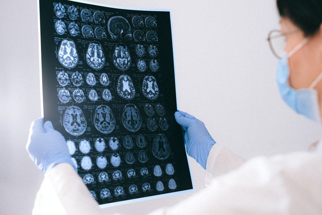
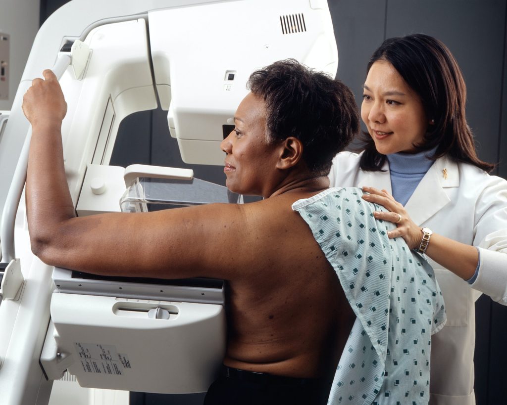
Category: Lab Tests and Imaging

MRI Scan Combination Could Detect Hypertrophic Cardiomyopathy Early
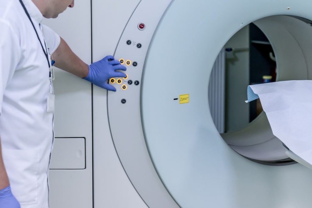
Credit: Pixabay CC0

Combining two types of heart scan techniques could help detect hypertrophic cardiomyopathy (HCM) before symptoms and signs on conventional tests appear, according to a new study led by UCL researchers. To do this, they used two cutting-edge heart scanning techniques: cardiac diffusion tensor imaging (cDTI), which shows the heart’s microstructure and cardiac MRI perfusion (perfusion CMR), which reveals microvascular disease. Their findings, published in Circulation, will help doctors select appropriate treatments.

HCM is a disease which affects around 1 in 500 in the UK, causing thickening of heart muscle and can lead to heart failure and cardiac arrest.

Researchers studied the hearts of three groups: healthy people, people who already had HCM, and people with an HCM-causing genetic mutation but no overt signs of disease.

The scans showed that people with overt signs of HCM have very abnormal organisation of their heart muscle cells and a high rate and severity of microvascular disease compared to healthy volunteers, helping doctors more accurately spot the early signs of HCM.

Crucially, the scans were also able to identify abnormal microstructure and microvascular disease in the people who had a problematic gene but no symptoms or muscle thickening. They found that 28% had defects in their blood supply, compared to healthy volunteers. This meant that doctors were able to more accurately spot the early signs of HCM developing in patient’s hearts.

The first drug to slow HCM progression, mavacamten, has recently been approved for use in Europe and will allow doctors to reduce the severity of the disease once symptoms and muscle thickening have appeared. Genetic therapies are also in development which could prevent symptoms entirely by intercepting HCM development at an early stage.

Perfusion CMR is already being used in some clinics to help differentiate people with HCM from other causes of muscle thickening. The researchers think that these revolutionary new therapies, combined with cDTI and perfusion CMR scans, give doctors the best ever chance of treating people at risk of HCM early enough that the condition never develops.

Dr George Joy, who led the research with Professor James Moon and Dr Luis Lopes (all UCL Institute of Cardiovascular Science), said: “The ability to detect early signs of HCM could be crucial in trials testing treatments aimed at preventing early disease from progressing or correcting genetic mutations. The scans could also enable treatment to start earlier than we previously thought possible.

“We now want to see if we can use the scans to identify which patients without symptoms or heart muscle thickening are most at risk of developing severe HCM and its life-changing complications. The information provided from scans could therefore help doctors make better decisions on how best to care for each patient.”

Dr Luis Lopes (UCL Institute of Cardiovascular Science), senior author of the study, said: “By linking advanced imaging to our cohort of HCM patients (and relatives) with extensive genetic testing, this study detected microstructural abnormalities in vivo in mutation carriers for the first time and was the first to compare these parameters in HCM patients with and without a causal mutation.

“The findings allow us to understand more about the early subclinical manifestations of this serious condition but also provide additional clinical tools for screening, monitoring and hopefully in the near future for therapeutic decision-making.”

Source: University College London

Plasma Protein Biomarkers Could Detect Early Mental Health Problems in Adolescents

Photo by Andrew Neel on Unsplash

Researchers at the University of Eastern Finland have identified plasma protein-based biomarkers capable of identifying adolescents at risk of developing mental health issues. Such biomarkers could revolutionise early detection and prevention of mental health problems in young people.The results were published in Nature Mental Health.

Some 10–20% of adolescents struggle with mental health conditions, with the majority going undiagnosed and untreated. This points to a need for new, early indicators of mental health problems to catch these cases and intervene with treatment before the conditions progress.

In the study carried out in the research group of Professor Katja Kanninen, the researchers used self-reported Strengths and Difficulties Questionnaire (SDQ) scores to evaluate mental health risk in participants aged between 11 and 16 years. Blood sample analyses showed that 58 proteins were significantly associated with the SDQ score. Bioinformatic analyses were used to identify the biological processes and pathways linked with the identified plasma protein biomarker candidates. Key enriched pathways related to these proteins included immune responses, blood coagulation, neurogenesis, and neuronal degeneration. The study employed a novel symbolic regression algorithm to create predictive models that best separate low and high SDQ score groups.

According to Professor Kanninen, plasma biomarker studies in mental disorders are an emerging field.

“Alterations in plasma proteins have been previously associated with various mental health disorders, such as depression, schizophrenia, psychotic disorders, and bipolar disorders. Our study supports these earlier findings and further revealed that specific plasma protein alterations could indicate a high risk for mental dysfunction in adolescents,” Professor Kanninen notes.

According to the researchers, this pilot study will be followed by more specific investigations of the potential biomarkers for identification of individuals at risk of mental health problems, opening a new avenue for advancements in adolescent mental health care.

Source: University of Eastern Finland

A Quick and Inexpensive Test for Osteoporosis

Photo by Mehmet Turgut Kirkgoz on Unsplash

In osteoporosis, treatment would be most effective with early detection – something not yet possible with current X-ray based osteoporosis diagnostic tests, which lack the requisite sensitivity. Now, researchers reporting in ACS Central Science have developed a biosensor that could someday help identify those most at risk for osteoporosis using less than a drop of blood.

Early intervention is critical to reducing the morbidity and mortality associated with osteoporosis. The most common technique used to measure changes in bone mineral density (BMD) – dual-energy X-ray absorptiometry – is not sensitive enough to detect BMD loss until a significant amount of damage has already occurred. Several genomic studies, however, have reported genetic variations known as single nucleotide polymorphisms (SNPs) that are associated with increased risk for osteoporosis. Using this information, Ciara K. O’Sullivan and colleagues wanted to develop a portable electrochemical device that would allow them to quickly detect five of these SNPs in finger-prick blood samples in a step toward early diagnosis.

The device involves an electrode array to which DNA fragments for each SNP are attached. When lysed whole blood is applied to the array, any DNA matching the SNPs binds the sequences and is amplified with recombinase polymerase that incorporates ferrocene, a label that facilitates electrochemical detection. Using this platform, the researchers detected osteoporosis-associated SNPs in 15 human blood samples, confirming their results with other methods.

As the DNA does not have to be purified from the blood, the analysis can be performed quickly (about 15 minutes) and inexpensively (< $0.5 per SNP). Furthermore, because the equipment and reagents are readily accessible and portable, the researchers say that the device offers great potential for use at point-of-care settings, rather than being limited to a centralised laboratory. The technology is also versatile and can be readily adapted to detect other SNPs, as the researchers showed previously when identifying drug resistance in Tuberculosis mycobacterium from sputum and cardiomyopathy risk from blood. Although the device does not diagnose osteoporosis itself, it might help physicians identify people whom they should monitor more closely.

Source: Chemical Society

Researchers Discover a Lipid Biomarker that can Identify Preeclampsia Risk

Photo by Shvets Production on Pexels

University of Virginia School of Medicine researchers have discovered a lipid biomarker to identify pregnant women at risk of preeclampsia, complications from which are the second-leading cause of maternal death around the world. Their findings are published in the Journal of Lipid Research.

The UVA scientists, led by Charles E. Chalfant, PhD, say that their finding opens the door to simple blood tests to screen patients. Further, the approach worked regardless of whether the women were on aspirin therapy, which is commonly prescribed to women thought to be at risk.

“Clinicians have been seeking simple tests to predict risk of preeclampsia before symptoms appear. Although alterations in some blood lipid levels have been known to occur in preeclampsia, they have not been endorsed as useful biomarkers. Our study presents the first comprehensive analysis of lipid species, yielding a distinctive profile associated with the development of preeclampsia,” said Chalfant. “The lipid ‘signature’ we described could significantly improve the ability to identify patients needing preventative treatment, like aspirin, or more careful monitoring for early signs of disease so that treatment could be initiated in a timely fashion.”

Preeclampsia affects up to 7% of all pregnancies. Symptoms typically appear after 20 weeks and include high blood pressure, kidney problems and abnormalties in blood clotting. The condition is associated with dangerous complications such as kidney and liver dysfunction and seizures, as well as a lifelong increased risk of heart disease for the mothers. An estimated 70 000 women around the world die from preeclampsia and its complications each year.

Doctors commonly recommend low-dose aspirin for at-risk women, but it works for only about half of patients, and it needs to be started within the first 16 weeks of pregnancy – well before symptoms appear. That makes it all the more important to identify women at risk early on, and to better understand preeclampsia in general.

Chalfant and his team wanted to find ‘biomarkers’ in the blood of pregnant women that could reveal their risk of developing preeclampsia. They examined blood plasma samples collected from 57 women in their first 24 weeks of pregnancy, then looked at whether the women went on to develop preeclampsia. The researchers found significant differences in ‘bioactive’ lipids in the blood of women who developed preeclampsia and those who did not.

This, the researchers say, should allow doctors to stratify women’s risk of developing preeclampsia by measuring lipid changes in their blood. The changes represent an important ‘lipid fingerprint’, the scientists say, that could be a useful tool for identifying, preventing and better treating preeclampsia.

“The application of our comprehensive lipid profiling method to routine obstetrical care could significantly reduce maternal and neonatal morbidity and mortality,” Chalfant said. “It represents an example of how personalised medicine could address a significant public health challenge.”

Source: University of Virginia Health System

Gravity-powered Biomedical Devices Pull Droplets Through a Maze

Source: Unsplash CC0

Biomedical engineers at Duke University have developed an entirely new approach to building point-of-care diagnostic devices that only use gravity to transport, mix and otherwise manipulate the liquid droplets involved. The demonstration, in the journal Device, requires only commercially available materials and very little power to read results, making it a potentially attractive option for applications in low-resource settings.

“The elegance in this approach is all in its simplicity – you can use whatever tools you happen to have to make it work,” said Hamed Vahabi, a former postdoctoral researcher at Duke. “You could theoretically even just use a handsaw and cut the channels needed for the test into a piece of wood.”

The study was conducted in the laboratory of Ashutosh Chilkoti, the Alan L. Kaganov Distinguished Professor of Biomedical Engineering at Duke.

There is no shortage of need for simple, easy-to-use, point-of-care devices. Many demonstrations and commercial devices seek to make diagnoses or measure important biomarkers using only a few drops of liquid with as little power and expertise required as possible. Their goal is to improve health care for the billions of people living in low-resource settings far from traditional hospitals and trained clinicians.

All of these tests have the same basic requirements; they must move, mix and measure small droplets containing biological samples and the active ingredients that make measuring specific biomarkers possible. More expensive examples use tiny electrical pumps to drive these reactions. Others use the physics of liquids within microchannels (microfluidics) that create a sort of suction effect.

This is the first demonstration that only uses gravity. Each approach offers uniquely useful abilities as well as drawbacks.

“Most microfluidic devices need more than just capillary forces to operate,” Chilkoti said. “This approach is much simpler and also allows very complex fluid paths to be deigned and operated, which is not easy or cheap to do with microfluidics.”

The new gravity-driven approach relies on a set of nine commercially available surface coatings that can tweak the wettability and slipperiness at any given point on the device. That is, they can adjust how much droplets flatten down into pancakes or remain spherical while making it easier or harder for them to slide down an incline.

Used together in clever combinations, these surface coatings can create all the microfluidic elements needed in a point-of-care test. For example, if a given location is extremely slippery and a droplet is placed at an intersection where one side pulls liquid flat and the other pushes it into a ball, it will act like a pump and accelerate the droplet toward the former.

“We came up with many different elements to control the motion, interaction, timing and sequence of multiple droplets in the device,” Vahabi said. “All of these phenomena are well-known in the field, but nobody thought of using them to control the motion of droplets in a systematic way before.”

By combining these elements, the researchers created a prototype test to measure the levels of lactate dehydrogenase (LDH) in a sample of human serum. They carved channels within the test platform to create specific pathways for droplets to travel, each coated with a substance that stops the droplets from sticking along their journey. They also primed specific locations with dried reagents needed for the test, which are soaked up by droplets of simple buffer solution as they travel through.

The whole maze-like test is then capped with a lid containing a couple of holes where the sample and buffer solution are dripped in. Once loaded, the test is placed inside a box-like device with a handle that turns the test 90° to allow gravity to do its work. This device is also equipped with a simple LED and light detector that can quickly and easily detect the amount of blue, red, or green in the test results. This means that the researchers can tag three different biomarkers with different colours for various tests to measure.

In the case of this prototype LDH test, the biomarker is tagged with a blue molecule. A simple microcontroller measures how deep of a blue hue the test results become and how quickly it changes colour, which indicates the amount and concentration of LDH in the sample, to generate results.

“We could eventually also use a smart phone down the line to measure results, but that’s not something we explored in this specific paper,” said Jason Liu, a PhD candidate in the Chilkoti lab.

The demonstration provides a new approach for consideration when engineering inexpensive, low-power, point-of-care diagnostic devices. While the group plans to continue developing their idea, they also hope others will take notice and work on similar tests.

“While a well-designed microfluidic system can be fully automated and easy-to-use by passive means, the timing of discrete steps is usually programmed into the design of the device itself, making modifications to protocol more difficult,” added David Kinnamon, a PhD candidate in the Chilkoti group. “In this work, the user retains more control of the timing of steps while only modestly sacrificing ease-of-operation. Again, this is an advantage for more complex protocols.”

Source: Duke University

Gliomas Have Folates Receptors That Could be Targeted for Imaging

Photo by Anna Shvets on Pexels

University of Turku researchers have discovered that gliomas contain increased amount of folate receptor expression relative to adjacent brain tissue. This discovery is a new and significant finding in the field, which could allow folate-based radiopharmaceuticals can be used in positron emission tomography (PET) imaging to detect folate receptors in gliomas.

This phenomenon, which is described in Frontiers in Immunology, has been observed in both experimental models and human tumour samples.

“Prior to this discovery, the presence of folate receptors and their increased presence in gliomas had not been recognised, and thus they have not yet been used for imaging nor treatment purposes,” summarises Doctoral Researcher Maxwell Miner from the Turku PET Centre at the University of Turku in Finland.

According to research group leader and InFLAMES PI Professor Anne Roivainen this presents an especially exciting target for potential future treatments.

“Our results show an average of 100-fold increase in folate-based radiopharmaceutical accumulation in glioma tissue versus that of adjacent healthy brain tissue,” says Professor Roivainen.

Urgent need for new chemotherapy treatments

Glioma brain tumours originate from the non-neuronal glial cells in the brain, which outnumber neurons in quantity. Gliomas comprise numerous subgroups, with even a high degree of morphological and receptor variability within a single cancerous lesion.

This exceptional cellular heterogeneity can make treatment difficult. There is an urgent need for new chemotherapy treatments particularly for the most malignant brain cancers as they often grow in an infiltrative web-like manner on their periphery making distinguishing the boundaries between glioma and non-glioma difficult. The researchers at the Turku PET Centre hope that this recent discovery will lead to further investigation into folate-targeted brain tumour detection and treatment.

Source: University of Turku

US Task Force to Recommend Earlier Start to Breast Cancer Screening

Photo by National Cancer Institute on Unsplash

In a move bringing it closer in line with other organisations’ breast cancer screening guidelines, The United States Preventative Task Force (USPSTF) has released a draft statement recommending mammography every other year (biennially) from ages 40 to 74.

These recommendations are not applicable to women with a genetic marker or syndrome linked to increased breast cancer risk, a history of high-dose chest radiotherapy at a young age, or previous breast cancer or a high-risk breast lesion on previous biopsies.

According to the USPSTF, “new and more inclusive science about breast cancer in people younger than 50 has enabled us to expand our prior recommendation and encourage all women to get screened in their 40s. We have long known that screening for breast cancer saves lives, and the science now supports all women getting screened, every other year, starting at age 40.”

South African cancer screening guidelines typically closely follow American ones, according to an article by Lipschitz in the South African Journal of Radiology. Many countries had not recommended screening at the ages of 40–50 due to fears of overdiagnosis.

The UPSTF made particular attention the fact that black women are 40% more likely to die of breast cancer than white women, and have a high rate of aggressive cancers at young ages.

The recommendations are not without criticism. Biennial screenings are not seen as worth it by Desountis et al., as it leaving two years between tests leaves too much time for a tumour to grow.

Debra Monticciolo, MD, of Massachusetts General Hospital in Boston, and a member of the Society of Breast Imaging’s board of directors, told MedPage Today that she was “disappointed” with the decision to recommend biennial scans.

“Even if you look at their own data,” Monticciolo said, “annual screening results in more deaths averted, no matter what type of screening program you put in those models.”

The UPSTF has posted the new recommendations on its website for comment.

Regarding the ongoing debated about continued screening in women ages 75 and older, and supplemental screening for those with dense breasts, the UPSTF found there was not enough evidence for a recommendation.

Low Power Mode for MRIs Could Cut Energy Use by Half

Credit: Pixabay CC0

Medical centres could save energy and reduce expenses by turning off MRIs and putting them in the lowest power mode instead of idling them when not in use, according to a new Radiology study.

Health care is responsible for up to 4.4% of global carbon emissions, and imaging contributes an outsized share due to its energy-intensive devices, especially MRI. A 2020 study found that three CTs and four MRIs used the same amount of energy per year as a town of 852 people, for example.

Though turning a machine off is better than idling, a substantial amount of MRI energy consumption occurs in “off” mode, which still draws a constant level of power for cooling. To address this, a new “power save” mode was developed that saves even more energy than the “off” mode by cycling cooling components on and off.

UC San Francisco researchers sought to compare energy consumption in the “idle,” “off” and “power save” modes. The researchers found that turning off MRIs overnight for 12 hours reduced their energy use by 25–33%, and that enabling the additional “power save” mode while the machine was off lowered power use by an additional 22–28%. Switching from idle directly to “power save” decreases energy use by 46–51%.

While just one company currently offers the “power save” mode while machines are off, it’s a design strategy worth replicating, the study noted.

“The results of this study demonstrate the potential energy and cost savings any radiology practice can obtain by using these simple power-down methods,” said assistant professor Sean Woolen, MD, first author on the study. “Our goal was to find ways for radiology departments worldwide to reduce their collective environmental footprint.”

Imaging has become increasingly central to medical decision-making, so it’s imperative to evaluate the design and operations of these machines in order to decarbonise health care, added Woolen.

Health Care Industry Would Save Millions

The study was made possible thanks to an academic-industry partnership comprising UCSF, Siemens Healthineers, Siemens USA, and Siemens Smart Infrastructure. Siemens provided technology and funding to equip MRI machines with power meters and install power monitoring software, and UCSF performed data collection and analysis.

The researchers equipped four outpatient MRI scanners from three different vendors with power meters and examined data over 39 days. They calculated energy output, costs (assuming a mean cost of $0.14 per kilowatt hour), and carbon use.

On an annual basis, switching a scanner from “idle” mode to “off” mode for 12 hours saved 12.3 to 21 megawatt hours (MWh) of electricity, where a megawatt is equal to 1000 kilowatts of electricity used continuously for an hour. This translated to annual savings of $1717 to $2943, and 8.7 to 14.9tonnes of C02-equivalent (MTCO2eq), a metric used to compare emissions of greenhouse gases based on their potential to contribute to global warming.

Switching from off to “power save” mode reduced energy use by an additional 8.8 to 11.4MWh and saved $1226 to $1594 and 6.2 to 8.1 MTC02eq per year.

“Often when we talk about how to decarbonise, solutions seem out of reach, but this initiative is proof that innovators everywhere can have impact,” added Barbara Humpton, CEO of Siemens USA. “The technology to decarbonise is here and ours is hard at work, helping industries like health care uncover ways to be more efficient and take concrete action to meet their carbon-reduction targets.”

The potential impact of adopting this technique as an industry standard would not impact patient care and would be an effective strategy to reduce cost and carbon emissions in health care, added Woolen.

Measuring Tissue Stiffness with Ultrasound Yields Sharper Images

Researchers have developed a new ultrasound method that for the first time can measure the level of tension in human tissue – a key indicator of disease. The breakthrough, published in the journal Science Advances, could be used to build new ultrasound machines that are able to better discriminate between abnormal tissue, scarring, and cancer.

Images produced by the current techniques ultrasound used in healthcare aren’t usually enough to diagnose whether tissues are abnormal. To improve diagnosis, the researchers developed a way to measure forces such as tension by using an ultrasound machine. Tension is generated in all living tissue, so measuring it can indicate whether tissue is functioning properly or if it’s affected by disease.

The researchers harnessed a technique from a rail project at the University of Sheffield, which uses sound waves to measure tension along railway lines. The technique, used both for rail and medical ultrasound, relies on a simple principle: the greater the tension, the faster sound waves propagate. Using this principle, the researchers developed a method that sends two sound waves in different directions. The tension is then related to the speed of the waves by using mathematical theories developed by the researchers.

Previous ultrasound methods have struggled to show the difference between stiff tissue or tissue under tension. The developed technique is the first capable of measuring tension for any type of soft tissue, and without knowing anything about it. In this new paper, the researchers describe the new method and demonstrate how they used it to measure tension inside a muscle.

Study leader Dr Artur Gower, Lecturer in Dynamics at the University of Sheffield, said: “When you go to the hospital, a doctor might use an ultrasound device to create an image of an organ, such as your liver, or another part of your body, such as the gut, to help them explore what the cause of a problem might be. One of the limitations of ultrasounds used in healthcare now is that the image alone is not enough to diagnose whether any of your tissues are abnormal.

“What we’ve done in our research is develop a new way of using ultrasound to measure the level of tension in tissue. This level of detail can tell us whether tissues are abnormal or if they are affected by scarring or disease. This technique is the first time that ultrasound can be used to measure forces inside tissue, and it could now be used to build new ultrasound machines capable of diagnosing abnormal tissue and disease earlier.”

Source: University of Sheffield

Sex Differences in Brain Glycogen After a Stroke may Yield New Treatments

Credit: American Heart Association

Although males and females are equally impacted by stroke, there are differences in recovery. Since oestrogen and progesterone have known neuroprotective effects, it is important to gauge their effects in stroke recovert. In a paper published in IBRO Neuroscience Reports, researchers have discovered differences between biomarkers such as glycogen levels in the brains of male and female mice.

“A stroke is caused by a loss of blood flow to brain cells. Without urgent intervention this may cause those cells to die because they constantly need energy and nutrients from the blood,” said Prof Nicole Sylvain, clinical research coordinator and lab manager at the University of Saskatchewan.

Sylvain and her colleagues are looking at treatments for post-stroke recovery that help supplement these energy losses. Using the Canadian Light Source (CLS) at the University of Saskatchewan (USask), the team was able to identify energy biomarkers in the brain, which could eventually inform clinicians about the effects of potential stroke treatments on brain recovery after a stroke.

The group’s recent study examined post-stroke differences between male and female mice, and found that female mice have higher amounts of glycogen in their brains. When the supply of glycogen is disrupted by stroke, the brain is severely impacted.

Most pre-clinical stroke research has been performed using male lab animals, with results usually generalised to both sexes. In clinical stoke cases, females have a higher incidence of ischaemic stroke and poorer outcomes, compared to males.

“We found that, for the most part, male data can be generalised for females, however, some of the metabolic markers we measured were actually different,” Sylvain said. “It’s really important to do the research on both sexes.”

It would be impossible for the team to detect the biomarkers without to the Mid-IR beamline.

“The only way to detect them in such an accurate way across the brain is with infrared imaging, so the CLS has been absolutely vital to our research.”

Source: University of Saskatchewan