Category: Lab Tests and Imaging

A Stool Sample Could Detect Some Parkinson’s Cases Early

Old man with magnifying glass
Image by Mar Lezhava on Unsplash

One early indicator of Parkinson’s disease (PD) is isolated REM-sleep behaviour disorder. Researchers have shown that a greater concentration of α-synuclein aggregates can be detected in the stool samples of patients. In the scientific journal npj Parkinson’s Disease, they now present a method for detecting these aggregates.

There are two forms of PD. In 70% of cases, it originates in the central nervous system. However, in around 30% of cases it originates in the nervous system of the intestine (“enteric nervous system”). The latter form is referred to as “body-first Parkinson’s disease” (for short: body-first PD) and the characteristic deposits of aggregates of the body’s own α-synuclein protein are formed in the neurons in the intestine.

A preliminary form of body-first PD is the so-called isolated REM-sleep behaviour disorder (for short: iBRD). It causes in part complex movements during REM-sleep insofar as the patient experiences vivid and disturbing dreams. These movements can endanger the sufferer themselves or others.

A research team headed by Professor Erdem Gültekin Tamgüney from the Institute of Physical Biology at HHU now reports that it is possible to detect an elevated level of α-synuclein aggregates in the stool samples of patients. To achieve this, the team used a new surface-based fluorescence intensity distribution analysis (sFIDA) to detect and quantify individual particles of α-synuclein aggregates.

Professor Tamgüney: “We are the first to prove the presence of α-synuclein aggregates in stool samples. Our results show a significantly higher level of α-synuclein aggregates in iRBD patients compared with healthy individuals or patients with Parkinson’s. These findings could lead to a non-invasive diagnostic tool for prodromal synucleinopathies — including Parkinson’s — which could in turn enable therapies to be initiated at an early stage before symptoms occur.” However, more research is required before the process can find its way into clinical practice, for example investigation into why the level is lower in Parkinson’s patients.

The study was conducted in a collaboration to establish a biobank with stool samples from patients and control subjects, and to develop the test procedure and conduct the tests on the samples, and to eventually commercialise the technique.

Background

In body-first PD, the deposits of fibrils of the body’s own α-synuclein protein, which are characteristic of Parkinson’s, are first formed in the neurons of the enteric nervous system, which serves the gastrointestinal tract. The aggregates then spread to the central nervous system in a way similar to prions, i.e. an existing aggregate combines individual α-synuclein proteins in its vicinity into further aggregates in a nucleation process; these aggregates then spread further through the body.

The influence of what happens in the gastrointestinal tract on the brain is referred to as the “gut-brain axis.” The gastrointestinal tract is exposed to the environment and it is possible that harmful substances such as chemicals, bacteria or viruses ingested directly with food or via interaction with the microbiome of the gastrointestinal tract may trigger the pathological formation of α-synuclein aggregates.

Source: Heinrich-Heine University Duesseldorf

Patients ‘Don’t Need to be Checked for Everything’, Recommendation Says

Blood samples
Photo by National Cancer Institute on Unsplash

Commonly ordered tests can provide early warning of underlying disease, but could also create unnecessary risks of false positive results, provoking anxiety in the patient, wasted time and money and risks of invasive testing.

Therefore, to combat commonly ordered – but not always necessary – procedures and tests, the Society of General Internal Medicine (SGIM) on Tuesday released its revised list of recommendations on five primary care procedures and tests that patients and physicians should question.

Northwestern University’s Dr Jeffrey A. Linder and David Liss, who have previously published research on the benefits of primary care checkups, helped revise the list.

For instance, the age-old idea of getting an annual physical exam with “routine blood tests” from a primary care doctor is a misconception because a person’s age and other risk factors should influence how frequently they should see their doctor, Linder said.

“We often have patients come in asking us to ‘check me for everything,’ but this is a potentially anxiety-provoking, dangerous thing for patients because the more testing we do, the more stuff we find, and the more we need to follow up,” said Linder, chief of the division of general internal medicine at Northwestern University Feinberg School of Medicine and a Northwestern Medicine physician. “In someone who is asymptomatic, an ‘abnormality’ is much more likely to be a false positive or of no clinical significance than for us to catch early disease.

“False positives can expose patients to all of the anxiety, costs, hassle and time commitment, and danger from sometimes invasive testing, with a very low likelihood that it is going to improve their health.”

This isn’t to say nobody should get a checkup every year. For instance, patients who have overdue preventive services, rarely see their primary care physician, have low self-rated health and/or are aged 65 or older should get an annual checkup, the scientists said.

The newly revised list is part of SGIM’s Choosing Wisely campaign, which is an initiative of the American Board of Internal Medicine Foundation. SGIM members originally selected the topics in 2013 and later updated the list in 2017.

The list generated controversy when it was first developed in 2013, recalls Linder.

“The list was widely misinterpreted as ‘specialty society says you don’t need to see your doctor,’ but that was not what it said,” Linder said.

Time and downstream financial costs also are issues of these commonly ordered but oftentimes unnecessary tests and procedures, Liss said.

“Patients and care teams often spend valuable time on low-value checkups that could have been devoted to high-need patients,” said Liss, research associate professor of general internal medicine at Feinberg. “There also is the overall increase in costs to the health system. And even if annual checkups are covered by most insurance, patients often have copays for services like blood draws and other diagnostic tests.”

The revised list was developed after months of careful consideration and review, using the most current evidence about management and treatment options. Linder and Liss served as ad hoc members of the SGIM’s Choosing Wisely Working Group.

Here are the five recommendations, based on a review of the most recent studies in the field:

  1. Don’t recommend daily home glucose monitoring in patients with Type 2 diabetes mellitus not using insulin.
  2. Don’t perform routine annual checkups unless patients are likely to benefit; the frequency of checkups should be based on individual risk factors and preferences. During checkups, don’t conduct comprehensive physical exams or routine lab testing.
  3. Don’t perform routine pre-operative testing before low-risk surgical procedures.
  4. Don’t recommend cancer screening in adults with life expectancy of less than 10 years.
  5. Don’t place, or leave in place, peripherally inserted central catheters for patient or provider convenience.

Source: Northwestern University

A Severe Form of Dementia may in Fact be Caused by a Cerebrospinal Fluid Leak

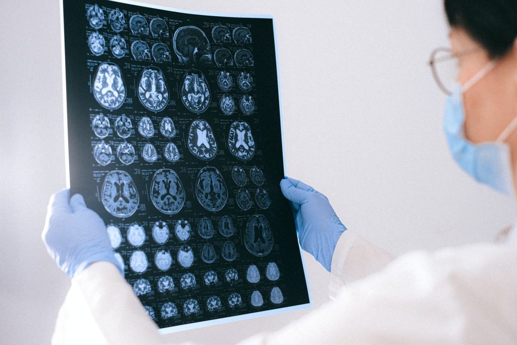
MRI images of the brain
Photo by Anna Shvets on Pexels

A new study suggests that some patients diagnosed with behavioural-variant frontotemporal dementia (bvFTD) – a presently incurable, mentally debilitating condition – may instead have a cerebrospinal fluid leak, which is detectable on MRI scans and often treatable. The researchers say these findings, published in the peer-reviewed journal Alzheimer’s & Dementia: Translational Research and Clinical Interventions, could lead to a cure.

“Many of these patients experience cognitive, behavioural and personality changes so severe that they are arrested or placed in nursing homes,” said Wouter Schievink, MD, professor of Neurosurgery at Cedars-Sinai. “If they have behavioural-variant frontotemporal dementia with an unknown cause, then no treatment is available. But our study shows that patients with cerebrospinal fluid leaks can be cured if we can find the source of the leak.”

When cerebrospinal fluid (CSF) leaks into the body, the brain can sag, causing dementia symptoms. Schievink said many patients with brain sagging, detectable in MRI, go undiagnosed, and he advises clinicians to take a second look at patients with telltale symptoms.

“A knowledgeable radiologist, neurosurgeon or neurologist should check the patient’s MRI again to make sure there is no evidence for brain sagging,” Schievink said.

Clinicians can also ask about a history of severe headaches that improve when the patient lies down, significant sleepiness even after adequate night-time sleep, and whether the patient has ever been diagnosed with a Chiari brain malformation, a condition in which brain tissue extends into the spinal canal. Brain sagging, Schievink said, is often mistaken for a Chiari malformation.

Even when brain sagging is detected, the source of a CSF leak can be difficult to locate. When the fluid leaks through a tear or cyst in the surrounding membrane, it is visible on CT myelogram imaging with the aid of contrast medium.

Schievink and his team recently discovered an additional cause of CSF leak: the CSF-venous fistula. In these cases, the fluid leaks into a vein, making it difficult to see on a routine CT myelogram. To detect these leaks, technicians must use a specialized CT scan and observe the contrast medium in motion as it flows through the cerebrospinal fluid.

In this study, investigators used this imaging technique on 21 patients with brain sagging and symptoms of bvFTD, and they discovered CSF-venous fistulas in nine of those patients. All nine patients had their fistulas surgically closed, and their brain sagging and accompanying symptoms were completely reversed.

“This is a rapidly evolving field of study, and advances in imaging technology have greatly improved our ability to detect sources of CSF leak, especially CSF-venous fistula,” said Keith L. Black, MD, chair of the department of Neurosurgery at Cedars-Sinai. “This specialised imaging is not widely available, and this study suggests the need for further research to improve detection and cure rates for patients.”

The remaining 12 study participants, whose leaks could not be identified, were treated with nontargeted therapies designed to relieve brain sagging, such as implantable systems for infusing the patient with CSF. However, only three of these patients experienced relief from their symptoms.

“Great efforts need to be made to improve the detection rate of CSF leak in these patients,” Schievink said. “We have developed nontargeted treatments for patients where no leak can be detected, but as our study shows, these treatments are much less effective than targeted, surgical correction of the leak.”

Source: Cedars-Sinai Medical Center

A Quick Scan Can Pinpoint Hypertension-causing Adrenal Nodules

Stethoscope
Photo by Hush Naidoo on Unsplash

Doctors have demonstrated a new type of CT scan that lights up tiny nodules in the adrenal glands which give rise to hypertension in about 5% of hypertensive patients. enabling hypertension to be cured by their removal. The nodules are discovered in about 5% of hypertensive patients.

Published in The Journal of Hypertension, this work solves a 60-year problem of how to detect the hormone-producing nodules without a difficult and failure-prone catheter study that is available in only a few hospitals. The research also found that, when combined with a urine test, the scan detects a group of patients who come off all their blood pressure medicines after treatment.

The study, led by doctors at Queen Mary University of London and Barts Hospital, and Cambridge University Hospital, involved 128 participants for whom hypertension was found to be caused by aldosterone. The scan found that in two thirds of patients with elevated aldosterone secretion, this is coming from a benign nodule in just one of the adrenal glands, which can then be safely removed. The scan uses a very short-acting dose of metomidate, a radioactive dye that sticks only to the aldosterone-producing nodule.

The scan was as accurate as the old catheter test, but quick, painless and technically successful in every patient. Until now, the catheter test was unable to predict which patients would be completely cured of hypertension by surgical removal of the gland. By contrast, the combination of a ‘hot nodule’ on the scan and urine steroid test detected 18 of the 24 patients who achieved a normal blood pressure off all their drugs.

Professor Morris Brown, co-senior author of the study and Professor of Endocrine Hypertension at Queen Mary University of London, said: “These aldosterone-producing nodules are very small and easily overlooked on a regular CT scan. When they glow for a few minutes after our injection, they are revealed as the obvious cause of hypertension, which can often then be cured. Until now, 99% are never diagnosed because of the difficulty and unavailability of tests. Hopefully this is about to change.”

In most people with hypertension, the cause is unknown, and the condition requires life-long treatment by drugs. Previous research by the group at Queen Mary University discovered that in 5–10% of people with hypertension the cause is a gene mutation in the adrenal glands, which results in excessive amounts of the steroid hormone, aldosterone, being produced. Aldosterone causes salt retention, driving up blood pressure. Patients with excessive aldosterone levels in the blood are resistant to treatment with standard antihypertensives, and at increased risk of cardiovascular disease.

Source: Queen Mary University of London

Global Medical Isotope Shortage to Ease with Renewed Production

Radiation warning sign
Photo by Vladyslav Cherkasenko on Unsplash

Amid the ongoing global shortage of medical isotopes, there is at least some good news: two European research reactors have been fired up again and will be delivering molybdenum-99 and iodine-131 isotopes. In addition, a new reactor to produce Mo-99 through a new method has also been completed in the US and is awaiting testing and certification.

Mo-99 is the world’s most important medical diagnostic radioisotope precursor, and is the parent isotope of technetium-99m (Tc-99m). Tc-99m is used in more than 40-million diagnostic procedures each year. The production of this isotope is acutely vulnerable to supply chain disruption and much of the machinery used to produce it is ageing. South African nuclear corporation NTP also produces a small amount of the isotope locally at its Pelindaba facility.

Nuclear Medicine Europe (NMEU) was notified that the LVR-15 reactor resumed operations on Friday morning November 18 and the first irradiated targets from it are being processed today November 23rd. In addition, NMEU was notified that the HFR reactor resumed operations on November 23 and achieved full power operation at 14:30 CET.

The Mo-99 global supply situation will largely return to normal within the next 7-10 days with the I-131 supply situation returning to normal within two weeks, according to NMEU’s predictions. NMEU will provide further communication to the nuclear medicine community as developments warrant.

At the new production facility in the US, the isotope manufacturer NorthStar will produce Mo-99 through a new method, based on irradiation of molybdenum-100 targets using electron accelerators. This will be the first facility in the world to produce commercial-scale Mo-99 using this technology. The facility also includes new, high-capacity equipment for processing and packaging Mo-99 for distribution to radiopharmacies and hospitals.

What’s Really in that Tattoo Ink?

Photo by benjamin lehman on Unsplash

After testing nearly 100 tattoo inks, researchers reported that, even when the ink bottles had ingredient labels, those ingredients listed on them were often inaccurate. The team also detected small particles that could be harmful to cells.

In the US, the Food and Drug Administration regulates tattoo ink, but in South Africa, tattoo ink [PDF] is imported largely unregulated.

The researchers presented their findings at a meeting of the American Chemical Society (ACS). 

“The idea for this project initially came about because I was interested in what happens when laser light is used to remove tattoos,” said lead researcher John Swierk, PhD. “But then I realised that very little is actually known about the composition of tattoo inks, so we started analysing popular brands.”

Tattoo artists interviewed to see what they knew about the inks they use on their customers could quickly identify a brand they preferred, but they didn’t know much about its contents. “Surprisingly, no dye shop makes pigment specific for tattoo ink,” Dr Swierk explained. “Big companies manufacture pigments for everything, such as paint and textiles. These same pigments are used in tattoo inks.” He also notes that tattoo artists must be licensed in the locales where they operate for safety reasons, yet no federal or local agency regulates the contents of the inks themselves.

Tattoo inks are made up of a pigment and a carrier solution. The pigment could be a molecular compound such as a blue pigment; a solid compound such as titanium dioxide, which is white; or a combination of the two compound types such as light blue ink, which contains both the molecular blue pigment and titanium dioxide. The carrier solution transports the pigment to the middle layer of skin and typically helps make the pigment more soluble, sometimes controlling the viscosity of the ink solution and perhaps containing an anti-inflammatory ingredient.

Dr Swierk’s team has been studying the particle size and molecular composition of tattoo pigments. From these analyses, they have confirmed the presence of ingredients that aren’t listed on some labels. For example, in one case ethanol was not listed, but the chemical analysis showed it was present in the ink. The team has also been able to identify what specific pigments are present in some inks.

“Every time we looked at one of the inks, we found something that gave me pause,” Dr Swierk said. “For example, 23 of 56 different inks analysed to date suggest an azo-containing dye is present.” Although many azo pigments are not health concerns when they are chemically intact, bacteria or UV light can degrade them into another nitrogen-based compound that is a potential carcinogen, according to the Joint Research Centre, which provides independent scientific advice to the European Union.

In addition, the team has analysed 16 inks using electron microscopy, and about half contained particles under 100nm. “That’s a concerning size range,” said Dr Swierk. “Particles of this size can get through the cell membrane and potentially cause harm.”

After the researchers run a few more tests and have the data peer reviewed, they will add the information to their website “What’s in My Ink?” “With these data, we want consumers and artists to make informed decisions and understand how accurate the provided information is,” said Dr Swierk.

Source: American Chemical Society

Low Serum Urate Increases Sarcopenia Risk

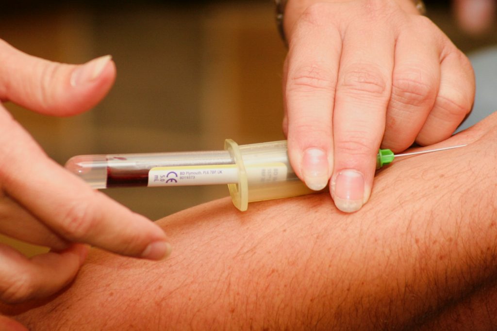
Blood sample being drawn
Photo by Hush Naidoo Jade Photography on Unsplash

Adults with low blood levels of urate, the end-product of the purine metabolism in humans, may be at higher risk of sarcopenia and may face a higher risk of early death, according to a new study published in Arthritis & Rheumatology.

Whether or nor low serum urate (SU) levels contribute to adverse outcomes has been the subject of controversy.  The study involved 13 979 participants aged 20 years and older, sourced from the National Health and Nutrition Examination Survey from 1999–2006.

Low serum urate concentrations (<2.5 mg/dL in women; <3.5 mg/dL in men) were associated with low lean mass, underweight BMI (<18.5 kg/m2), and higher rates of weight loss. While low SU was associated with increased mortality (61%) before adjusting for body composition, its effect was reduced and non-significant after adjustment for body composition and weight loss.

“These observations support what many have intuited, namely that people with low serum urate levels have higher mortality and worse outcomes not because low urate is bad for health, but rather that low urate levels tend to occur among sicker people, who have lost weight and have adverse body composition,” explained lead author Joshua F. Baker, MD, MSCE, of the University of Pennsylvania. “While this observational study doesn’t disprove a causal association, it does suggest that great care is needed in interpreting epidemiologic associations between urate levels and health outcomes.”

Source: Wiley

Shortage of Blood Test Tubes Prompts Saving Efforts

Blood sample being drawn
Photo by Hush Naidoo Jade Photography on Unsplash

A pandemic-related shortage of a mundane item, ‘blue top’ blood test tubes used toollect blood samples from patients, has caused headaches for health systems worldwide.

But it may also have a silver lining: A lesson in how to reduce unneeded medical tests, whether or not there’s a shortage, according to a new study published in JAMA Internal Medicine.

The shortage gave researchers a chance to see if alerting doctors at the moment they’re placing an order could encourage them to seek a test only when results will immediately affect care.

In the new study, an alert led to a nearly immediate 29% drop in orders for one common test. The reduced level persisted for months.

“This shows that small interventions can make a big difference, and suggests the potential for other types of low-value care to benefit from a similar intervention,” says lead author Madison Breeden, MD, who conducted the study during her year as chief resident of Quality and Patient Safety. She’s already exploring if the approach might reduce unnecessary antibiotic prescriptionse

Breeden and her colleagues describe what happened in spring 2021 when University of Michigan Health supply chain and pathology experts began worrying about a potential shortage of ‘blue top’ tubes. The pandemic had created very high demand for the chemical the tubes contain: sodium citrate, which stabilises blood samples until a laboratory team can analyse three blood clotting-related properties, called PT, INR and PTT.

After emailing all providers, U-M Health added a ‘best practices alert’ to doctors’ test-ordering electronic system. They could still order PT/INR/PTT tests, but were asked for “thoughtful restraint in reflexive ordering.”

The alert began popping up a month before the FDA issued an official shortage notice and the issue got widespread attention. The shortage continues today and has grown to other types of tests.

The researchers looked at what happened for six months after the alert began appearing at U-M Health, and compared it with data from six months before.

“There are very important reasons to order this test in some patients, for instance before an operation or when managing certain conditions and treatments,” Breeden explains. “But it may also be part of a standard order set that’s put in during an emergency department visit and continues to be ordered repeatedly after the patient is admitted to the hospital, even though the results won’t change their care.” For such patients, a one-time test might be indicated, but not repeated testing.

Busy doctors entering orders for tests don’t tend to think about the supplies and people power needed to carry out those tests, Breeden notes. In the face of a shortage, or of strong evidence that a test is often over-ordered, an alert could help prioritize the tests for those who need them most.

Canadian experts have actually flagged PT/INR/PTT tests as a target for reducing unnecessary care, through the Choosing Wisely program. So has the American Society for Clinical Laboratory Science, a medical professional group.

AI Picks up Incidental Pulmonary Embolism on Chest CT

Photo by Kanasi on Unsplash

According to a study published in the American Journal of Roentgenology, an AI tool for detection of incidental pulmonary embolus (iPE) on conventional contrast-enhanced chest CT examinations had high false negative and moderate false positive rates for detection, and was even able to pick up some iPEs missed by radiologists.

“Potential applications of the AI tool include serving as a second reader to help detect additional iPEs or as a worklist triage tool to allow earlier iPE detection and intervention,” wrote lead investigator Kiran Batra from the University of Texas Southwestern Medical Center in Dallas. “Various explanations of misclassifications by the AI tool (both false positives and false negatives) were identified, to provide targets for model improvement.”

Batra and colleagues’ retrospective study included 2,555 patients (1,340 women, 1,215 men; mean age, 53.6 years) who underwent 3,003 conventional contrast-enhanced chest CT examinations between September 2019 and February 2020 at Parkland Health in Dallas, TX. Using an FDA-approved, commercially available AI tool (Aidoc) to detect acute iPE on the images, a vendor-supplied natural language processing algorithm was then applied to the clinical reports to identify examinations interpreted as positive for iPE.

Ultimately, the commercial AI tool had NPV of 99.8% and PPV of 86.7% for detection of iPE on conventional contrast-enhanced chest CT examinations (ie, not using CT pulmonary angiography protocols). Of 40 iPEs present in the team’s study sample, 7 were detected only by the clinical reports, and 4 were detected only by AI.

Noting that both the AI tool and clinical reports detected iPEs missed by the other method, “the diagnostic performance of the AI tool did not show significant variation across study subgroups,” the authors added.

Source: American Roentgen Ray Society

Nuclear Stress Testing Identifies Candidates Most in Need of Angioplasty

Photo by Robina Weermeijer on Unsplash

Patients identified by nuclear stress testing as having severe stress-induced myocardial ischaemia may benefit from angioplasty, while those with mild or no ischaemia will not, according to a new study reported in the Journal of the American College of Cardiology.

Following stress testing, coronary revascularisation restores blood flow to blocked arteries. For patients with severe ischaemia, early revascularisation saw a more than 30% reduction in mortality compared to patients with severe ischaemia who were treated with medication, but there was no benefit for the other groups.

Conducted by the Icahn School of Medicine at Mount Sinai, this is the first large-scale study to investigate stress testing in patient management when applied to the full spectrum of patients who have both varying degrees of myocardial ischaemia and heart function. This new study can help guide physicians on how to manage caring for patients with suspected heart disease.

Stress tests are indicated when physicians suspect that a patient’s chest pain or other clinical symptoms are from coronary artery disease (CAD). These help determine if a patient has obstructive CAD which leads to significant ischaemia. If the ischaemia due to obstructive CAD is severe, adequate blood flow can be restored with coronary artery bypass grafting surgery or percutaneous coronary intervention (PCI), where a catheter is used to place stents in the blocked coronary arteries. Nuclear stress testing is the most common stress test used to detect myocardial ischaemia.

“There is keen interest in assessing how measurement of myocardial ischaemia during stress testing can help shape physicians’ decision to refer patients for coronary revascularisation procedures, but this issue has not been well studied among patients who have underlying heart damage,” explains lead author Alan Rozanski, MD. “Our study, which evaluated a large number of patients with pre-existing heart damage who underwent cardiac stress testing, finally addresses this clinical void.”

The researchers analysed records of more than 43 000 patients who underwent nuclear stress testing with suspected CAD between 1998 and 2017 with a median 11-year follow-up for mortality/survival. The investigators grouped patients according to both their level of myocardial ischaemia during stress testing as well as their left ventricular ejection fraction (LVEF). Low LVEF measurements indicate prior heart damage that could be from scarring of the heart due to a prior heart attack.

The study provides two important clinical insights. First, the study showed that the frequency of myocardial ischemia during stress testing varies according to patients’ heart function. Of the 39 883 patients with normal heart function (LVEF > 55%), fewer than 8% of them had ischaemia. However, among the 3560 patients with reduced heart function (LVEF less than 45%, which indicates prior heart damage), more than 40% of them had myocardial ischaemia. The study also showed that the presence of myocardial ischaemia increases the risk of death in patients with normal and reduced heart function. Among both groups of patients, performing bypass or PCI procedures was not associated with improved survival among patients with either no or only mild ischaemia during the cardiac stress test. Among patients with severe ischaemia, coronary procedures were associated with more than 30% higher survival rates compared to those managed with medication only. This was the case for patients with and without heart damage.

“These results confirm the benefits of stress testing for clinical management. What you want from any test when considering coronary revascularisation procedures is that the test will identify a large percentage of patients who are at low clinical risk and do so correctly, while identifying only a small percentage of patients who are at high clinical risk and do so correctly. That is what we found with nuclear stress testing in this study,” explains Dr Rozanski. “Importantly, the presence of severe ischaemia does not necessarily mean that coronary revascularisation should be applied. New data from a large clinical trial suggests that when medical therapy is optimised it may be as effective as coronary revascularisation in such patients. But regardless, the presence of severe ischaemia indicates high clinical risk which then requires aggressive management to reduce clinical risk.”

Source: The Mount Sinai Hospital / Mount Sinai School of Medicine