Category: Diseases, Syndromes and Conditions

After an Infection, Brain Inflammation Triggers Muscle Weakness

Photo by Andrea Piacquadio

Infections and neurodegenerative diseases cause inflammation in the brain. But for unknown reasons, patients with brain inflammation often develop muscle problems that seem to be independent of the central nervous system. Now, researchers at Washington University School of Medicine in St. Louis have revealed how brain inflammation releases a specific protein that travels from the brain to the muscles and causes a loss of muscle function.

The study, published in Science Immunology, also identified ways to block this process, which could have implications for treating or preventing the muscle wasting sometimes associated with inflammatory diseases, including bacterial infections, Alzheimer’s disease and long COVID.

“We are interested in understanding the very deep muscle fatigue that is associated with some common illnesses,” said senior author Aaron Johnson, PhD, an associate professor of developmental biology. “Our study suggests that when we get sick, messenger proteins from the brain travel through the bloodstream and reduce energy levels in skeletal muscle. This is more than a lack of motivation to move because we don’t feel well. These processes reduce energy levels in skeletal muscle, decreasing the capacity to move and function normally.”

Fruit fly and mouse models

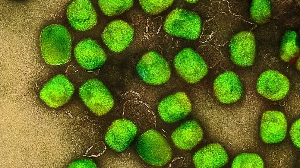
To investigate the effects of brain inflammation on muscle function, the researchers modelled three different types of diseases – an E. coli bacterial infection, a SARS-CoV-2 viral infection and Alzheimer’s. When the brain is exposed to inflammatory proteins characteristic of these diseases, damaging chemicals called reactive oxygen species build up. The reactive oxygen species cause brain cells to produce an immune-related molecule called interleukin-6 (IL-6), which travels throughout the body via the bloodstream. The researchers found that IL-6 in mice – and the corresponding protein in fruit flies – reduced energy production in muscles’ mitochondria, the energy factories of cells.

“Flies and mice that had COVID-associated proteins in the brain showed reduced motor function – the flies didn’t climb as well as they should have, and the mice didn’t run as well or as much as control mice,” Johnson said. “We saw similar effects on muscle function when the brain was exposed to bacterial-associated proteins and the Alzheimer’s protein amyloid beta. We also see evidence that this effect can become chronic. Even if an infection is cleared quickly, the reduced muscle performance remains many days longer in our experiments.”

Johnson, along with collaborators at the University of Florida and first author Shuo Yang, PhD (who did this work as a postdoctoral researcher in Johnson’s lab) make the case that the same processes are likely relevant in people. The bacterial brain infection meningitis is known to increase IL-6 levels and can be associated with muscle issues in some patients, for instance. Among COVID-19 patients, inflammatory SARS-CoV-2 proteins have been found in the brain during autopsy, and many long COVID patients report extreme fatigue and muscle weakness even long after the initial infection has cleared. Patients with Alzheimer’s disease also show increased levels of IL-6 in the blood as well as muscle weakness.

Potential treatment targets

The study pinpoints potential targets for preventing or treating muscle weakness related to brain inflammation. The researchers found that IL-6 activates what is called the JAK-STAT pathway in muscle, and this is what causes the reduced energy production of mitochondria. Several therapeutics already approved by the Food and Drug Administration for other diseases can block this pathway. JAK inhibitors as well as several monoclonal antibodies against IL-6 are approved to treat various types of arthritis and manage other inflammatory conditions.

“We’re not sure why the brain produces a protein signal that is so damaging to muscle function across so many different disease categories,” Johnson said. “If we want to speculate about possible reasons this process has stayed with us over the course of human evolution, despite the damage it does, it could be a way for the brain to reallocate resources to itself as it fights off disease. We need more research to better understand this process and its consequences throughout the body.

“In the meantime, we hope our study encourages more clinical research into this pathway and whether existing treatments that block various parts of it can help the many patients who experience this type of debilitating muscle fatigue,” he said.

Source: Washington University School of Medicine

Debunking Myths About Mpox

Mpox (monkeypox) virus. Source: NIH

Myths are widely held beliefs about various issues, including illness and disease. They come about through frequent storytelling and retelling. Dr Themba Hadebe, Clinical Executive at Bonitas Medical Fund, helps debunks myths about monkeypox (mpox).

Myth 1: Mpox (formerly monkeypox) is a new disease created in a lab

Fact: The mpox virus was discovered in Denmark (1958) in a colony of monkeys at a laboratory kept for research.  The first reported human case was in 1970 in the DRC. Mpox is a zoonotic disease, meaning it can be spread between animals and people. It is found regularly in parts of Central and West Africa and can spread from person to person or occasionally from animals to people.  

Myth 2: Mpox comes from monkeys

Fact: Despite its name, monkeypox does not come from monkeys. The disease earned the name when the ‘pox like’ outbreaks happened in the research monkeys. While monkeys can get mpox, they are not the reservoir (where a disease typically grows and multiplies). The reservoir appears to be rodents.

Myth 3:  Only a handful of people have contracted mpox

Fact: Globally, more than 97 000 cases and 186 deaths were reported across 117 countries in the first four months of 2024. South Africa is among the countries currently experiencing an outbreak.  On the 5 July, it was reported that the number of mpox cases in the country has risen to 20. This after four more cases have been confirmed in Gauteng and KwaZulu-Natal in the last few days.

15 patients have, however been given a clean bill of health.

Myth 4:  It is easy to diagnose mpox

Fact: It is easy to mistake mpox for something else. While the rash can be mistaken for chickenpox, shingles or herpes, there are differences between these rashes. Symptoms of mpox include fever, sore throat, headache, muscle aches, back pain, low energy and swollen lymph nodes. Fever, muscle aches and a sore throat appear first. The rash begins on the face and spreads over the body, extending to the palms of the hands and soles of the feet and develops over 2-4 weeks in stages. The ‘pox’ dip in the centre before crusting over.

Laboratory confirmation is required. A sample of one of the sores is diagnosed by a PCR test for the virus (MPXV).

Myth 5: Mpox is easily treated

Fact: ‘Currently,’ says the National Institute for Communicable Diseases (NICD), ‘there is no registered treatment for mpox in South Africa. However, the World Health Organization (WHO) recommends the use of TPOXX for treatment of severe cases, in immunocompromised people’. However, the Department of Health (DoH) has only obtained this treatment, with approval on a compassionate use basis, for the five known patients with severe disease.

There is no mpox vaccine currently available in South Africa.

Myth 6: You can get mpox from being in a crowd or from a public toilet seat

Fact: Mpox is not like Covid-19 which is highly contagious. It spreads through direct contact via blood, bodily fluid, skin or mucous lesions or respiratory droplets.

It can also spread though bites and scratches. Studies have shown that the virus can stay on surfaces but it is not spreading in that way or in a public setting. The risk of airborne transmission appears low.

Myth 7: Mpox is deadly

Fact: While mpox lesions can look similar to smallpox lesions, mpox infections are much milder and are rarely fatal. That said, symptoms can be severe in some patients, needing hospitalisation and, in rare cases, result in death. It is, however, painful and very unpleasant. So, it is important to avoid infection.

Myth 8: Mpox is sexually transmitted

Fact: You can become infected though close, direct contact with the lesions, rash, scabs or certain bodily fluids of someone who has mpox. Even though this could imply transmission though sexual activity, it is not limited to that.  You can also be exposed if you are in close physical proximity to infected people, such as spouses or young children who sleep in the same bed.

Myth 9: I can’t protect myself from getting Mpox

Fact:  You can take precautions: Avoid handling clothes, sheets, blankets or other materials that have been in contact with an infected animal or person. Wash your hands well with soap and water after any contact with an infected person or animal and clean and disinfect surfaces. Practice safe sex and use personal protective equipment (PPE) when caring for someone infected with the virus.

Myth 10:  You can’t stop other people being infected by you

Fact: You may not protect them by 100% but you can isolate. Also, alert people who have had recent contact with you.  Wash your hands regularly with soap and water or use hand sanitiser, especially before or after touching sore and disinfected shared spaces.  Cover lesions when around other people, keep skin dry and uncovered (unless in a room with someone else).

Mpox is a notifiable medical condition but is treatable, if you are concerned, call the DoH toll free number of 0800 029 999 but remember, your GP is your first port of call for all your healthcare needs.

Dengue Linked to Heightened Short- and Long-term Risk of Depression in Taiwan

New study also uncovers short-term links with sleep disorders

Photo by Ekamelev on Unsplash

Analysis of the medical records of nearly 50 000 people who experienced dengue fever in Taiwan suggests that this disease is associated with elevated short- and long-term risk of depression. Hsin-I Shih and colleagues of National Cheng Kung University and National Health Research Institutes, Taiwan present these findings in the open-access journal PLOS Neglected Tropical Diseases.

People may develop dengue fever after being bitten by a mosquito carrying the dengue virus. Dengue fever can be mild, but it can also progress to life-threatening severity, and some people may have long-term health effects. Prior research has uncovered links between active dengue fever and psychiatric disorders, such as depression and anxiety. However, few studies have examined the long-term risk of such disorders after a dengue infection.

To address this knowledge gap, Shih and colleagues analysed the medical records of 45 334 dengue patients in Taiwan and, for comparison, 226 670 patients who did not experience dengue. Covering the years 2002 to 2015, the researchers examined whether dengue patients were more likely to develop anxiety, depressive disorders, and sleep disorders at various time points after infection. To help account for other factors that could influence mental health, the dengue patients were grouped with demographically similar non-dengue patients for statistical analysis.

The researchers found that the dengue patients had a greater likelihood of developing a depressive order across all timeframes, including less than three months, three to 12 months, and more than 12 months after their infection. Sleep disorders were only elevated within three to 12 months post-infection, and there was no observable elevated risk of anxiety.

Taking a closer look at patients whose dengue was severe enough for them to be hospitalized, the researchers found an elevated risk of anxiety disorders within the first three months of infection, as well as elevated risk of sleep disorders in the first 12 months. This subgroup also had elevated risk of depression across timeframes.

These findings suggest a potential link between dengue fever and subsequent depressive disorder. However, further research is needed to determine whether dengue contributes directly to development of depression, or if the association is due to some indirect mechanism.

The authors add: “This study highlights a significant association between dengue fever and an elevated risk of depression in both the short and long term, underscoring the need for further research into the mental health impacts of dengue infection.”

Provided by PLOS

In Knee Osteoarthritis, Inactivity may be more Complex than Believed

Photo by Towfiqu barbhuiya

Knee osteoarthritis (OA) is a common cause of pain and joint stiffness. And while physical activity is known to ease symptoms, only one in 10 people regularly exercise. Understanding what contributes to patients’ inactivity is the focus of a world first study from the University of South Australia. Here, researchers have found that people with knee OA unconsciously believe that activity may be dangerous to their condition, despite medical advice telling them otherwise.

The study, published in PAIN, found that of those surveyed, 69% of people with knee pain had stronger implicit (unconscious) beliefs that exercise was dangerous than the average person without pain. It’s an interesting finding that not only highlights the conflicted nature of pain and exercise, but also that what people say and what people think, deep down, may be entirely different things.

Lead researcher, and UniSA PhD candidate based at SAHMRIBrian Pulling, says the research provides valuable insights for clinicians treating people with knee OA.

“Research shows that physical activity is good for people with knee OA, but most people with this condition do not move enough to support joint or general health,” Pulling says.

“To understand why people with OA might not be active, research studies typically use questionnaires to assess fear of moving. But unfortunately, questionnaires are limited – what we feel deep down (and how our system naturally reacts to something that is threatening) may be different to what we report. And we still know that many people are avoiding exercise, so we wanted to know why.”

To assess this, the researchers developed a tool that can detect and evaluate people’s implicit beliefs about exercise; that is, whether they unconsciously think activity is dangerous for their condition.

“We found that that even among those who said they were not fearful about exercise, they held unconscious beliefs that movement was dangerous,” Pulling says.

“Our research shows that people have complicated beliefs about exercise, and that they sometimes say one thing if asked directly yet hold a completely different implicit belief.

“People are not aware that what they say doesn’t match what they choose on the new task; they are not misrepresenting their beliefs.

“This research suggests that to fully understand how someone feels about an activity, we must go beyond just asking directly, because their implicit beliefs can sometimes be a better predictor of actual behaviour than what people report. That’s where our tool is useful.”

The online implicit association test presents a series of words and images to which a participant must quickly associate with being either safe or dangerous. The tool intentionally promotes instant responses to avoid deliberation and other influencing factors (such as responding how they think they should respond).

Associate Professor Tasha Stanton says that the new tool has the potential to identify a group of people who may have challenges increasing their activity levels and undertaking exercise.

“What people say and what people do are often two different things, Assoc Prof Stanton says.

“Having access to more accurate and insightful information will help health professionals better support their patients to engage with activity and exercise. It may also open opportunities for pain science education, exposure-based therapy, or cognitive functional therapy…things that would not usually be considered for someone who said that they were not scared to exercise.”

Researchers are now looking to see if implicit beliefs are directly associated with behaviour and are asking for people to complete the Implicit Association Test (takes seven minutes). At the end of the test participants are given their results in comparison to the rest of the population.

To take the test, please click here: https://unisasurveys.qualtrics.com/jfe/form/SV_0OZKUqzBNtiKGF0

Source: University of South Australia

Researchers Develop Nanoparticle Therapeutic for Fibrosis

Credit: Scientific Animations CC4.0

Researchers at The University of Texas at El Paso are developing a new therapeutic approach that uses nanoparticles for the treatment of skin and lung fibrosis, conditions that can result in severe damage to the body’s tissues.

Md Nurunnabi, PhD, is an associate professor in UTEP’s School of Pharmacy and the lead researcher on two studies published this June in the Journal of Controlled Release; one study focuses on skin fibrosis and the other on lung fibrosis.

“We are closer than ever to developing a safe, effective and reliable approach to treating fibrosis,” Nurunnabi said.

Fibrosis is a condition in which the tissues in an organ become thicker and stiffer, Nurunnabi says. This can have multiple damaging effects, such as the lungs not being able to hold enough oxygen or blood vessels becoming narrower, leading to high blood pressure.

“I studied fibrosis during my postdoctoral training but became interested in focusing on it in my lab during the COVID-19 pandemic,” Nurunnabi said. “I observed that many people were passing away not because of COVID itself, but because of the inflammation and fibrosis caused by the viral infection in the lungs. Our lab focuses on developing nanotechnology that can target specific cells.”

Fibrosis can occur as a side effect of chemotherapy or the result of a viral infection or autoimmune disease, a condition in which the body’s immune system attacks its own cells. For example, with an autoimmune condition, the body kills fibroblasts, the cells that help form connective tissue. The body then produces more collagen than it needs, which leads to fibrosis.

Nurunnabi’s team focused on designing a nanoparticle that could target the cells that are responsible for fibrosis development and progression without disturbing the “good” cells necessary for the body’s healthy functioning. Rather than killing the ”bad” cells, the team was successful in modifying them so that they no longer produced excess collagen, in effect rehabilitating the cells. The studies were conducted in the test tube and in mice.  

“Dr Nurunnabi’s research into skin and lung fibrosis sheds light on the devastating impact of these conditions, whether acute or chronic,” said José Rivera, PharmD, founding dean of the School of Pharmacy. “His findings offer hope for improved treatments that could significantly increase life expectancy and enhance the quality of life for affected individuals.” 

Source: University of Texas at El Paso

New Human Monoclonal Antibodies could Fight Influenza B

Creative artwork featuring colourised 3D prints of influenza virus (surface glycoprotein hemagglutinin is blue and neuraminidase is orange; the viral membrane is a darker orange). Note: Not to scale. Credit: NIAID

Researchers at Vanderbilt University Medical Center have isolated human monoclonal antibodies against influenza B, a significant public health threat that disproportionately affects children, the elderly and other immunocompromised individuals, as they report in the journal Immunity.

Seasonal flu vaccines cover influenza B and the more common influenza A but do not stimulate the broadest possible range of immune responses against both viruses.

In addition, people whose immune systems have been weakened by age or illness may not respond effectively to the flu shot.

Small-molecule drugs that block neuraminidase, a major surface glycoprotein of the influenza virus, can help treat early infection, but they provide limited benefit when the infection is more severe, and they are generally less effective in treating influenza B infections. Thus, another way to combat this virus is needed.

The VUMC researchers describe how, from the bone marrow of an individual previously vaccinated against influenza, they isolated two groups of monoclonal antibodies that bound to distinct parts of the neuraminidase glycoprotein on the surface of influenza B.

One of the antibodies, FluB-400, broadly inhibited virus replication in laboratory cultures of human respiratory epithelial cells. It also protected against influenza B in animal models when given by injection or through the nostrils.

Intranasal antibody administration may be more effective and have fewer systemic side effects than more typical routes – intravenous infusion or intramuscular injection – partly because intranasal antibodies may “trap” the virus in the nasal mucus, thereby preventing infection of the underlying epithelial surface, the researchers suggested.

These findings support the development of FluB-400 for the prevention and treatment of influenza B and will help guide efforts to develop a universal influenza vaccine, they said.

“Antibodies increasingly have become an interesting medical tool to prevent or treat viral infections,” said the paper’s corresponding author, James Crowe Jr, MD. “We set out to find antibodies for the type B influenza virus, which continues to be a medical problem, and we were happy to find such especially powerful molecules in our search.”

Source: Vanderbilt University Medical Center

New Approach to Epstein-Barr Virus and Resulting Disease

An electron micrograph showing three Epstein-Barr virus (EBV) particles colourised red-orange. Credit: NIAID

The Epstein-Barr virus can cause a spectrum of diseases, including a range of cancers. Emerging data now show that inhibition of a specific metabolic pathway in infected cells can diminish latent infection and therefore the risk of downstream disease, as reported by researchers from the University of Basel and the University Hospital Basel in the journal Science.

Exactly 60 years ago, pathologist Anthony Epstein and virologist Yvonne Barr announced the discovery of a virus that has carried their names ever since. The Epstein-Barr virus (EBV) made scientific history as the first virus proven to cause cancer in humans. Epstein and Barr isolated the pathogen, which is part of the herpesvirus family, from tumour tissue and demonstrated its cancer-causing potential in subsequent experiments.

Most people are carriers of EBV: 90% of the adult population are infected with the virus, usually experiencing no symptoms and no resulting illness. Around 50% become infected before the age of five, but many people don’t catch it until adolescence. Acute infection with the virus can cause glandular fever – also known as “kissing disease” – and can put infected individuals out of action for several months. In addition to its cancerogenic properties, the pathogen is also suspected to be involved in the development of autoimmune diseases such as multiple sclerosis.

As yet, no drug or approved vaccination can specifically thwart EBV within the body. Now, a research group from the University of Basel and the University Hospital Basel has reported a promising starting point for putting the brakes on EBV. Their results have been published in the journal Science.

EBV hijacks the metabolism of infected cells

Researchers led by Professor Christoph Hess have deciphered how the immune cells infected with EBV – the so-called B cells – are reprogrammed. Known as “transformation,” this process is necessary for the infection to become chronic and cause subsequent diseases such as cancer. Specifically, the team discovered that the virus triggers the infected cell to ramp up the production of an enzyme known as IDO1. This ultimately leads to greater energy production by the power plants of infected cells: the mitochondria. In turn, this additional energy is needed for the increased metabolism and the rapid proliferation of B cells reprogrammed by EBV in this way.

Clinically, the researchers focused on a group of patients who had developed EBV-triggered blood cancer following organ transplantation. To prevent a transplanted organ from being rejected, it is necessary to weaken the immune system using medications. This, in turn, makes it easier for EBV to gain the upper hand and cause blood cancer, referred to as post-transplant lymphoma.

In the paper, which has now been published, the researchers were able to show that EBV upregulates the enzyme IDO1 already months before post-transplant lymphoma is diagnosed. This finding may help to develop biomarkers for the disease.

Second chance for a failed drug

“Previously, IDO1 inhibitors have been developed in the hope that they could help to treat established cancer – which has unfortunately turned out not to be the case. In other words, there are already clinically tested inhibitors against this enzyme,” explains Christoph Hess. Accordingly, this class of drugs might now receive a second chance in applications aimed at dampening EBV infection and thereby tackling EBV-associated diseases. Indeed, in experiments with mice, IDO1 inhibition with these drugs reduced the transformation of B cells and therefore the viral load and the development of lymphoma.

“In transplant patients, it’s standard practice to use drugs against various viruses. Until now, there’s been nothing specific for preventing or treating Epstein-Barr virus associated disease,” says Hess.

Source: University of Basel

Body Lice may be Bigger Plague Spreaders than Previously Thought

Study could challenge widespread view that fleas, rats are the only contributors to outbreaks

Fluorescent image of a human body louse with Yersinia pestis infection (orange/red) in the Pawlowsky glands.
Image Credit: David M. Bland (CC0)

A new laboratory study suggests that human body lice are more efficient at transmitting Yersinia pestis, the bacterium that causes plague, than previously thought, supporting the possibility that they may have contributed to past pandemics. David Bland and colleagues at the United States’ National Institute of Allergy and Infectious Diseases present these findings in the open-access journal PLOS Biology on May 21st.

Y. pestis has been the culprit behind numerous pandemics, including the Black Death of the Middle Ages that killed millions of people in Europe. It naturally cycles between rodents and fleas, and fleas sometimes infect humans through bites; thus, fleas and rats are thought to be the primary drivers of plague pandemics. Body lice – which feed on human blood – can also carry Y. pestis, but are widely considered to be too inefficient at spreading it to contribute substantially to outbreaks. However, the few studies that have addressed lice transmission efficiency have disagreed considerably.

To help clarify the potential role of body lice in plague transmission, Bland and colleagues conducted a series of laboratory experiments in which body lice fed on blood samples containing Y. pestis. These experiments involved the use of membrane feeders, which simulate warm human skin, enabling scientists to study transmission potential in a laboratory setting.

They found that the body lice became infected with Y. pestis and were capable of routinely transmitting it after feeding on blood containing levels of the pathogen similar to those found in actual human plague cases.

They also found that Y. pestis can infect a pair of salivary glands found in body lice known as the Pawlowsky glands, and lice with infected Pawlowsky glands transmitted the pathogen more consistently than lice whose infection was limited to their digestive tract. It is thought that Pawlowsky glands secrete lubricant onto the lice’s mouthparts, leading the researchers to hypothesise that, in infected lice, such secretions may contaminate mouthparts with Y. pestis, which may then spread to humans when bitten.

These findings suggest that body lice may be more efficient spreaders of Y. pestis than previously thought, and they could have played a role in past plague outbreaks.

The authors add, “We have found that human body lice are better at transmitting Yersinia pestis than once appreciated and achieve this in more than one way.  We describe a new bite-based mechanism in which a set of accessory salivary glands unique to lice, termed the Pawlowsky glands, become infected with Y. pestis and secrete lubricant containing plague bacilli onto the insect’s mouthparts prior to blood feeding.”

Provided by PLOS

“Silent Disease” Outed at African Hepatitis Convention

Many South Africans with hepatitis go undiagnosed

By Liezl Human for GroundUp

The African Viral Hepatitis Convention, held in Cape Town, has put a spotlight on the need to eliminate from the African continent hepatitis B and C, the “silent disease”.

The World Health Organisation(WHO) says Africa “accounts for 63% of new hepatitis B infections, and yet only 18% of newborns in the region receive the hepatitis B birth-dose vaccination”.

In South Africa, 2.8-million people are infected with hepatitis B and 240 000 have chronic hepatitis C. Of those with hepatitis B, only about 23% have been diagnosed.

The convention, hosted by The Gastroenterology and Hepatology Association of sub-Saharan Africa (GHASSA) in conjunction with the International Hepato-Pancreato Biliary Association (IHPBA), took place over several days.

On the last day, a declaration was adopted, demanding the “immediate prioritisation of national elimination plans”, allocation of resources domestically, and the political commitment to eliminate hepatitis.

“As a community of people living with viral hepatitis, advocates for those living with viral hepatitis, healthcare workers, academics and those who simply care, we say no more … All the tools to eliminate viral hepatitis are available and are uncomplicated interventions,” the declaration read.

Hepatitis B

– Liver infection caused by the Hepatitis B virus

– Usually transmitted from mother to child, as well as between children under the age of five, and via injection drug use and sex in adults

Source: Wikipedia

Hepatitis C

– Liver infection caused by the Hepatitis C virus

– Usually transmitted by injection drug use, poorly sterilised medical equipment, needlestick injuries, and transfusions

Source: Wikipedia

The convention follows a WHO 2024 global hepatitis report that says globally deaths are on the rise and that 1.3 million people died of viral hepatitis in 2022, with hepatitis B causing 83% and hepatitis C causing 17% of deaths.

In Africa, 300,000 people died from hepatitis B and C. This is despite having the “knowledge and tools to prevent, diagnose and treat viral hepatitis”.

There are vaccines available for hepatitis B, and hepatitis C can be cured with medication. Hepatitis B is spread through blood and bodily fluids.

Hepatitis-related liver cancer rates and deaths are also on the rise, according to the WHO report.

At the convention Mark Sonderup, a hepatologist at Groote Schuur Hospital, said, “Inaction now results in a bigger problem later.”

Danjuma Adda, former president of the World Hepatitis Alliance, spoke about stigma as barriers to receiving care.

“Because of high stigma we have low testing because people are not motivated to be tested … We need to change the narrative,” he said.

Anban Pillay, the deputy director-general of the National Department of Health, said that at a national level, guidelines around hepatitis education and treatment can be created, but there “has to be advocacy at a local level” too. He also stressed the importance that voices of patients on the challenges they face be heard at a national and provincial level.

Pillay said that the conference had highlighted “gaps in our programme” and that it will identify and implement interventions that have worked in other countries.

At the end of the last session of the hepatitis convention, the declaration was read and signed by those in attendance.

Republished from GroundUp under a Creative Commons Attribution-NoDerivatives 4.0 International License.

Source: GroundUp

First Test of Drug in a Patient with Rare Blood Clotting Disorder is a Success

Source: Wikimedia CC0

A team led by investigators from Massachusetts General Hospital, a founding member of the Mass General Brigham healthcare system, used a new drug to save the life of a patient with immune thrombotic thrombocytopenic purpura (iTTP), a rare disorder characterised by uncontrolled clotting throughout the small blood vessels. The group describes the first clinical use of the drug for iTTP in the New England Journal of Medicine.

“The drug is a genetically engineered version of the missing enzyme in iTTP, and we showed that it was able to reverse the disease process in a patient with an extremely severe form of this condition,” said lead author Pavan K. Bendapudi, MD, an investigator in the Division of Hematology and Blood Transfusion Service at Massachusetts General Hospital and an assistant professor of Medicine at Harvard Medical School.

iTTP results from an autoimmune attack against an enzyme called ADAMTS13 that is responsible for cleaving a large protein involved in blood clotting. The current mainstay of therapy for this life-threatening blood disorder is plasma exchange, which removes the harmful autoantibodies and provides extra ADAMTS13. Plasma exchange induces a clinical response in most patients but can restore at best only about half of normal ADAMTS13 activity. By contrast, a recombinant form of human ADAMTS13 (rADAMTS13) offers the possibility of greatly increased ADAMTS13 delivery.

rADAMTS13 was recently approved for patients with congenital thrombotic thrombocytopenic purpura, which occurs in patients born with complete loss of the ADAMTS13 gene. It’s questionable whether rADAMTS13 could be effective in iTTP given the presence of inhibitory anti-ADAMTS13 autoantibodies, but Bendapudi and his colleagues received permission from the US Food and Drug Administration to utilize rADAMTS13 donated from the manufacturer under a compassionate use protocol in a dying patient with treatment-resistant iTTP.

“We found that rADAMTS13 rapidly reversed this patient’s disease process despite the current dogma that inhibitory autoantibodies against ADAMTS13 would render the drug useless in this condition,” said Bendapudi. “We were the first physicians to use rADAMTS13 to treat iTTP in the United States, and in this case it helped to save the life of a young mother.”

Bendapudi noted that the infused rADAMTS13 overwhelmed the inhibitory autoantibodies in the patient and reversed the thrombotic effects of iTTP. This impact was observed almost immediately upon administration of rADAMTS13, after daily plasma exchange had failed to induce remission.

“I think rADAMTS13 has the potential to replace the current standard of care in acute iTTP. We will need larger, well-designed trials to evaluate this possibility,” said Bendapudi.

A phase 2b randomized clinical trial of rADAMTS13 in iTTP was recently initiated.

Source: Massachusetts General Hospital