Category: Cardiovascular Disease

Parkinson’s Drug Improved BP in Young T1D Patients

Young people with Type 1 diabetes (T1D) who took bromocriptine, a medication used to treat Parkinson’s disease and Type 2 diabetes, had lower blood pressure and less stiff arteries after one month of treatment compared to taking placebo, according to a small study published today in Hypertension.

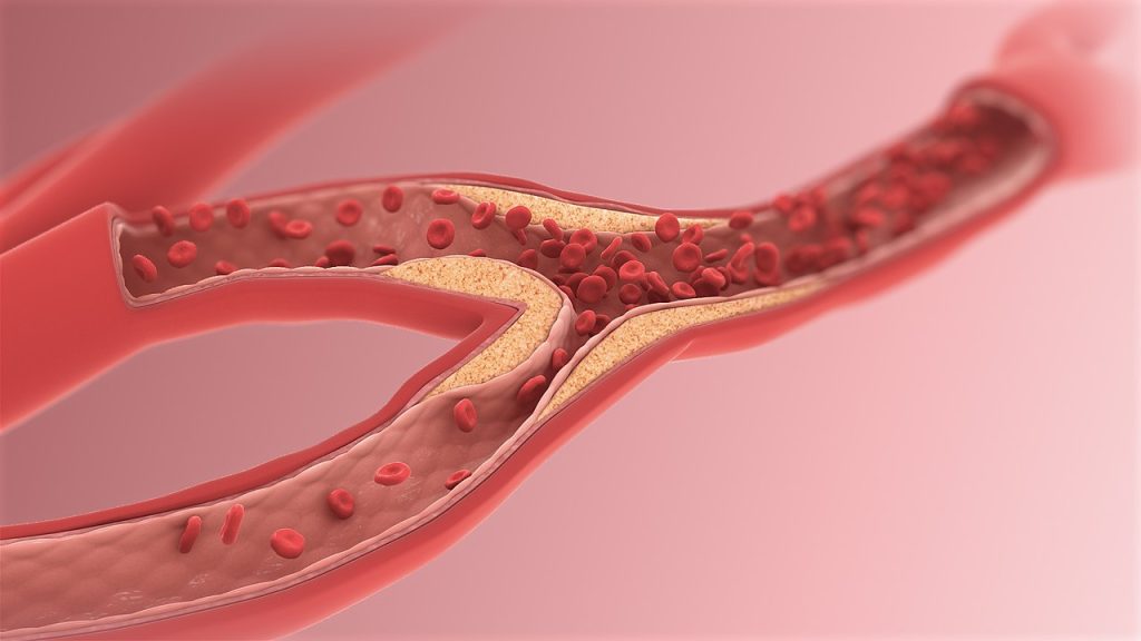
Hypertension and stiff arteries contribute to the development of heart disease, for which those with T1D are at higher risk. Those diagnosed with T1D as children have even higher risks for heart disease than people diagnosed in adulthood. Therefore, researchers are interested in ways to slow down the onset of vascular disease in children with T1D.

“We know that abnormalities in the large vessels around the heart, the aorta and its primary branches, begin to develop in early childhood in people with Type 1 diabetes,” said lead study author Michal Schäfer, PhD, a researcher and fourth-year medical student at the University of Colorado School of Medicine. “We found that bromocriptine has the potential to slow down the development of those abnormalities and decrease the risk for cardiovascular disease in this population.”

The multidisciplinary team conducted this study to examine the impact of bromocriptine on blood pressure and aortic stiffness compared with a placebo in adolescents with Type 1 diabetes. Bromocriptine is in a class of medications called dopamine receptor agonists. It increases levels of dopamine, a chemical in the brain, which leads to an increase in the body’s responsiveness to insulin, called insulin sensitivity. Bromocriptine has been FDA-approved since 2009 to treat adults with Type 2 diabetes due to its effect on insulin sensitivity.

The study included 34 participants (13 male, 21 female) aged 12 to 21 years who had been diagnosed with Type 1 diabetes for at least a year, and their HbA1c was 12% or less. An HbA1c level of 6.5% or higher indicates diabetes. They were randomly divided into two groups of 17, with one group receiving bromocriptine quick-release therapy and the other receiving a placebo once daily. The study was conducted in two phases. Participants took the first treatment or placebo for 4 weeks in phase 1, then had no treatment for a 4-week “wash-out” period, followed by phase 2 with 4 weeks on the opposite treatment. In this “crossover” design, each participant served as their own control for comparison.

Blood pressure and aortic stiffness were measured at the start of the study and at the end of each phase. Aortic stiffness was determined by assessing the large arteries with cardiovascular magnetic resonance imaging (MRI) and a measurement of the velocity of the blood pressure pulse called pulse wave velocity.

The study found:

  • Compared to placebo, blood pressure was significantly decreased with bromocriptine. On average, bromocriptine therapy resulted in a systolic blood pressure decrease of 5 mm Hg and a diastolic blood pressure decrease of 2 mm Hg at the end of 4 weeks of treatment.
  • Aortic stiffness was also reduced with bromocriptine therapy. The improvement in aortic stiffness was most pronounced in the ascending aorta with a lowered pulse wave velocity of about 0.4 meters/second, and an increase in distensibility, or elasticity, of 8%. In the thoraco-abdominal aorta, bromocriptine was associated with a lowered pulse wave velocity of about 0.2 meters/second, with a 5% increase in distensibility.

“A stiff aorta predisposes a patient to other health issues, such as organ dysfunction or atherosclerosis and higher stress or strain on cardiac muscle,” Schäfer said. “We were able to take it a notch further and show, using more sophisticated metrics, that these central large arteries are impaired, and impairment among adolescents and young adults with Type 1 diabetes may be decelerated with this drug.”

The study’s small size is a limitation. However, the researchers note that further research into bromocriptine’s impact on vascular health in a greater number of people with Type 1 diabetes is warranted; they are planning larger trials.

Source: American Heart Association

Exploring the Mysteries of Sudden Cardiac Death

Photo by Freestocksorg on Pexels

In August 2007, a soccer match took place that fans of the club Sevilla FC will not forget: the 22-year-old Antonio Puerta suffered a cardiac arrest and collapsed on the field, passing away in hospital a few days later. It was later discovered that the player was affected from a condition named arrhythmogenic cardiomyopathy.

Arrhythmogenic cardiomyopathy can lead to sudden death, and it particularly affects young athletes. Using genetically modified mice, which develop a similar disease to humans, researchers identified previously unknown mechanisms and potential therapeutic targets. Their results were published in the journal Circulation.

This inherited disease is estimated to occur at 1 in every 5000, with men being more commonly affected than women. “Arrhythmogenic cardiomyopathy leads to arrhythmia with a loss of cardiac muscle cells, deposits of connective tissue and fat within the cardiac muscle. This can cause sudden cardiac death, often during exercise,” says Volker Spindler, anatomist and head of the Cell Adhesion group at the University of Basel’s Department of Biomedicine.

Today, a range of gene mutations are known to trigger the condition. However, even with an early diagnosis there is no cure, only options for the management of symptoms are available. “Patients are advised to avoid any competitive or endurance sports and have to take medications such as beta blockers. Where appropriate, a catheter ablation may be performed or an implantable defibrillator may be used” says the cardiologist Gabriela Kuster, who heads the Myocardial Research group at the Department of Biomedicine. Sometimes the only option is a heart transplant.

Cardiac muscle cells become less ‘sticky’

The starting point for the project was the notion that many of the mutations affect structures known as the desmosomes. These are protein clusters on the surface of cardiac muscle cells that ensure a tight connection between the cells. “You can imagine these clusters to act like a piece of Velcro,” says the physician Dr Camilla Schinner, first author of the study. This led to the theory that the mutations reduce adhesion between the cells, thus weakening the cardiac muscle.

To test this hypothesis, Spindler’s team introduced a mutation similar to that found in patients into the genome of mice. The cardiac function of these animals was then examined by Kuster’s group. The result: the genetically modified animals showed a heart disease with arrhythmia that resembled arrhythmogenic cardiomyopathy in humans. In addition, microscopic and biochemical analysis indeed showed reduced adhesion between the cardiac muscle cells. The researchers also observed the scarring of the cardiac muscle typical for this disease.

Preventing cardiac tissue damage

Their next step was to investigate how diseased cardiac muscle differed from healthy conditions at the molecular level. Mice with the mutation showed an increased amount of a particular protein at the Velcro-like structures of the heart muscle cells. This leads, via a series of events, to connective tissue deposition and scarring of the heart. The addition of a substance which blocks this cascade prevented disease progression – which is why Spindler here sees a potential new treatment approach.

“Nevertheless, there is still a long way to go until an application in humans may be considered,” he points out. “But we now have better options to study the disease in more detail to improve our understanding of the underlying mechanisms.”

Source: University of Basel

Difficulty Sleeping Linked to Indicators of Poor Cardiometabolic Health

Sleeping man
Photo by Mert Kahveci on Unsplash

In the first study of its kind, University of South Australia researchers report that people who reported trouble sleeping were on average more likely to have indicators of poor cardiometabolic health – inflammatory markers, cholesterol and body weight – which can contribute to type 2 diabetes. The study was published in The Science of Diabetes Self-Management and Care.

Type 2 diabetes affects more than 422 million people around the globe.

As the Christmas season starts to ramp up, the UniSA researchers are reminding people to prioritise a good night’s sleep as new research shows that a troubled sleep may be associated with risk factors for type 2 diabetes.

UniSA researcher Dr Lisa Matricciani says different aspects of sleep are associated with risk factors for diabetes.

“Everyone knows that sleep is important. But when we think about sleep, we mainly focus on how many hours of sleep we get, when we should also be looking at our sleep experience as a whole,” Dr Matricciani says.

“How soundly we sleep, when we go to bed and get up, and how regular our sleep habits are, may be just as important as sleep duration.”

“In this study, we examined the association of different aspects of sleep, and risk factors for diabetes, and found a connection between those who had troubled sleep and those who were at risk of type 2 diabetes.”

The study assessed more than 1000 Australian adults* with a median age of 44.8 years. Researchers examined a range of sleep characteristics: self-report trouble sleeping, duration, timing, efficiency, and day-to-day sleep length variability.

“People who reported having trouble sleeping were also more likely to have a higher body mass index, as well as blood markers of cholesterol and inflammation,” Dr Matricciani says.

“When it comes down to the crunch, we know we must prioritise our sleep to help stay in good health. More research is needed, but as this study shows, it’s important to think about sleep as a whole, not just as one aspect.”

Notes

  • *Most participants (87%) were mothers.
  • 48% of all participants reported that they never had troubled sleep.

Source: University of South Australia

Protecting Patients with Automatic External Defibrillators (AEDs)

Automated external defibrillators (AEDs) are an important lifesaving technology and may have a role to play in treating workplace cardiac arrest. Most sudden cardiac deaths occur outside of the hospital, and many patients visiting doctors’ practices are already at risk for cardiovascular events, many going there because they are already feeling unwell as a precursor to a cardiac arrest.

It is estimated that 5% or less of victims of sudden cardiac deaths are successfully resuscitated and discharged alive from the hospital. 

In a study on public access defibrillation (PAD), communities with volunteers trained in CPR and the use of AEDs had twice as many victims survive compared to communities with volunteers trained only in CPR.

There are potential risks that come with the improper use of these devices. It is therefore essential to do thorough research before purchasing a specific product.

Why install an AED in my practice?

The primary purpose of an AED is to assist in the detection of heart arrhythmias. When an arrhythmia is identified with the use of an AED, a shock can be delivered to the heart to help normalise the function and rhythm of the patient’s heart.

In addition to being a tool used in the treatment of heart arrhythmia, another critical purpose that an AED device serves is to help restore a heartbeat in cases where a patient had suffered a sudden cardiac arrest (SCA), where patients may be saved if a shock is delivered inside a three minute window.

A 2021 study of out-of-hospital cardiac arrests in Cape Town found a rate of 23.2 per 100 000 population – likely an underestimate. Less than one in 10 cases had resuscitation attempts, and the average time for arrival of emergency services was 26 minutes. Thus, placing automated external defibrillators (AEDs) in the workplace can mean the difference between life and death.

AED use within the first three minutes after a patient experiences a cardiac arrest has an efficacy rate of about 80%. Performing CPR together with an AED is proven to be a significant improvement of efficacy rate compared to the CPR method alone. Therefore, it is necessary to install AEDs in the workplace, which will likely be granted the highest level of protection and make employees rest assured.

When does an AED shock?

The most common abnormal rhythm that causes cardiac arrest is ventricular fibrillation (VF). Electric shock can prevent ventricular fibrillation and restore the heart to its natural rhythm. An AED is applied to the casualty using two chest pads. The machine delivers electrical shocks to the heart muscle through the chest pad, which double as sensors to detect electrical activity. If the machine senses electrical activity with a heartbeat, it will not give an electric shock. Therefore, AEDs are very safe because they do not produce shock unless required. Products such as the AED7000 also feature locks preventing unintentional defibrillation.

Is it safe to use an AED?

Many associate AEDs with their specialised in-hospital equivalents, which require specialist training for safe use. Use of an AED device is usually considered exceptionally safe: the mechanism underlying the procedure is designed to ensure the patient will not be harmed when the device is use appropriately. Many newer models, such as the AED7000 series or the i-PAD NF1200, come with a smart system, which guides the rescuers through the procedure, ensuring the safety of both the patient and the person conducting the defibrillation.

Is it legal to use an AED on a patient?

There have been some concerns about the use of an AED device on patients of cardiac arrest. The concerns are primarily related to the fear of being sued by the patient when these devices are used. While South Africa lacks a Good Samaritan law to protect bystanders when assisting a patient, there is also no legal obligation for a member of the public to assist. However, doctors are legally required to render assistance in an emergency if they are able to do so.

Generally speaking, an AED is considered a safe device. Nevertheless, there are cases where the use of these devices could be considered inappropriate, for example, when the patient’s heart stops due to problems other than ventricular fibrillation, and thus cannot be saved by defibrillation. In these cases, there may be certain legalities involved when someone does decide to use an AED on such a patient.

The good news is that most AEDs will tell you whether or when it is appropriate to give a shock when you apply the pads on the patient’s chest. However, it’s always best to provide non-medically trained staff with AED knowledge and CPR training.

A Cup of Tea a Day Keeps the Doctor Away

Photo by Joanna Kosinska on Unsplash

Tea has long been known to have many health benefits, but now a study of 881 elderly women found they were far less likely to have extensive build-up of abdominal aortic calcification (AAC) if they consumed a high level of flavonoids in their diet – found in black and green tea, apples, nuts, citrus fruit, berries.

AAC is the calcification of the abdominal aorta and is a predictor of cardiovascular risk such as heart attack and stroke. It has also been found to be a reliable predictor for late-life dementia.

Edith Cowan University researcher and study lead Ben Parmenter said while there were many dietary sources of flavonoids, some had particularly high amounts.

“In most populations, a small group of foods and beverages – uniquely high in flavonoids – contribute the bulk of total dietary flavonoid intake,” he said.

“The main contributors are usually black or green tea, blueberries, strawberries, oranges, red wine, apples, raisins/grapes and dark chocolate.”

The flavonoid family

There are many different types of flavonoids, such as flavan-3-ols and flavonols, which the study indicated appear to also have a relationship with AAC.

Study participants who had a higher intake of total flavonoids, flavan-3-ols and flavonols were 36–39% less likely to have extensive AAC.

Black tea was the study cohort’s main source of total flavonoids and was also associated with significantly lower odds of extensive AAC.

Compared with respondents who didn’t drink tea, participants who had two-to-six cups per day had 16–42% less chance of having extensive AAC.

However, some other dietary sources of flavonoids such as fruit juice, red wine and chocolate, did not show a significant beneficial association with AAC.

Not just tea

Though black tea was the main source of flavonoids in the study – likely due to the age of the participants – Mr Parmenter said people could still benefit from flavonoids without putting the kettle on.

“Out of the women who don’t drink black tea, higher total non-tea flavonoid intake also appears to protect against extensive calcification of the arteries,” he said.

“This implies flavonoids from sources other than black tea may be protective against AAC when tea is not consumed.”

Mr Parmenter said this was important as it allows non-tea drinkers to still benefit from flavonoids in their diet.

“In other populations or groups of people, such as young men or people from other countries, black tea might not be the main source of flavonoids,” he said.

“AAC is a major predictor of vascular disease events, and this study shows intake of flavonoids, that could protect against AAC, are easily achievable in most people’s diets.”

Source: Edith Cowan University

‘Good Cholesterol’ may not be Such a Good Cardiovascular Risk Indicator

Source: Wikimedia CC0

A study published in the Journal of the American College of Cardiology found that high-density lipoprotein (HDL) cholesterol, often called ‘good cholesterol’, may not be as effective as scientists once believed in uniformly predicting cardiovascular disease risk among adults of different racial and ethnic backgrounds.

The research found that while low levels of HDL cholesterol predicted an increased risk of heart attacks or related deaths for White adults – a long-accepted association – the same was not true for Black adults. Additionally, higher HDL cholesterol levels were not associated with reduced cardiovascular disease risk for either group.

“The goal was to understand this long-established link that labels HDL as the beneficial cholesterol, and if that’s true for all ethnicities,” said senior author Nathalie Pamir, PhD, an associate professor at Oregon Health & Science University, Portland. “It’s been well accepted that low HDL cholesterol levels are detrimental, regardless of race. Our research tested those assumptions.”

Pamir and colleagues reviewed data from 23 901 participants from the Reasons for Geographic and Racial Differences in Stroke Study (REGARDS). Previous studies that shaped perceptions about ‘good’ cholesterol levels and heart health were conducted in the 1970s through research with a majority of white adult study participants. For the current study, researchers were able to look at how cholesterol levels from Black and White middle-aged adults without heart disease who lived throughout the country overlapped with future cardiovascular events.

Study participants enrolled in REGARDS between 2003–2007 and researchers analysed information collected throughout a 10- to 11-year period. Black and white study participants shared similar characteristics, such as age, cholesterol levels, and underlying risk factors for heart disease, including having diabetes, high blood pressure, or smoking. During this time, 664 Black adults and 951 White adults experienced a heart attack or heart attack-related death. Adults with increased levels of LDL cholesterol and triglycerides had modestly increased risks for cardiovascular disease, which aligned with findings from previous research.

However, the study was the first to find that lower HDL cholesterol levels only predicted increased cardiovascular disease risk for white adults. It also expands on findings from other studies showing that high HDL cholesterol levels are not always associated with reduced cardiovascular events. The REGARDS analysis was the largest US study to show that this was true for both Black and White adults, suggesting that higher than optimal amounts of ‘good’ cholesterol may not provide cardiovascular benefits for either group.

“What I hope this type of research establishes is the need to revisit the risk-predicting algorithm for cardiovascular disease,” Pamir said. “It could mean that in the future we don’t get a pat on the back by our doctors for having higher HDL cholesterol levels.”

Pamir explained that as researchers study HDL cholesterol’s role in supporting heart health, they are exploring different theories. One is quality over quantity. That is, instead of having more HDL, the quality of HDL’s function – in picking up and transporting excess cholesterol from the body – may be more important for supporting cardiovascular health.

The authors conclude that in addition to supporting ongoing and future research with diverse populations to explore these connections, the findings suggest that cardiovascular disease risk calculators using HDL cholesterol could lead to inaccurate predictions for Black adults.

“When it comes to risk factors for heart disease, they cannot be limited to one race or ethnicity,” said Pamir. “They need to apply to everyone.”

Source: NIH/National Heart, Lung and Blood Institute

Flu Vaccine Reduces Early Mortality in HF Patients

In a study published in The Lancet Global Health, an international team of researchers has found that influenza vaccines greatly reduce both pneumonia and cardiovascular complications in people with heart failure.

“If you have heart failure, you should get your flu shot because it can save your life – that is what we found in this study,” said the study’s principal investigator Mark Loeb. “It is underappreciated that influenza vaccine can save people from cardiovascular death.”

The study showed that over the entire year the influenza vaccine reduced pneumonia by 40% and hospitalisation by 15% in patients with HF. During influenza season in autumn and winter, the influenza vaccine reduced deaths by 20% in these patients.

Data gathered during flu season also showed the vaccine helped protect against cardiovascular complications, such as heart attacks and strokes.

Trial investigators tracked more than 5000 patients with HF in 10 countries across Africa, Asia and the Middle East, where few people have regular influenza vaccination. They received either an influenza vaccine or a placebo annually between June 2015 and November 2021.

While the flu has long been associated with an increased risk of life-threatening cardiovascular events, Loeb said that people with heart failure are already vulnerable to poor health outcomes. Patients with the condition have a 50% chance of dying within five years, while 20% are hospitalised for cardiovascular complications every year.

“Importantly, we looked at low and middle-income countries where 80 per cent of cardiovascular disease occurs and where flu vaccination rates are low.”

Salim Yusuf, executive director of PHRI and an author of the study said: “The flu shot should be part of the standard practise in people with heart failure given how simple, inexpensive and safe it is. Avoiding one sixth of deaths from heart disease and preventing hospitalizations makes it very cost effective and that can have an important public health and clinical impact.”

The study from McMaster University and partners marks the first clinical trial of the flu vaccine’s effectiveness in patients with HF.

Source: McMaster University

Bariatric Surgery Slashes Risk of Cardiovascular Events

Obesity
Image source: Pixabay CC0

A study of obese adults with nonalcoholic fatty liver disease (NAFLD) and morbid obesity has shown that those who underwent bariatric surgery suffered far fewer extreme cardiovascular events subsequently.

Reporting their results in JAMA Network Open, the researchers, reported that these obese patients (BMI > 40) undergoing bariatric surgery had a 49% lower risk of developing adverse cardiovascular events.

“The findings provide evidence in support of bariatric surgery as an effective therapeutic tool to lower elevated risk of cardiovascular disease for select individuals with obesity and NAFLD,” said Vinod K. Rustgi, profesor at Rutgers Robert Wood Johnson Medical School. “These finding are tremendously impactful for many reasons.”

NAFLD, and a more advanced form known as NASH, are rapidly increasing causes of liver disease which occur because of excessive fat storage in the liver. As such it is common in obesity and type 2 diabetes.

In the study, researchers analysed outcomes data, using a medical insurance database, from 2007 to 2017. Of 230 million covered individuals, 86 964 adults between the ages of 18 and 64 who had obesity and NAFLD were identified. Of those, 68% were female, 35% underwent bariatric surgery and 65% received nonsurgical care.

Bariatric surgery patients experienced a 49% decrease in the risk of developing major cardiovascular events such as heart attacks, heart failure or ischemic strokes. They were also far less likely to experience angina, atherosclerotic events or arterial blood clots.

The association between bariatric surgery and risk reduction of developing cardiovascular disease has not been studied to this level of detail before, the researchers said.

There is growing evidence that bariatric surgery, because of the weight reduction it brings about in patients, offers definitive health benefits. A study conducted by Rustgi and colleagues, published in the journal Gastroenterology in March 2021, showed that bariatric surgery can also significantly reduce the risk of cancer, especially obesity-related, in obese individuals with NAFLD. Importantly, these cancers included colorectal, pancreatic, endometrial, thyroid cancer, multiple myeloma and hepatocellular carcinoma.

“Although bariatric surgery is a more aggressive approach than lifestyle modifications, it may be associated with other benefits, such as improved quality of life and decreased long-term health care burden,” Rustgi said.

Source: Rutgers University

Smartwatch Equals Treadmill Test in Detecting HF

A smartwatch ECG can accurately detect heart failure (HF) in nonclinical environments, according to a study published in Nature Medicine. Researchers analysed Apple Watch ECG recordings with AI to identify patients with ventricular dysfunction. Study participants were able to remotely record their smartwatch ECGs at any time, with the data automatically and securely uploaded to their electronic health records via a smartphone app.

“Currently, we diagnose ventricular dysfunction – a weak heart pump – through an echocardiogram, CT scan or an MRI, but these are expensive, time consuming and at times inaccessible. The ability to diagnose a weak heart pump remotely, from an ECG that a person records using a consumer device, such as a smartwatch, allows a timely identification of this potentially life-threatening disease at massive scale,” says senior study author Paul Friedman, MD, chair of the Department of Cardiovascular Medicine at Mayo Clinic.

Ventricular dysfunction might not cause symptoms, but affects about 2% of the population and 9% of people over 60. Symptoms may develop with a low ejection fraction, including shortness of breath, a rapid heart rate and swelling in the legs. Early diagnosis is important because once identified, there are numerous treatments to improve quality of life and decrease the risks of heart failure and death.

Mayo researchers interpreted Apple Watch single-lead ECGs by modifying an earlier algorithm developed for 12-lead ECGs that is proven to detect a low ejection fraction.

While the data are early, the modified AI algorithm using single-lead ECG data had an area under the curve of 0.88 to detect low ejection fraction. By comparison, this measure of accuracy is as good as or slightly better than a medical treadmill diagnostic test.

“These data are encouraging because they show that digital tools allow convenient, inexpensive, scalable screening for important conditions. Through technology, we can remotely gather useful information about a patient’s heart in an accessible way that can meet the needs of people where they are,” says first author Zachi Attia, PhD, the lead AI scientist in the Department of Cardiovascular Medicine at Mayo Clinic.

“Building the capability to ingest data from wearable consumer electronics and provide analytic capabilities to prevent disease or improve health remotely in the manner demonstrated by this study can revolutionize health care. Solutions like this not only enable prediction and prevention of problems, but also will eventually help diminish health disparities and the burden on health systems and clinicians,” says co-author Bradley Leibovich, MD, the medical director for the Mayo Clinic Center for Digital Health.

Approximately 420 of the 2454 participants had an echocardiogram within 30 days of logging an Apple Watch ECG in the app. Of those, 16 patients had low ejection fraction confirmed by the echocardiogram, which provided a comparison for accuracy.

Source: Mayo Clinic

Cryoablation may be a Better First-stage Atrial Fibrillation Treatment

Photo by Piron Guillaume on Unsplash

A recent study comparing atrial fibrillation (AF) treatments, published in The New England Journal of Medicine, shows that early intervention with cryoballoon catheter ablation (cryoablation) is more effective at reducing the risk of serious long-term health impacts, when compared to the current first step in treatment, antiarrhythmic drugs.

“By treating patients with cryoablation right from the start, we see fewer people advancing to persistent, more life-threatening forms of atrial fibrillation,” says Dr Jason Andrade, an associate professor of medicine at University of British Columbia. “In the short term, this can mean less recurrences of arrhythmia, improved quality of life and fewer visits to the hospital. In the long run, this can translate into a reduced risk of stroke and other serious heart problems.”

When used for AF, cryoablation is a minimally invasive procedure that involves guiding a small tube into the heart to kill problematic tissue by freezing. Historically, the procedure has been reserved as a secondary treatment when patients don’t respond to antiarrhythmic drugs.

“This study adds to the growing body of evidence that early intervention with cryoablation may be a more effective initial therapy in the appropriate patients,” says Dr Andrade.

Early intervention halts disease progression

AF affects approximately 3% of the population, and while the condition starts as an isolated electrical disorder, each recurring incident can cause electrical and structural changes in the heart that can lead to persistent AF, where episodes last more than seven days.

“Atrial fibrillation is like a snowball rolling down a hill. With each atrial fibrillation episode there are progressive changes in the heart, and the heart rhythm problem gets worse,” explains Dr Andrade.

The new findings, stemming from a multi-site clinical trial, show that cryoablation can stop this snowball effect.

Researchers enrolled 303 patients with AF in Canada. Half of the patients were randomised to receive antiarrhythmic drugs, while the other half were treated with cryoablation. All patients received an implantable monitoring device that recorded their cardiac activity throughout the study period.

At three years follow-up, patients in the cryoablation group were less likely to progress to persistent AF compared to patients treated with antiarrhythmic drugs. Over the follow-up period, the cryoablation patients also had lower hospitalisation rates and experienced fewer serious adverse health events that resulted in death, functional disability or prolonged hospitalisation.

Addressing the root cause

Because cryoablation targets and destroys the cells that initiate and perpetuate AF, the researchers say it can lead to longer-lasting benefits.

“With cryoablation, we’re treating the cause of the condition, instead of using medications to cover-up the symptoms,” says Dr Andrade. “If we start with cryoablation, we may be able to fix atrial fibrillation early in its course.”

The new study builds on previous work by Dr Andrade and colleagues demonstrating that cryoablation was more effective than antiarrhythmic drugs at reducing the short-term recurrence of atrial fibrillation.