Category: Cardiovascular Disease

Commonly Used Arm Positions can Greatly Overestimate BP Readings

Photo by Hush Naidoo on Unsplash

A study led by Johns Hopkins Medicine researchers concludes that commonly used ways of positioning the patient’s arm during blood pressure (BP) screenings can substantially overestimate test results and may lead to a misdiagnosis of hypertension.

In a report on the study, published in JAMA Internal Medicine, investigators examined the effects of three different arm positions: an arm supported on a desk, arm supported on a lap, and an unsupported arm hanging at the patient’s side. Researchers found that lap support overestimated systolic pressure by nearly 4mmHg, and an unsupported arm hanging at the side overestimated systolic pressure by nearly 7mmHg.

The findings confirm that arm position makes a “huge difference” when it comes to an accurate blood pressure measurement, says Tammy Brady, MD, PhD, senior author of the study. And they underscore the importance of adhering to clinical guidelines calling for firm support on a desk or other surface when measuring blood pressure, the investigators add.

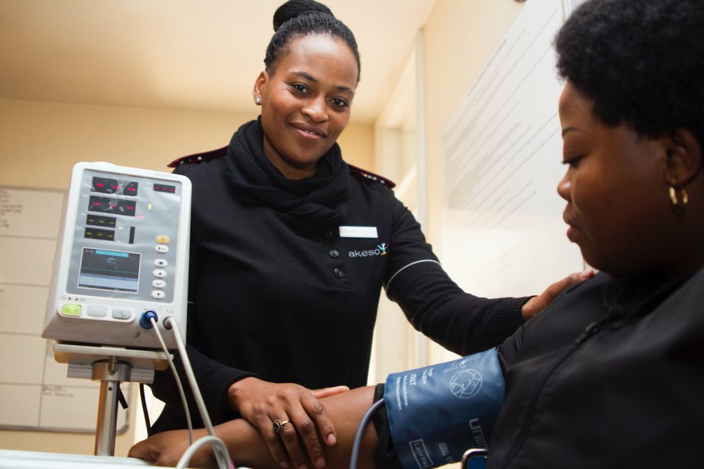
The latest clinical practice guidelines from the American Heart Association emphasise several key steps for an accurate measurement – including appropriate cuff size, back support, feet flat on the floor with legs uncrossed, and an appropriate arm position, in which the middle of an adjustable BP cuff is positioned at mid-heart level on an arm supported on a desk or table.

Despite these recommendations, the researchers say BP is too often measured with patients seated on an exam table without any, or inadequate, arm support. In some cases, a clinician holds the arm, or the patient holds an arm in their lap. In the new Johns Hopkins study, the researchers recruited 133 adult participants (78% Black, 52% female) between Aug. 9, 2022, and June 1, 2023. Study participants, who ranged from age 18 to 80, were sorted at random into one of six possible groups that differed by order of the three seated arm positions. Measurements were taken during a single visit between 9 a.m. and 6 p.m. Before BP measures were taken, all participants first emptied their bladders and then walked for two minutes to mimic a typical clinical scenario in which people walk into a clinic or office before screening takes place. They then underwent a five-minute, seated rest period with their backs and feet supported. Each person, wearing an upper arm BP cuff selected and sized based on their upper arm size, had three sets of triplicate measurements taken with a digital blood pressure device 30 seconds apart.

Upon completion of each set of three measurements, the cuff was removed, participants walked for two minutes and rested for five minutes. In the same visit, they then underwent a fourth set of triplicate measurements with their arm supported on a desk, a set used to account for well-known variations in BP readings. All of the measurements were conducted in a quiet and private space, and participants were asked not to talk to researchers or use their phones during the screening.

Researchers found that BP measurements obtained with arm positions frequently used in clinical practice – an arm on the lap or unsupported at the side – were markedly higher than those obtained when the arm was supported on a desk, the standard, recommended arm position. Supporting the arm on the lap overestimated systolic and diastolic BP by 3.9mmHg and 4.0mmHg, respectively. An unsupported arm at the side overestimated systolic by 6.5mmHg and diastolic by 4.4mmHg.

“If you are consistently measuring blood pressure with an unsupported arm, and that gives you an overestimated BP of 6.5mmHg, that’s a potential difference between a systolic BP of 123 and 130, or 133 and 140 – which is considered stage 2 hypertension,” says study author Sherry Liu, MHS, an epidemiology research coordinator at Johns Hopkins Bloomberg School of Public Health.

Investigators caution that their study results may only apply during screenings with automated BP devices, and may not apply to readings done with other BP devices.

However, Brady says, the findings suggest that clinicians need to pay better attention to best practice guidelines, and that patients “must advocate for themselves in the clinical setting and when measuring their BP at home.”

Source: Johns Hopkins Medicine

A New Heart Failure Treatment Targets Abnormal Hormone Activity

Right side heart failure. Credit: Scientific Animations CC4.0

Scientists have discovered a potential new treatment for heart failure with preserved ejection fraction (HFpEF), a type of heart disease that is notoriously difficult to treat. The diseased heart cells were found to have high levels of glucagon activity, a pancreatic hormone that raises blood glucose levels. The scientists then demonstrated that a drug that blocks the hormone’s activity can significantly improve heart function.

In heart failure, which is considered a global pandemic, the heart can no longer pump blood effectively. Globally, an estimated 64 million people live with this condition with HFpEF accounting for around half of the cases.

In HFpEF, the heart can pump normally but its muscles are too stiff to relax to re-fill the chambers with blood properly. It is often seen in older adults and people with multiple risk factors including high blood pressure (hypertension), obesity and diabetes. They typically have symptoms such as shortness of breath, fatigue and reduced ability to exercise. This is unlike heart failure with reduced ejection fraction (HFrEF), where heart muscle is weakened and pumping volume reduced.

There have been studies on how the heart is stressed by hypertension and metabolic diseases associated with obesity, such as diabetes, but these have been done in isolation of each other. This latest study, which was published in Circulation Researchaddresses this gap by taking into account both stressors, revealing for the first time, the molecular pathway that contributes to HFpEF progression.

In pre-clinical studies, the team of scientists, which included collaborators from the University of Cincinnati College of Medicine, University of California Los Angeles, University of Toronto and University of North Carolina School of Medicine, investigated how stress from hypertension affected lean hearts versus diabetic/obese ones. In their findings, the lean models developed heart failure with reduced ejection fraction (HFrEF), typically observed in hypertensive patients. The obese models however, developed heart failure with preserved ejection fraction (HFpEF), proving that a combination of stressors give rise to the disease and providing a good model for further studies.

Using advanced single-cell RNA-sequencing technologies, the scientists were then able to study the expression of every detected gene in every single heart cell, allowing them to uncover specific genetic variations in cells associated with HFpEF. The scientists found that in the obese models, the most active genes were the ones driving the activity of glucagon.

Professor Wang Yibin, Director of the Cardiovascular & Metabolic Disorders Programme at Duke-NUS and senior author of the study, said:

“Under stress conditions such as high blood pressure and metabolic disorders like obesity and diabetes, we found that glucagon signalling becomes excessively active in heart cells. This heightened activity contributes to the development of heart failure with preserved ejection fraction (HFpEF) by increasing heart stiffness and impairing its ability to relax and fill with blood.”

The team then tested a drug that blocks the glucagon receptor in a pre-clinical model of HFpEF and found significant improvements in heart function, including reduced heart stiffness, enhanced relaxation, improved blood filling capacity and overall better heart performance.

Assistant Professor Chen Gao from the Department of Pharmacology, Physiology and Neurobiology at the University of Cincinnati College of Medicine; and the study’s first author, said:

“Our study shows strong evidence that a glucagon receptor blocker could work well to treat HFpEF. Repurposing this drug, which is already being tested in clinical trials for diabetes, could bypass the lengthy drug development process and provide quicker and more effective relief to millions of heart patients.”

Professor Patrick Tan, Senior Vice-Dean for Research at Duke-NUS, commented:

“With our ageing population, there will likely be more patients with multiple conditions, including heart failure, diabetes and hypertension, presenting a significant challenge to health systems. Uncovering the synergistic impact of such illnesses and their underlying mechanisms is key to better understanding the complex process of heart failure and developing an effective treatment for the disease.”  

The researchers hope to work with clinical partners to conduct clinical trials to test the glucagon receptor blocker in humans with HFpEF. If these succeed, it could become one of the first effective treatments for this challenging condition, significantly improving the quality of life for millions worldwide.

Source: Duke University

Heart Defects in Babies Born after Fertility Treatment are More Common

Intracytoplasmic Sperm Injection (ICSI) procedure. Credit: Scientific Animations CC4.0

The risk of being born with a major heart defect is 36% higher in babies who were conceived after assisted reproductive technology, such as in vitro fertilisation (IVF), according to results of a very large study published in the European Heart Journal.

Researchers say the finding is important since congenital heart defects are the most common form of birth defects, and some of them are associated with life threatening complications.

The study also shows that the increase in risk is particularly associated with multiple births which are more common in assisted reproduction.

 The study was led by Professor Ulla-Britt Wennerholm from the University of Gothenburg in Sweden. She said: “Previous research shows that there are increased risks for babies conceived with the help of assisted reproductive technology. These include preterm birth and low birth weight. We wanted to investigate whether the risk of heart defects was higher for babies born following assisted reproduction.”

The research included all liveborn children born in Denmark between 1994 and 2014, all children born in Finland between 1990 and 2014, those born in Norway between 1984 and 2015 and those born in Sweden from 1987 to 2015; more than 7.7 million in total.

Researchers compared data on babies born following assisted reproduction, including IVF, intracytoplasmic sperm injection (ICSI) and embryo freezing, with data on babies conceived naturally.

They assessed how many liveborn children in each group were diagnosed with a major heart defect or with a serious heart defect either in the womb or in the first year of life. They took into account other factors that can increase the risk of congenital heart defects, such as child’s year of birth, country of birth, mother’s age at delivery, if the mother smoked during pregnancy, or if the mother had diabetes or heart defects.

This showed that heart defects were around 36% more common in babies born after assisted reproduction, compared to babies conceived without such treatment (absolute risk 1.84% vs 1.15%). This risk was similar regardless of the type of assisted reproduction used (IVF or ICSI, fresh or frozen embryos). However, the risk was greater for multiple births following assisted reproduction compared to singleton births following assisted reproduction (2.47% vs 1.62%).

Professor Wennerholm said: “We already know that babies born after assisted reproductive technology have a higher risk of birth defects in general however, we have found a higher risk also in congenital heart defects, the most common major birth defect.

“The fact that the risk of heart defects is similar regardless of the type of assisted reproduction used may indicate that there is some common factor underlying infertility in parents and congenital heart disease in their babies.

“Congenital heart defects can be extremely serious requiring specialist surgery when babies are very young, so knowing which babies are at the greatest risk can help us diagnose heart defects as early as possible and ensure the right care and treatment are given. More and more people are conceiving with the help of assisted reproductive technology, so we might expect to see increases in cases of congenital heart defects worldwide.”

In an accompanying editorial, Dr Nathalie Auger from University of Montreal Hospital Research Centre in Canada and colleagues said: “Assisted reproductive technology is a popular intervention in reproductive medicine, with these procedures accounting for 2% to 8% of births depending on the country. While most neonates born after assisted reproductive technology are healthy, these procedures are not without risks.

“In one of the largest studies to date, the researchers found that assisted reproductive technology was associated with the risk of major heart defects diagnosed prenatally or up to one year of age.

“Patients who use assisted reproductive technology tend to differ from the general population. These patients may have underlying morbidities that affect both fertility and the risk of heart defects.”

Source: European Society of Cardiology

The Prevalence of Atrial Fibrillation has been Greatly Underestimated

Human heart. Credit: Scientific Animations CC4.0

Atrial fibrillation, a rapid, irregular heartbeat that can lead to stroke or sudden death, is three times more common than previously thought, affecting nearly 5% of the population, according to new estimates from UC San Francisco.

A-Fib, as the condition is commonly known, has been on the rise for at least the past decade, driven by the aging of the population, along with increasing rates of hypertension, diabetes and obesity. Earlier projections had estimated that 3.3 million U.S. adults had atrial fibrillation, but these have not been updated in more than two decades.

“Atrial fibrillation doubles the risk of mortality, is one of the most common causes of stroke, increases risks of heart failure, myocardial infarction, chronic kidney disease and dementia, and results in lower quality of life,” said first author Jean Jacques Noubiap, MD, PhD, a postdoctoral scholar at UCSF with a specialty in global cardiovascular health.

“Fortunately, atrial fibrillation is preventable, and early detection and appropriate treatment can substantially reduce its adverse outcomes,” he said.

Rising numbers reflect need for better prevention and treatment

UCSF investigators reviewed the medical records of nearly 30 million adult patients who received some form of acute or procedural care in California from 2005 to 2019. About 2 million of these people had been diagnosed with A-Fib, and the numbers grew over time, rising from 4.49% of the patients treated between 2005 and 2009 to 6.82% of the patients treated between 2015 and 2019.

The data were standardised to reflect the entire country, and researchers estimated the current national prevalence to be at least 10.55 million. They also found that during the study timeframe, A-Fib patients skewed younger, were less likely to be female and more likely to have hypertension and diabetes.

A-Fib has a broad spectrum of complications from shortness of breath and light-headedness to blood clots, stroke and even heart failure. Studies have shown that people with A-Fib are up to five times more likely to have a stroke. The authors said that by outlining the scope of the problem, these new estimates can help guide health care planning, resource allocation and public health interventions.

“Physicians recognise that atrial fibrillation is often encountered in essentially every field of practice,” said senior and corresponding author Gregory M. Marcus, MD, MAS, a cardiologist and electrophysiologist at UCSF Health. “These data provide objective evidence to demonstrate that prior projections severely underestimated how common it truly is.”

Digital technologies may reveal it is even more common than the current analysis indicates.

“With the growing use of consumer wearables designed to detect atrial fibrillation combined with safer and more effective means to treat it, this current prevalence of atrial fibrillation in health care settings may soon be dwarfed by future health care utilisation that will occur due to the disease,” Marcus said.

Source: University of California San Francisco

Sleep Apnoea May Increase Risk of Abdominal Aortic Aneurysms

Photo by Mert Kahveci on Unsplash

Obstructive sleep apnoea may be a risk factor for developing abdominal aortic aneurysms, according to researchers from the University of Missouri School of Medicine and NextGen Precision Health.

Abdominal aortic aneurysms occur when the aorta swells and potentially ruptures, causing life-threatening internal bleeding. Obstructive sleep apnoea is characterised by episodes of a complete or partial airway collapse with an associated drop in oxygen saturation or arousal from sleep. It can increase the risk of developing cardiovascular problems. Citing studies that indicate a higher prevalence of abdominal aortic aneurysms in patients with obstructive sleep apnoea, MU researchers examined the link between the two using mouse models.

The research team found that intermittent hypoxia caused by obstructive sleep apnoea increased the susceptibility of mice to develop abdominal aortic aneurysms.

“Chronic intermittent hypoxia by itself is not enough to cause abdominal aortic aneurysms, but for a patient with obstructive sleep apnoea who also has additional metabolic problems like obesity, our findings suggest it may help degrade aortic structures and promote aneurysm development,” said Luis Martinez-Lemus, study author and a professor of medical pharmacology and physiology.

Intermittent hypoxia happens during obstructive sleep apnoea when throat muscles relax and block the flow of air into the lungs. According to the research, the loss of oxygen triggers certain enzymes called MMPs. The increased enzyme activity can degrade the extracellular matrix, which acts like a cell scaffolding network, weakening the aorta.

“Patients with abdominal aortic aneurysms usually don’t notice any symptoms, except for some back and belly pain, until the aneurysm bursts. Once that happens, it’s crucial to get the patient to surgery quickly so doctors can repair the aorta,” said Neekun Sharma, the lead author of the study. “Learning how these aneurysms develop can help us find ways to monitor or slow down their progression, especially for patients who have obstructive sleep apnoea.”

Source: University of Missouri-Columbia

New Approach to Defibrillation may Improve Cardiac Arrest Outcomes

Photo by Mikhail Nilov

Joshua Lupton, MD, has no memory of his own cardiac arrest in 2016. He only knows that first responders resuscitated his heart with a shock from a defibrillator, ultimately leading to his complete recovery and putting him among fewer than one in 10 people nationwide who survive cardiac arrest outside of a hospital.

He attributes his survival to the rapid defibrillation he received from first responders – but not everybody is so fortunate.

Now, as lead author on an observational study published in JAMA Network Open, he and co-authors from Oregon Health & Science University say the study suggests the position in which responders initially place the two defibrillator pads on the body may make a significant difference in returning spontaneous blood circulation after shock from a defibrillator.

“The less time that you’re in cardiac arrest, the better,” said Lupton, assistant professor of emergency medicine in the OHSU School of Medicine. “The longer your brain has low blood flow, the lower your chances of having a good outcome.”

Researchers used data from the Portland Cardiac Arrest Epidemiologic Registry, which comprehensively recorded the placement position of defibrillation pads from July 1, 2019, through June 30, 2023. For purposes of the study, researchers reviewed 255 cases treated by Tualatin Valley Fire & Rescue, where the two pads were placed either at the front and side or front and back.

They found placing the pads in front and back had 2.64-fold greater odds of returning spontaneous blood circulation, compared with placing the pads on the person’s front and side.

The current common knowledge among health care professionals is that pad placement – whether front and side, or front and back – is equally beneficial in cardiac arrest. The researchers cautioned that their new study is only observational and not a definitive clinical trial. Yet, given the crucial importance of reviving the heartbeat as quickly as possible, the results do suggest a benefit from placing the pads on the front and back rather than the front and side.

“The key is, you want energy that goes from one pad to the other through the heart,” said senior author Mohamud Daya, MD, professor of emergency medicine in the OHSU School of Medicine.

Placing the pads in the front and back may effectively “sandwich” the heart, raising the possibility that the electrical current will be delivered more comprehensively to the organ. 

However, that’s not readily possible in many cases. For example, the patient may be overweight or positioned in such a way that they can’t be easily moved.

“It can be hard to roll people,” said Daya, who also serves as medical director for Tualatin Valley Fire & Rescue. “Emergency medical responders can often do it, but the lay public may not be able to move a person. It’s also important to deliver the electrical current as quickly as possible.”

In that respect, pad placement is only one factor among many in successfully treating cardiac arrest.

Lupton survived his cardiac arrest and went on to complete medical school at the very hospital where he spent several days recovering in the intensive care unit – Johns Hopkins University in Baltimore. The episode led him to alter the focus of his research so that he could examine ways to optimise early care for cardiac arrest patients.

The results of the new study surprised him.

“I didn’t expect to see such a big difference,” he said. “The fact that we did may light a fire in the medical community to fund some additional research to learn more.”

Source: Oregon Health & Science University

SA Heart Congress Unites Cardiologists for Better Care

The SA Heart Annual Congress will take place from 8–10 November at the Sandton Convention Centre, Johannesburg. The three-day Congress, themed ‘Cardiology Connections,’ will promote collaboration and dialogue among local and international Cardiology professionals. The congress offers a unique platform for experts, practitioners, and researchers worldwide to share insights on the latest advancements and challenges in cardiovascular medicine.

The dynamic programme includes keynote speeches, panel discussions, workshops, and networking sessions. The agenda covers a comprehensive range of cardiology topics, designed to provide practical knowledge and inspire innovation in the field. Attendees will gain critical insights into the latest developments that have the potential to enhance patient care.

“We are excited to welcome a distinguished international and local faculty,” says Dr Ahmed Vachiat, SA Heart Congress Convenor. “At the core of SA Heart is the mission to advance cardiovascular care through education, research, and advocacy. By connecting healthcare professionals from across sectors, this Congress will drive forward our vision of improving cardiovascular care for all in South Africa. We are also grateful for the invaluable support of our local experts, whose contributions consistently uphold international standards of excellence.”

A significant focus this year is strengthening connections among various special interest groups, including the Society of Cardiovascular Interventions (SASCI), Cardiovascular Imaging Society of South Africa (CISSA), Cardiovascular Arrhythmia Society of South Africa (CASSA), Heart Failure Association of South Africa (HEFFSA), Intervention Society of Cardiovascular Allied Professionals (ISCAP), South African Society of Cardiovascular Research (SASCAR), and the Paediatric Society of Cardiology (PCSSA).

Joint sessions and interdisciplinary programmes will enable these groups to work together to enhance healthcare delivery for all patients in need of cardiac intervention and treatment. Workshops and scientific sessions will feature innovative learning approaches aimed at facilitating knowledge exchange and professional growth.

A cardiovascular team from the Mayo Clinic – Prof Vuyi Nkomo (Imaging Cardiologist), Prof Sorin Pislaru (Chair, Structural Heart Disease), and Dr Juan Crestanello (Chair, Cardiothoracic Surgery) – will conduct an echocardiography workshop and contribute to various specialist workshops on Friday morning, November 8th.

Dr Thomas Alexander, a respected interventional cardiologist based in India, will share insights on establishing STEMI networks in South Africa. Prof Stylianos Pyxaras from Germany and Dr Andrew Ludwiniec from the UK will discuss chronic total occlusions and complex coronary interventions. Prof Azfar Zaman and Prof Roy Gardner also from the UK and leaders in their field, as well as Prof Thierry Lefevre from France, will join esteemed local experts in addressing important cardiovascular topics.

A new addition to this year’s programme is the Imbizo on Rheumatology and Cardiac diseases. Over 40 Abstracts have been submitted and research sessions guided by SASCAR will be keeping delegates up to date with the latest in the field of Cardiology.

In addition, an excellent parallel paediatric programme will feature global leaders, Prof Krishna Kumar, from India and Prof McDaniel from the USA, with a pre-congress workshop and highly interactive sessions that will incorporate insights from local experts.

“This year, a Heartbeat Stage will feature insightful talks, engaging presentations, and a special networking address,” says Dr Vachiat. “We are honoured to have Dr Imtiaz Sooliman from Gift of the Givers, who will share his thoughts on ‘Connecting Hearts and Social Responsibility’.”

For more information, visit SA Heart.

Moderate Coffee and Caffeine Link to Lower Risk for Cardiometabolic Diseases

Photo by Mike Kenneally on Unsplash

Consuming moderate amounts of coffee and caffeine regularly may offer a protective effect against developing multiple cardiometabolic diseases, including type 2 diabetes, coronary heart disease and stroke, according to new research published in the Endocrine Society’s Journal of Clinical Endocrinology & Metabolism.

Researchers found that regular coffee or caffeine intake, especially at moderate levels, was associated with a lower risk of new-onset cardiometabolic multimorbidity (CM), which refers to the coexistence of at least two cardiometabolic diseases.

The prevalence of individuals with multiple cardiometabolic diseases, or CM, is becoming an increasing public health concern as populations age around the world, notes the study.

Coffee and caffeine consumption could play an important protective role in almost all phases of CM development, researchers found.

“Consuming three cups of coffee, or 200-300 mg caffeine, per day might help to reduce the risk of developing cardiometabolic multimorbidity in individuals without any cardiometabolic disease,” said lead author Chaofu Ke, MD, PhD, at Suzhou Medical College of Soochow University, in Suzhou, China.

The study found that compared with non-consumers or consumers of less than 100mg caffeine per day, consumers of moderate amount of coffee (3 drinks per day) or caffeine (200-300 mg per day) had a 48.1% or 40.7% reduced risk for new-onset CM.

Ke and his colleagues based their findings on data from the UK Biobank, a large and detailed longitudinal dietary study with over 500 000 participants aged 37-73 years. The study excluded individuals who had ambiguous information on caffeine intake. The resulting pool of participants included a total of 172 315 individuals who were free of any cardiometabolic diseases at baseline for the analyses of caffeine, and a corresponding 188 091 individuals for the analyses of coffee and tea consumption.

The participants’ cardiometabolic diseases outcomes were identified from self-reported medical conditions, primary care data, linked inpatient hospital data and death registry records linked to the UK Biobank.

Coffee and caffeine intake at all levels were inversely associated with the risk of new-onset CM in participants without cardiometabolic diseases. Those who reported moderate coffee or caffeine intake had the lowest risk, the study found. Moderate coffee or caffeine intake was inversely associated with almost all developmental stages of CM.

“The findings highlight that promoting moderate amounts of coffee or caffeine intake as a dietary habit to healthy people might have far-reaching benefits for the prevention of CM,” Ke said.

Addressing a research gap

Numerous epidemiological studies have revealed the protective effects of coffee, tea and caffeine consumption on morbidity of single cardiometabolic diseases. However, the potential effects of these beverages on the development of CM were largely unknown.

The authors reviewed the available research on this topic and found people with single cardiometabolic disease may have a two-fold higher all-cause mortality risk than those free of any cardiometabolic diseases. By contrast, the researchers found individuals with CM may have an almost 4 to 7 times higher risk of all-cause mortality. The researchers also noted that CM may present higher risks of loss of physical function and mental stress than those with single diseases.

Source: Endocrine Society

Unhealthy Behaviours Contribute to More Coronary Artery Disease Deaths in the Poor

Four behaviours explain a majority of the socioeconomic disparities observed in the disease

Source: Wikimedia CC0

Lower socioeconomic status is associated with higher rates of death from coronary artery disease compared to higher socioeconomic status, and more than half of the disparities can be explained by four unhealthy behaviours. Dr Yachen Zhu of the Alcohol Research Group, US, and Dr Charlotte Probst of the Centre for Addiction and Mental Health, Canada, report these findings in a new study published September 17th in the open-access journal PLOS Medicine.

Coronary artery disease, also known as coronary heart disease or ischaemic heart disease, occurs when the arteries supplying the heart cannot deliver enough oxygen-rich blood due to plaque buildup, and is a major cause of death in the US. The condition poses a greater risk to people with lower socioeconomic status, but previous studies have reported conflicting results on whether certain unhealthy behaviours, like smoking, are primarily responsible for the observed disparities in deaths from the disease.

In the new study, researchers used data from 524 035 people aged 25 years and older whose mortality statuses were recorded in the National Death Index and who answered the National Health Interview Survey on demographics and health behaviours. The team used education as the primary indicator for socioeconomic status, and investigated four behavioural risk factors: smoking, alcohol use, physical inactivity and BMI. The four factors together explained 74% of the differences in mortality risk from coronary artery disease in men belonging to different socioeconomic levels and 61% in women.

The researchers conclude that their results highlight the need for effective public health policies and interventions that address each of these behaviours – both separately and together – because unhealthy behaviours often cluster among individuals from low socioeconomic backgrounds. They urge public health campaigns to raise awareness about heart health with messaging and outreach efforts customised for male and female audiences. The authors add, “These efforts are crucial to reducing the socioeconomic disparities in deaths from coronary artery disease in the US.”

Provided by PLOS

A Cuffless Smartphone App that Can Measure Blood Pressure

Photo by Ivan Samkov on Pexels

Researchers at the University of Pittsburgh are pioneering a new approach to blood pressure monitoring, using the devices we carry with us every day. Ramakrishna Mukkamala, professor of bioengineering at Pitt’s Swanson School of Engineering, is passionate about developing accessible blood pressure (BP) detection tools. Instead of designing a new medical device to monitor BP, Mukkamala decided to take advantage of the sensors readily available in smartphones and figure out how to detect blood pressure with them. 

“The most significant thing you can do to reduce your risk of cardiovascular disease is to lower high blood pressure through lifestyle changes, but in underserved populations, many people don’t have access to blood pressure cuffs, regular doctor’s appointments, or even know it’s a problem,” Mukkamala said. “But they do have smartphones.”

Mukkamala’s team harnessed tools already built into most smartphones, like motion-sensing accelerometers, front cameras, and touch sensors to build an Android smartphone application that can measure an individual’s pulse pressure. The user performs a hand-raising motion while holding the smartphone to make a measurement. The results of the project, published in Scientific Reports, demonstrate a promising new technology that could uniquely help reduce the burden of systolic hypertension globally, particularly in underserved populations. 

Designing blood pressure technology for a touchscreen 

Turning a smartphone into a monitoring device is no easy task, as Vishaal Dhamotharan, graduate student in the Cardiovascular Health Tech Laboratory, found out through multiple iterations of app development. Because smartphones don’t have force sensing tools, a crucial element of the project was figuring out how to replicate the effects of a traditional blood pressure exam using only a cell phone, which the team solved by using a familiar force – gravity.

“Because of gravity, there’s a hydrostatic pressure change in your thumb when you raise your hands up above your heart, and using the phone’s accelerometer, you’re able to convert that into the relative change in pressure.” Dhamotharan said. 

By pairing this hand-raising motion with guided thumb maneuvers on the smartphone, the team was able to calculate each participant’s pulse pressure, the difference between systolic and diastolic numbers. For example, an individual with a BP measurement of 120/80 has a pulse pressure of 40. For Sanjeev Shroff, collaborator and bioengineering department chair, this publication is a promising advancement for blood pressure measurement devices. 

“Development of a cuffless blood pressure measurement device that does not require any external calibration is the holy grail – such a device currently does not exist,” Shroff said. “The research work reported in this publication is an important step in the right direction, and is also encouraging for additional work aimed at obtaining systolic, diastolic, and mean pressures.”

Although pulse pressure isn’t typically used in cardiovascular disease monitoring, the study revealed its significance as a metric for detecting hypertension, according to Céderick Landry, assistant professor at the University of Sherbrooke and former postdoctoral researcher in the lab. 

“Guidelines typically require doctors to measure both systolic and diastolic blood pressure, and pulse pressure is just the difference between the two.” Landry said. “We showed that if you only have access to pulse pressure, it’s still very correlated with hypertension, so part of our challenge now is changing the mentality on how to best measure things.”

Hypertension management within reach

This app could bring blood pressure monitoring software to any smartphone owner, enabling consistent self-monitoring and easy sharing of results with healthcare providers. This innovation is especially promising for managing hypertension, which can often be lowered through lifestyle changes such as reducing salt intake, quitting smoking and exercising regularly. 

“This app would be really useful in low-income settings where people may not even have existing access to blood pressure tools.” Dhamotharan said. “Being able to measure blood pressure more frequently would allow an individual to track any significant changes in blood pressure, monitor for hypertension, and be able to manage their conditions with that knowledge.” 

“The research is here – we just need some help making the technology better.” Landry said. “This is the first method of its kind, and even better, it’s something that we can start implementing right now.”

Source: University of Pittsburgh