Category: Cancer

At-home Melanoma Testing with Skin Patch Test

A microneedle patch captures cancer biomarkers in the top-most layer of skin to detect melanoma in animal tissue samples

The newly designed ExoPatch being removed from a sample of mouse skin successfully distinguished melanoma from healthy skin in mice. A gel coating the microneedles picks up cancer indicators from the top-most layer of the skin. Dissolving the gel releases exosomes into a solution, which is then used on a two-lined test strip, similar to an at-home COVID-19 test. Image credit: Jeremy Little, Michigan Engineering.

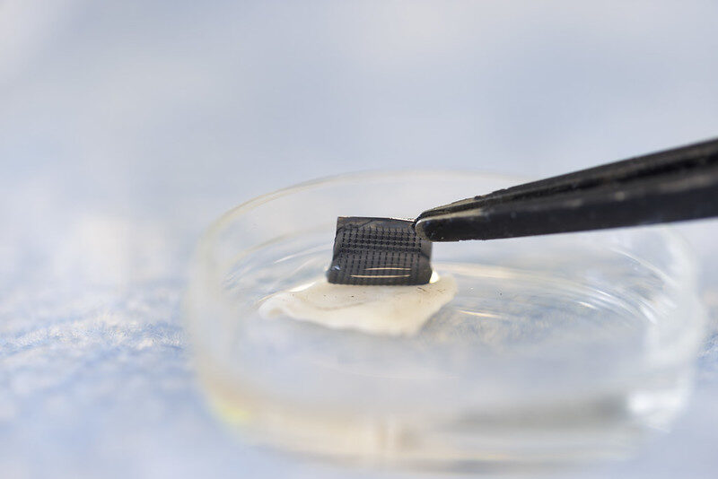
Melanoma testing could one day be done at home with a skin patch and test strip with two lines, similar to COVID-19 home tests, according to University of Michigan researchers. Developed with funding from the National Institutes of Health, the new silicone patch with star-shaped microneedles, called the ExoPatch, distinguished melanoma from healthy skin in mice.

The patch and test move toward rapid at-home melanoma testing, helping patients catch the most aggressive form of skin cancer early without a biopsy or blood draw.

“The star-shaped needles make puncture easier and less painful, but they are so small that they only go through the top-most layer of the skin, the epidermis, and do not draw blood,” said Sunitha Nagrath, the Dwight F. Benton Professor of Chemical Engineering at U-M and co-corresponding author of the study published in Biosensors and Bioelectronics.

The ExoPatch microneedles, at just 0.6mm long with a width of less than 100 nm (0.0001 mm) at the tip, are coated with a gel that picks up exosomes, tiny packages released by cells, from the interstitial fluid that fills the spaces between cells in the epidermis.

Once thought to be trash ejected from cells for cleanup, exosomes actually contain DNA and RNA fragments that cells use to communicate with each other. Cancer cell exosomes can help tumours spread by preparing tissues to accept tumour cells before arrival, and detecting them can catch cancer earlier than past methods.

The gel that coats the ExoPatch contains a protein called Annexin V that attracts and sticks exosomes to the microneedles’ surface. Once removed from the skin, placing the patch in an acid dissolves the gel, which releases the exosomes into a solution. After dipping a test strip into the solution, two lines form if the sample contains melanoma exosomes, and one line forms for a negative test – the same way an at-home COVID-19 test strip works.

“A fair-skinned person with moles must go to the doctor about every six months to send off a biopsy to see if they’re malignant or benign. With this test, they could instead test at home, get the results right away and follow up with a dermatologist for a positive result,” Nagrath said.

As a first step in the proof-of-concept study, the researchers tested the ExoPatch on a tissue sample of pig skin, which closely resembles human skin in thickness and composition. Using a microscope, they found the microneedles penetrated about 350 to 600nm into the skin. For scale, the epidermis on the human forearm is about 18 300nm thick.

To test whether the ExoPatch could capture melanoma exosomes from skin tissue, the research team tested tissue samples of mouse skin, half from healthy mice and half from mice injected with a fragment of a human melanoma tumour. After a 15-minute application, the ExoPatch was placed under a powerful microscope.

“When looking at microscopy images, I was happy to see how nicely the exosomes adhered to the microneedles and were within the 30 to 150 nanometre size range we expect,” said Scott Smith, U-M doctoral student of chemical engineering and co-lead author of the study.

After confirming the exosomes stuck to the ExoPatch, the researchers dissolved the gel and ran the sample through the test strips. The test successfully distinguished between melanoma and healthy tissues with a 3.5-fold darker line in melanoma samples.

The ExoPatch isolated 11.5 times more exosomal protein from melanoma tissue samples compared to healthy tissue, showing it can specifically target cancerous exosomes.

A pilot study in humans followed by a series of clinical trials will be the next steps to move the technology toward use. Beyond melanoma, the ExoPatch gel coating could be modified to detect exosomes released by other cancers with a solid tumour, including lung, breast, colon, prostate and brain cancer.

“This is the first patch designed to capture disease-specific exosomes from fluid under the skin. The potential applications are huge,” said Nagrath.

Source: University of Michigan

Combination of Diet and Medication Reprograms Paediatric Neuroblastoma

Credit: National Cancer Institute

Researchers at Children’s Hospital of Philadelphia (CHOP) found that combining a specialised diet with an approved medication interrupts the growth of high-risk neuroblastoma, a deadly paediatric cancer, by reprogramming tumour behaviour. The findings were published in the journal Nature.

Neuroblastoma originates from primitive cells meant to form nerve tissues but that remain “undifferentiated,” indicating cancer cells that haven’t specialized, often suggesting a more aggressive and unfavourable prognosis. These tumours rely on a steady supply of chemicals called polyamines that are essential for rapid cell growth and tumour progression. A medicine called difluoromethylornithine (DFMO) was approved by the Food and Drug Administration (FDA) to treat children with high-risk neuroblastoma, as DFMO blocks polyamine production. However, researchers sought to improve the effectiveness of the drug by using it at high doses and combining it with a diet that is depleted of the nutrients used by the body to make polyamines (arginine). This two-step approach was anticipated to lower polyamines substantially more than low dose DFMO alone.

“Our findings show that this treatment reduced polyamines in tumours to roughly 10% of their usual levels. This reduction greatly slowed tumour growth, and in many cases, completely eliminated the tumours,” said Michael D. Hogarty, MD, a lead author and an Attending Physician in the Division of Oncology at Children’s Hospital of Philadelphia. “Notably, the treatment altered the way the tumour cells make proteins, making it harder for them to grow and easier for them to mature, or differentiate.”

Hogarty and his team used a preclinical model to mimic MYCN-driven neuroblastoma, directly addressing the strong association between extra MYCN gene copies and aggressive neuroblastoma with poor prognosis. Animal models with tumours were divided into groups: one fed a normal diet and the other lacking amino acids for polyamine production. Each group either received DFMO in their drinking water or did not. The special diet or DFMO alone partially lowered polyamines and extended survival, but the combination had the most significant impact on tumours due to the profound polyamine depletion it caused.

The researchers plan to conduct additional preclinical studies, followed hopefully by clinical trials in children to determine the safety and efficacy of targeting this specific metabolic dependency of neuroblastoma cells. By complementing existing treatments, they hope to substantially improve patient outcomes, and because the therapy targets polyamines it may be effective in many other types of cancer that have frequent MYC gene activation. 

Source: Children’s Hospital of Philadelphia

Study Explains How Lymphoma Rewires Human Genome

Source: Pixabay CC0

Translocations are chromosomal “cut and paste” errors that drive many lymphomas, a type of blood cancer and the sixth most common form of cancer overall. This includes mantle cell lymphoma, a rare but aggressive subtype diagnosed in about one in every 100 000 people each year.

Translocations are known to spark cancer by altering the activity of the genes near the breakpoints where chromosomes snap and rejoin. For example, a translocation can accidentally cut a gene in half, silencing its activity, or create new hybrid proteins that help promote cancer.

A study published today in Nucleic Acids Research shows a new way translocations promote cancer. The translocation most typically found in mantle cell lymphoma drags a powerful regulatory element into a new area of the human genome, where its new position allows it to boost the activity of not just one but 50 genes at once.

The discovery of this genome rewiring mechanism shows the traditional focus on the handful of genes at chromosomal breakpoints is too narrow. The study also greatly expands the list of potential drug targets for mantle cell lymphoma, for which there is no known cure.

“We did not expect to see a single translocation boosting the expression of almost 7% of all genes on a single chromosome. The ripples of disruption are much bigger than expected, and also identify new cancer driver genes, each of which represents a new potential therapeutic target,” says Dr. Renée Beekman, corresponding author of the study and researcher at the Centre for Genomic Regulation (CRG) in Barcelona.

In mantle cell lymphoma, a piece of chromosome 14 swaps places with a piece of chromosome 11. A gene regulatory element called the IGH enhancer, which normally boosts the activity of antibody production in healthy B cells, lands right beside CCND1, a gene which helps cells divide. The enhancer treats CCND1 as if it were a gene encoding for antibodies, boosting its activity and fuelling the disease.

Previous research has shown that boosting CCND1 expression alone is insufficient to kickstart the formation of mantle cell lymphoma. To understand why, the scientists first created translocations in cells in a dish. They used CRISPR to replicate the exact chromosome break seen in patients.

“We built a system to generate translocations in healthy B cells. Because these are engineered cells, we can carry out experiments that are technically or ethically unfeasible with patient tissues, making it a really useful early disease model,” explains Dr. Roser Zaurin, co-author of the study.

The experiments revealed that over fifty genes along the entire chromosome 11 were much more active after the translocation took place. The translocation affected gene activity across 50 million base pairs, a significantly larger space than previously thought.

How DNA folds inside the engineered cells revealed why the translocation affects so many genes at once. “DNA loops inside cells. It’s what brings two segments of DNA that are far away from each other in two-dimensional space closer together in three-dimensional space. The translocation drags the strong IGH enhancer into a preexisting loop, placing it in a privileged position of control, enabling it to have a widespread impact on dozens of genes at the same time,” explains Dr. Anna Oncins, first author of the study.

Intriguingly, most of the genes affected by the enhancer were not silent to begin with. The IGH enhancer simply dials their activity up. This biological nuance may explain why the same translocation can have different consequences in different cell types or stages of development. Only genes which were already active are boosted.

The findings could lead to new strategies for the early-stage detection of mantle cell lymphomas. “Because the enhancer mainly supercharges genes that were already active in the very first B cell that acquires the swap, epigenetic profiling of at-risk cells could spot dangerous combinations before a mantle cell lymphoma appears,” explains Dr. Beekman.

The authors of the study next plan on studying exactly how the newly identified genes contribute to the initiation and progression of lymphoma. Understanding and eventually interrupting the effects of the chromosomal translocation could yield broader, more durable therapies for mantle-cell lymphoma and other types of cancers driven by chromosome swaps.

Source: Centre for Genomic Regulation

Sugary Drinks May Increase Risk of Metastasis in Advanced Colorectal Cancer

Photo by Breakingpic on Pexels

A new study from researchers at The University of Texas MD Anderson Cancer Center shows that the glucose-fructose mix found in sugary drinks directly fuels metastasis in preclinical models of advanced colorectal cancer. The study was published in Nature Metabolism.

A research team led by Jihye Yun, PhD, assistant professor of Genetics, studied how sugary drinks may affect late-stage colorectal cancer. Using laboratory cancer models, they compared the effects of the glucose-fructose mix found in most sugary drinks with those of glucose or fructose alone. Only the sugar mix made cancer cells more mobile, leading to faster spread to the liver – the most common site of colorectal cancer metastasis.

The sugar mix activated an enzyme called sorbitol dehydrogenase (SORD), which boosts glucose metabolism and triggers the cholesterol pathway, ultimately driving metastasis. This is the same pathway targeted by statins, common heart drugs that inhibit cholesterol production. Blocking SORD slowed metastasis, even with the sugar mix present. These findings suggest that targeting SORD could also offer an opportunity to block metastasis.

“Our findings highlight that daily diet matters not only for cancer risk but also for how the disease progresses once it has developed,” Yun said. “While these findings need further investigation, they suggest that reducing sugary drinks, targeting SORD or repurposing statins may benefit patients with colorectal cancer.”

The Yun Laboratory is interested in studying how diet affects the intestine and cancer development, and they have made important discoveries on the impacts of sugary drinks on colorectal cancer.

Sugar has long been indirectly linked to an increase in cancer risk through obesity. However, a previous study by Yun’s lab challenged that view, showing that even moderate intake of sugary drinks directly fuelled tumour growth in early-stage colorectal cancer, independent of obesity. The current study was done to determine how sugary drinks may impact later-stage disease.

While this study needs further clinical investigation, the results suggest that reducing sugary drinks and targeting the SORD enzyme may offer opportunities to reduce colorectal cancer metastasis. Additional studies are warranted to confirm these results outside of preclinical models.

Further, Yun explained it may be worthwhile to consider revisions to current dietary recommendations to reduce sugary drink consumption in this patient population. To meet nutritional needs, many patients with cancer are encouraged to have nutritional supplement drinks and concentrated juices that contain high glucose and fructose content.

Source: The University of Texas MD Anderson Cancer Center

Study Affirms Efficacy of Nicotinamide for Skin Cancer Prevention 

Overall, there was a 14% reduction in skin cancer risk. When nicotinamide was taken after a first skin cancer, the risk reduction rose to 54%.

3D structure of a melanoma cell derived by ion abrasion scanning electron microscopy. Credit: Sriram Subramaniam/ National Cancer Institute

The dietary supplement nicotinamide has been recommended by dermatologists for people with a history of skin cancer since 2015, when a clinical study with 386 participants showed that those who took the vitamin B3 derivative developed fewer new occurrences. 

But data to validate those findings in a larger study group has been lacking because nicotinamide can be purchased over the counter without being entered into patients’ medical records.

In a new study published in JAMA Dermatology, researchers found a way to get that data by analysing records from the Veterans Affairs Corporate Data Warehouse. Nicotinamide is on the VA’s official formulary, so the researchers checked the outcomes of 33 833 patients for their next skin cancer diagnosis following baseline treatment with 500 milligrams of nicotinamide twice daily for longer than 30 days. They looked for occurrences of basal cell carcinoma and cutaneous squamous cell carcinoma. 

The researchers compared 12 287 patients who received the treatment with 21 479 who did not. Overall, there was a 14% reduction in skin cancer risk. When nicotinamide was taken after a first skin cancer, the risk reduction rose to 54%, but the benefit declined with treatment initiation following subsequent skin cancers. The risk reduction was much larger for squamous cell carcinoma.  

“There are no guidelines for when to start treatment with nicotinamide for skin cancer prevention in the general population. These results would really shift our practice from starting it once patients have developed numerous skin cancers to starting it earlier. We still need to do a better job of identifying who will actually benefit, as roughly only half of patients will develop multiple skin cancers,” said the study’s corresponding author, Lee Wheless, MD, PhD, assistant professor of Dermatology and Medicine at Vanderbilt University Medical Center and a staff physician at VA Tennessee Valley Healthcare System. 

The researchers were also able to ascertain the outcomes of 1,334 patients who were immunocompromised due to having received solid organ transplants. Among solid organ transplant recipients, no overall significant risk reduction was observed, although early nicotinamide use was associated with reduced occurrences of cutaneous squamous cell carcinoma. 

Source: Vanderbilt University Medical Center

Magnesium Inhibits Colorectal Cancer Development – Mostly in Females

The findings from the current study suggest that magnesium also increases the gut synthesis of vitamin D, which does not go to the blood and takes effect locally.

Photo by Danilo Alvesd on Unsplash

Researchers from Vanderbilt University Medical Center have demonstrated in a precision-based clinical trial that a magnesium supplement increases gut bacteria in humans that have been shown to synthesise vitamin D and inhibit colorectal cancer carcinogenesis.

However, the effect was observed primarily in females – an outcome that the researchers surmised may be attributable to the role that oestrogen plays in shifting magnesium from circulation into cellular uptake.

Intestinal microbiome data and colonoscopy results were analysed from participants who were randomised by whether they had the TRPM7 genotype, which plays a crucial role in regulating magnesium and calcium uptake.

Previously, the investigators showed in the same randomised trial that magnesium enhances the synthesis of vitamin D and increases the blood levels of vitamin D. The findings from the current study suggest that magnesium also increases the gut synthesis of vitamin D, which does not go to the blood and takes effect locally.

These results from the trial were published in The American Journal of Clinical Nutrition.

“Our previous study showed magnesium supplementation increased blood levels of vitamin D when vitamin D levels were low,” said Qi Dai, MD, PhD, professor of Medicine. “The current study reveals that magnesium supplementation also increases the gut microbes which have been shown to synthesise vitamin D in the gut without sunlight and locally inhibit colorectal cancer development.”

The participants were divided into two arms, one that received the magnesium supplement and another that received a placebo. Their gut microbiome was analysed from stools, rectal swabs and rectal tissues. Among participants with adequate TRPM7 function, the magnesium supplement increased Carnobacterium maltaromaticum and Faecalibacterium prausnitzii, which were previously found to work synergistically to increase vitamin D and decrease colorectal carcinogenesis. Among those with inadequate TRPM7 function, the magnesium supplement reduced the abundance of F. prausnitzii in rectal mucosa.

Among 236 participants who all had a history of colorectal polyps, 124 underwent colonoscopies after completing the trial with a 3.5-year median follow-up time. A higher abundance of F. prausnitzii in rectal mucosa was associated with an almost threefold increase in developing additional polyps.

Source: Vanderbilt University Medical Center

Study Assesses Cancers in Children Exposed to Medical Imaging

Study of nearly 4 million children and adolescents finds that 10% of paediatric blood and bone marrow cancers may have stemmed from radiation exposure.

Credit: Pixabay CC0

A study led by UC San Francisco and UC Davis has concluded that radiation from medical imaging is associated with a higher risk of blood cancers in children.

For the study, which appears in NEJM, the researchers examined data from nearly 4 million children and estimated that 1 in 10 blood cancers – some 3000 cancers in all – may be attributable to radiation exposure from medical imaging. The risk increased proportionally based on the cumulative amount of radiation the children received.

The investigation is the first comprehensive assessment using data from children and adolescents in North America that quantifies the association between radiation exposure from medical imaging and blood and bone marrow cancers, such as leukaemia and lymphoma, which are the most common forms of cancer in children and adolescents.

Medical imaging saves lives by enabling timely diagnosis and effective treatment, but it also exposes patients to ionizing radiation, a known carcinogen, particularly through computed tomography (CT).

The authors caution that doctors and parents should avoid excessive radiation doses and minimize exposure when clinically feasible.

“Children are particularly vulnerable to radiation-induced cancer due to their heightened radiosensitivity and longer life expectancy,” said Rebecca Smith-Bindman, MD, a radiologist and professor of Epidemiology and Biostatistics, as well as Obstetrics, Gynecology and Reproductive Sciences at UCSF and the first author of the paper.

“While medical imaging can be lifesaving, our findings underscore the critical need to carefully evaluate and minimise radiation exposure during paediatric imaging to safeguard children’s long-term health,” said Smith-Bindman, who is also a member of the Philip R. Lee Institute for Health Policy Studies. “This involves ensuring that imaging is performed only when it provides essential information for the child’s care and, in cases such as CT scans, using the lowest possible radiation doses.”

Documenting risks in children

The study uses a retrospective cohort design, looking back at the complete imaging histories of 3.7 million children who were born between 1996 and 2016. The children were treated at six health care systems in the U.S. and Ontario, Canada. Investigators found a significant relationship between cumulative radiation dose and the risk of a hematologic malignancy, which includes tumours affecting the blood, bone marrow, lymph, and lymphatic system.

The risk of developing cancer varied significantly by imaging modality. CT, which is used to detect many abnormalities such as tumours, heart disease, and injuries of the spinal cord and brain, entails significant radiation exposure. But radiographs, which are used to diagnose both broken bones and pneumonia, expose children to much lower doses.

Among all the forms of medical imaging, the study found that chest radiography was the most common imaging exam that doctors performed. The most common form of CT was of the head and brain.

For children who underwent a head CT, the researchers attributed about a quarter of the children’s subsequent hematologic malignancies to radiation exposure. For those who had radiographs, by contrast, they estimated that only a small fraction of the children’s subsequent cancers were associated with radiation exposure.

Getting one or two head CTs was associated with a 1.8-fold increased risk of a cancer diagnosis, and this rose to 3.5 times for children who received more scans and were therefore exposed to more radiation.

Altogether, 2961 haematologic malignancies were diagnosed during the study period. Lymphoid malignancies accounted for 79.3%, while myeloid malignancies and acute leukaemia together accounted for 15.5%. About 58% of cancers occurred in males, and about half were diagnosed in children under 5.

The authors said that up to 10% of haematologic malignancies in children and adolescents could be prevented by reducing unnecessary imaging and optimising radiation doses. In many cases, the authors said, substituting non-ionising imaging modalities like ultrasound or MRI may be feasible without compromising diagnostic accuracy.

Benefits vs risks

The authors emphasised that while medical imaging remains an invaluable tool in paediatric care, their findings highlight the need to carefully balance its diagnostic benefits with potential long-term risks.

“This study provides robust, directly observed evidence of a clear dose–response relationship between radiation from medical imaging and hematologic malignancy risk in children and adolescents,” said Diana Miglioretti, PhD, UC Davis Health professor and chief of the Division of Biostatistics.

“Our findings align with international research highlighting that children are especially radiosensitive,” Miglioretti added. “It’s crucial for clinicians to weigh the immediate benefits of imaging against potential long-term health risks and to optimise imaging protocols to minimize radiation exposure.”

Source: University of California – San Francisco

New Study Reveals a Hidden Risk After Cervical Cancer

Cervical cancer. Credit: Scientific Animations CC4.0

For women who’ve overcome cervical cancer, new research from MUSC Hollings Cancer Center points to another health risk that may not be on their radar: anal cancer.

Led by Hollings researchers Haluk Damgacioglu, Ph.D., and Ashish Deshmukh, PhD, co-leader of the Cancer Prevention and Control Research Program, the study sheds light on an under-recognised risk facing women with a history of cervical cancer – and highlights the need for updated screening guidelines. The paper was published in JAMA Network Open.

While cervical cancer is one of the most preventable cancers, with a survival rate of over 90% when found early, clinical guidelines have not addressed what comes next for these patients, who may be at high risk for a related type of cancer.

“We’ve known for a long time that both cervical and anal cancers are caused by HPV, the human papillomavirus,” Deshmukh said. “But what hasn’t been well-understood is how that shared risk might connect the two diseases over a woman’s lifetime.”

Currently, anal cancer screening is recommended for certain high-risk groups, such as people living with HIV, organ transplant recipients and women with a history of vulvar cancer. But there are no clear screening guidelines for women with cervical cancer.

One problem has been a lack of long-term data on their risk and how that risk changes with age and over time. This study helps to fill that gap using high-quality, population-based data.

The researchers turned to the National Cancer Institute’s SEER (Surveillance, Epidemiology and End Results) program – a comprehensive set of registries that tracks cancer diagnoses across the US. They analysed data from more than 85 000 women diagnosed with cervical cancer, tracking them over two decades to see how many went on to develop anal cancer and when those diagnoses occurred.

What they found was striking. Compared with the general population, women with a history of cervical cancer had nearly twice the risk of developing anal cancer.

Anal cancer rates increased with age and over time, with the most diagnoses found in women ages 65 to 74 who were more than 15 years out from their original diagnosis. For women in this age group, the rate of anal cancer diagnoses surpassed a widely accepted threshold for recommending routine screening.

“Our study shows that the risk doesn’t go away – it actually increases with age and over time,” Damgacioglu said.

Why the delay? HPV-related cancers often take years, sometimes decades, to develop. In some cases, the virus may linger undetected or have spread from another part of the body.

“It’s a slow process,” Deshmukh said, “and that’s part of why it’s been so hard to detect. By the time symptoms show up, the cancer is often advanced.”

While anal cancer screening is not as routine as screening for other cancers, reliable methods do exist, including anal cytology (a kind of Pap screen) and anoscopy. Unfortunately, access to specialised screening remains limited. In South Carolina, for example, there is currently only one provider trained to perform high-resolution anoscopy.

That is why it is so important to identify and prioritise the highest-risk groups.

“These results tell us that women who had cervical cancer years ago should be considered for routine anal cancer screening,” Damgacioglu said. “Right now, that’s not happening.”

Deshmukh and his team are now working on a project to determine when and how often screening should happen.

Source: Medical University of South Carolina

Lymph Nodes Found to be Key to Successful Cancer Immunotherapy

Swollen lymph nodes. Credit: Scientific Animations CC0

New research has shown why preserving lymph nodes, often removed near tumours to prevent cancer spread, could improve patient outcomes and make immunotherapies more effective.

A team of researchers, led by the Peter Doherty Institute for Infection and Immunity (Doherty Institute), explored the cellular and molecular interactions revealing how lymph nodes play a crucial role in the fight against chronic infection and cancer.

The research, published across two papers in Nature Immunology (references and links below), showed that lymph nodes provide the right environment for stem-like T cells, an important type of immune cell, to survive, multiply and produce killer cells that can fight cancer or viruses. In other immune organs, such as the spleen, these cells don’t develop or proliferate as effectively, making lymph nodes essential for a strong immune response and successful immunotherapy.

The University of Melbourne’s Professor Axel Kallies, Laboratory Head at the Doherty Institute and senior author of both papers, said the findings have important implications for cancer therapy.

“Lymph nodes aren’t just passive waiting rooms for immune cells, they actively train and educate T cells, and send them off to do their job,” said Professor Kallies.

“Our research suggests that removing lymph nodes during cancer surgery, a common practice to prevent tumour spread, may inadvertently reduce the effectiveness of treatments, such as checkpoint blockade and CAR T cell therapies. Preserving lymph nodes could strengthen immune responses and increase the effectiveness of immunotherapy.”

This work may also help explain why some patients respond better to immunotherapy than others. The state and function of lymph nodes influence how well the immune system can produce cancer-fighting T cells, directly impacting the success of immunotherapy.

The University of Melbourne’s Dr Carlson Tsui, Postdoctoral Researcher at the Doherty Institute and first author of one of the papers, said the findings could help to develop new strategies to make immunotherapy more effective.

“Our research identifies molecular signals that are involved in the regulation of stem-like cells and in their capacity to produce effective killer cells. These findings could guide the development and refinement of immune-based treatments for cancer and chronic infection,” said Dr Tsui.

“Furthermore, our research shows that rather than only focusing on the tumour itself, therapies should also be designed to preserve and enhance lymph node function. By targeting these critical immune hubs, we could boost the body’s natural ability to fight cancer, increase the effectiveness of existing immunotherapies and help more patients respond to treatment.”

Together, the two peer-reviewed papers provide a deeper understanding of how lymph nodes shape immune responses. While they are based on work with animal models, they will guide future treatment strategies for chronic infection and cancer treatment.

Professor Shahneen Sandhu, Research Lead for the Melanoma Medical Oncology Service at the Peter MacCallum Cancer Centre, commented on the clinical implications of this work.

“While this research was done in the laboratory with pre-clinical models, we’re excited to study these findings in clinical samples from patients receiving immune checkpoint inhibitors, as part of an ongoing Melanoma Research Victoria collaboration with Professor Kallies,” Professor Sandhu said.

“Combining clinical and preclinical studies will help us translate these discoveries from bench to bedside and back, ultimately improving outcomes for cancer patients.”

  1. Tsui C, Heyden L, et al. Lymph nodes fuel KLF2-dependent effector CD8+ T cell differentiation during chronic infection and checkpoint blockade. Nature Immunology (2025). DOI: https://doi.org/10.1038/s41590-025-02276-7  
  2. Wijesinghe SKM, Rausch L, et al. Lymph-node-derived stem-like but not tumor-tissue-resident CD8+ T cells fuel anticancer immunity. Nature Immunology (2025). DOI: https://doi.org/10.1038/s41590-025-02219-2  

Source: Doherty Institute

Aspirin Found to Halve Recurrence for Certain Colorectal Cancers

Photo by cottonbro studio

A Swedish-led research team at Karolinska Institutet and Karolinska University Hospital has shown in a new randomised clinical trial that a low dose of the well-known medicine aspirin halves the risk of recurrence after surgery in patients with colon and rectal cancer with a certain type of genetic alteration in the tumour.

Every year, nearly two million people worldwide are diagnosed with colorectal cancer, and 20–40% develop metastases.

Previous observational studies have suggested that aspirin may reduce the risk of certain cancers and possibly also the risk of recurrence after surgery in patients with colorectal cancer harbouring mutations in genes within the PIK3 signaling pathway. These genes regulate key cellular processes such as growth and division. When mutated, these processes can become dysregulated, leading to uncontrolled cell proliferation and cancer development.

Randomised clinical trials were lacking

Prior findings have been inconsistent and no randomised clinical trials had previously confirmed the association. To address this gap, the ALASCCA trial was initiated, with the results now been published in The New England Journal of Medicine.

The current study included more than 3500 patients with colon and rectal cancer from 33 hospitals in Sweden, Norway, Denmark, and Finland. Patients whose tumours showed a specific genetic mutation in the PIK3 signalling pathway – a mutation found in approximately 40% of patients – were randomised to receive either 160mg of aspirin daily or a placebo for three years after surgery.

For patients with the genetic mutation in PIK3, the risk of recurrence was reduced by 55% in those who received aspirin compared with the placebo group.

“Aspirin is being tested here in a completely new context as a precision medicine treatment. This is a clear example of how we can use genetic information to personalise treatment and at the same time save both resources and suffering,” says first author Anna Martling, professor at the Department of Molecular Medicine and Surgery, Karolinska Institutet, and senior consultant surgeon at Karolinska University Hospital.

Less favourable environment for cancer

So how does aspirin reduce the risk of recurrence of colon and rectal cancer? The researchers believe that the effect is likely due to aspirin acting through several parallel mechanisms – it reduces inflammation, inhibits platelet function and tumour growth. This combination makes the environment less favourable for cancer.

“Although we do not yet fully understand all the molecular links, the findings strongly support the biological rationale and suggest that the treatment may be particularly effective in genetically defined subgroups of patients,” says Anna Martling.

The researchers believe that the results could have global significance and influence treatment guidelines for colon and rectal cancer worldwide. Anna Martling sees the fact that the drug is well established as a major advantage.

“Aspirin is a drug that is readily available globally and extremely inexpensive compared to many modern cancer drugs, which is very positive,” says Anna Martling.

Source: Karolinska Institutet