Category: Cancer

Why Cancer Cells Linger to Create Metastatic Cancer

Colon cancer cells. Source: National Cancer Institute on Unsplash

A major mystery in cancer research has been solved: How cancer cells remain dormant for years after leaving a tumour before awakening to create metastatic cancer.

According to findings by Mount Sinai researchers which were reported in Nature Cancer, the cells remain quiet by secreting a type of collagen, called type III collagen, in the environment around themselves, and only turn malignant once the level of collagen tapers off. The researchers found that by enriching the environment around the cells with this collagen, they could force the cells to remain in a dormant state and prevent tumour recurrence.

“Our findings have potential clinical implications and may lead to a novel biomarker to predict tumour recurrences, as well as a therapeutic intervention to reduce local and distant relapses,” said senior author Jose Javier Bravo-Cordero, PhD, Associate Professor of Medicine (Hematology and Medical Oncology) at The Tisch Cancer Institute at Mount Sinai. “This intervention aimed at preventing the awakening of dormant cells has been suggested as a therapeutic strategy to prevent metastatic outgrowth. As the biology of tumour dormancy gets uncovered and new specific drugs are developed, a combination of dormancy-inducing treatments with therapies that specifically target dormant cells will ultimately prevent local recurrence and metastasis and pave the way to cancer remission.”

Most cancer deaths result from metastases, which can occur several years after removal of a tumour. Previous work looked at how dispersed tumour cells awaken from dormancy; this new work showed how the cells remain dormant.

The study used high-resolution imaging techniques, including intravital two-photon microscopy, a technology that allows the visualisation of dormant cells in their environment in real time in a living animal. This technology allowed the researchers to track dormant tumour cells in mouse models using cancer cell lines. By using this technology, the researchers were able to visualise the changes in the architecture of the extracellular matrix as tumour cells became dormant and how it changed when these cells awoke.

The researchers demonstrated that an abundance of the collagen could potentially be used as a predictor of tumour recurrence and metastasis. In the mouse models, when type III collagen was increased around cancer cells that had left a tumour, cancer progression was interrupted and the disseminated cells were forced into a dormant state. Similar to wound treatment, in which collagen scaffolds have been proposed to treat complex skin wounds, this study suggests that by enriching the tumour microenvironment in type III collagen, metastasis may be prevented by sending tumour cells into a dormant state.

Source: The Mount Sinai Hospital / Mount Sinai School of Medicine

Metformin Ineffective in Most Breast Cancers

Breast cancer cells. Image source: National Cancer Institute on Unsplash

Researchers have found that the diabetes drug metformin, once hoped to hold enormous promise in treating breast cancer, does not prevent or stop the spread of the most common forms of the disease but may still have potential in HER2-positive breast cancer.

The randomised, double-blind trial enrolled 3600 patients who received two pills a day of either placebo or metformin. Overall, researchers found the addition of metformin to standard breast cancer treatments did not improve outcomes in the two most common types of breast cancer, hormone receptor-positive or negative.

“The results tell us that metformin is not effective against the most common types of breast cancer and any off-label use of this drug for the treatment of these common types of breast cancer should be stopped,” said Pamela Goodwin, a professor in the department of medicine at the University of Toronto’s Temerty Faculty of Medicine.

Prof Goodwin presented the findings at the 2021 San Antonio Breast Cancer Symposium.

While metformin was found not to be effective in treating the most common forms of breast cancer, there was evidence that use of metformin for five years might lead to a reduction in deaths from HER2-positive breast cancer, a less aggressive subtype which makes up about 20% of all breast cancers.

“Metformin is not beneficial for use in most common breast cancers, but in the cases of HER2 positive breast cancer, our findings suggest it may be beneficial,” said Prof Goodwin. “These results need to be replicated in future research before metformin is used as a breast cancer treatment, however, it could provide an additional treatment option for HER2-positive breast cancer,” she added

Previous studies suggested metformin may also reduce the risk of development and increase survival of some cancers, including breast cancer.

Metformin was theorised to slow breast cancer growth by improving patient metabolism, notably insulin levels, leading to reduced cancer cell growth, or that it might impact cancer cells directly.

Next steps would be to prospectively test the impact of metformin in patients with HER2-positive breast cancer in a randomised clinical trial. 

Source: University of Toronto

How Epithelial Cells Kick out Precancerous Neighbours

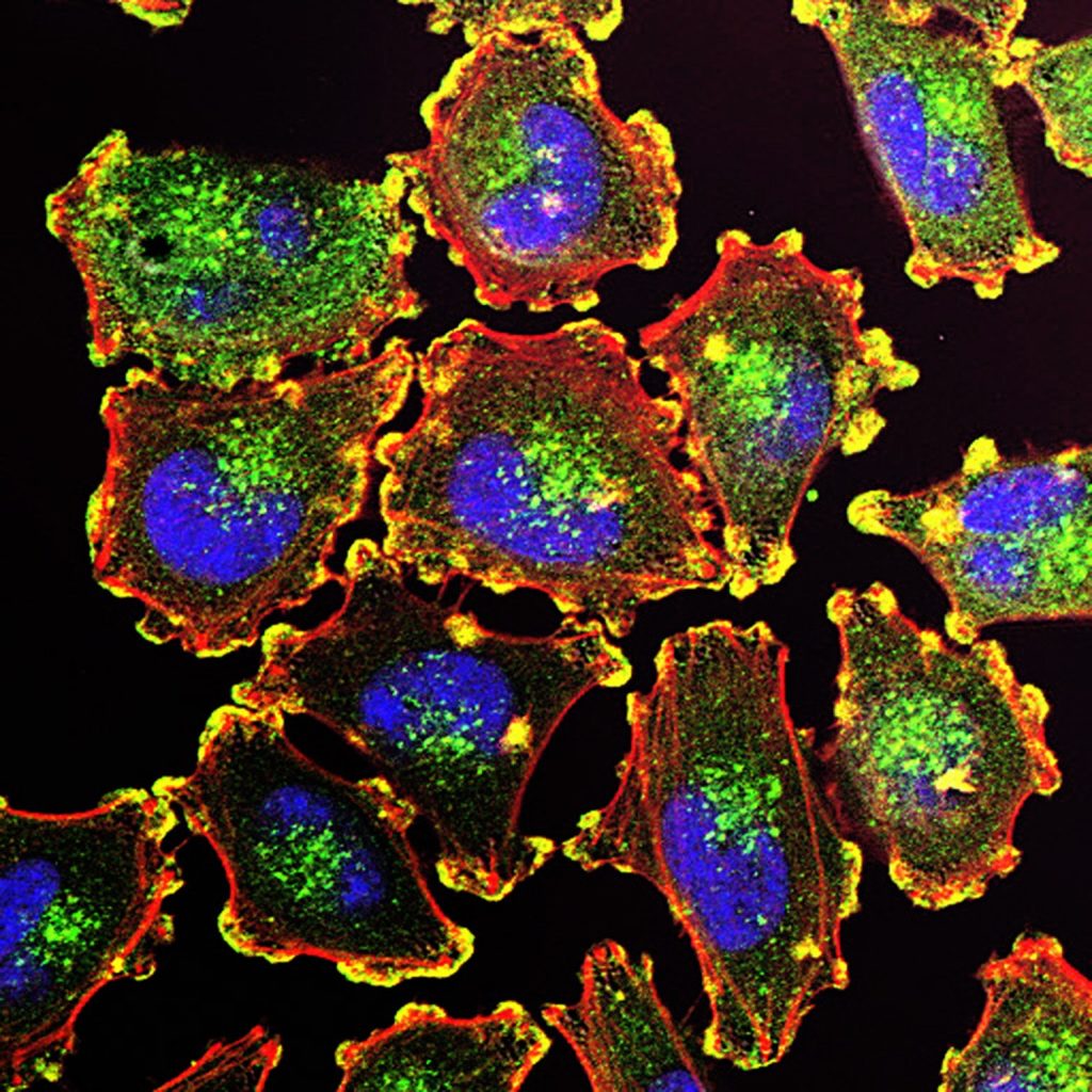
Melanoma cells. Source: National Cancer Institute.

Researchers have discovered the mechanism behind how normal epithelial cells push out precancerous ones present in the epithelium with  ‘cell competition’. Researchers have unravelled the interactions and cellular pathways leading to this extrusion, allowing them to identify a candidate for a therapeutic target for future cancer prevention research.

Recent studies have shown that the human body has defence mechanisms run by non-immune epithelial cells. These epithelial cells can recognise and extrude neighbouring precancerous cells from the epithelium, known as cell competition. This form of immune-like surveillance has garnered attention in recent years based on its potential for future immune-like therapeutic targets for cancer preventive treatment. However, it is still unknown what kind of ligand-receptor interactions are involved in the recognition of precancerous cells by normal epithelial cells.

Discussing the study, Professor Takeshi Maruyama, an Associate Professor at the Waseda Institute for Advanced Study at Waseda University, who led the research group, says, “During the process of cell competition, normal epithelial cells can be primed by contact with precancerous cells. However, it was previously unclear how neighbouring normal epithelial cells recognise precancerous cells to eliminate them.”

In this work, the researchers identified a plasma membrane protein, leukocyte immunoglobulin-like receptor B3 (LILRB3). AltR/LILRB3 interacts with major histocompatibility complex class I (MHC class I) that is expressed on precancerous epithelial cells.

MHC class I-AltR/LILRB3 interaction causes the activation of AltR/LILRB3, which triggers an intracellular SHP2–ROCK2 pathway. This SHP2–ROCK2 pathway leads to the “accumulation of cytoskeletal components”, creating a mechanical force to extrude precancerous cells, in the normal epithelial cells at the boundary with precancerous cells. This pushes the precancerous cells out of the epithelium to eliminate them from the body.

However, this occurs independently of natural killer or CD8+ T cell-mediated immune responses. “Our study describes a new immune-like mechanism by non-immune epithelial cells to suppress tumorigenesis,” said Prof Maruyama.

The researchers hope that these findings can be applied to cancer treatment. “The recombinant MHC-I-α3 protein used in this study enhances the elimination of precancerous cells and suppresses the formation of tumours and precancerous lesions,” added Prof Maruyama. “We hope that this biomolecule would contribute to a therapeutic candidate for cancer prevention by the elimination of precancerous cells.”

Source: Waseda University

Synthetic Progestogen in Utero Leads to Doubled Cancer Rate in Offspring

Photo by Shvets Productions on Pexels

In utero exposure to a synthetic progestogen used to prevent miscarriage can lead to an increased risk of developing cancer, according to a new study.

The study by researchers at The University of Texas Health Science Center at Houston (UTHealth Houston) was published in the American Journal of Obstetrics and Gynecology.

The drug, 17α-hydroxyprogesterone caproate (17-OHPC), is a synthetic progestogen frequently used by women in the 1950s and 1960s, and is still prescribed today to women to help prevent preterm birth. Progesterone helps the uterus grow during pregnancy and prevents early contractions that may lead to miscarriage.

“Children who were born to women who received the drug during pregnancy have double the rate of cancer across their lifetime compared to children born to women who did not take this drug,” said the study’s lead author, Caitlin C. Murphy, PhD, MPH, associate professor in the Department of Health Promotion and Behavioral Sciences at UTHealth School of Public Health in Houston. “We have seen cancers like colorectal cancer, pancreatic cancer, thyroid cancer, and many others increasing in people born in and after the 1960s, and no one really knows why.”

Researchers reviewed data from the Kaiser Foundation Health Plan on women who received prenatal care between June 1959 and June 1967, and the California Cancer Registry, which traced cancer in offspring through 2019.

Out of more than 18 751 live births, researchers discovered 1008 cancer diagnoses were made in offspring ages 0 to 58 years. Additionally, a total of 234 offspring were exposed to 17-OHPC during pregnancy. Offspring exposed in utero had cancer detected in adulthood at more than twice the rate of of those unexposed: 65% of cancers occurred in adults younger than 50.

“Our findings suggest taking this drug during pregnancy can disrupt early development, which may increase risk of cancer decades later,” Murphy said “With this drug, we are seeing the effects of a synthetic hormone. Things that happened to us in the womb, or exposures in utero, are important risk factors for developing cancer many decades after we’re born.”

A new randomised trial shows there is no benefit of taking 17-OHPC, and that it does not reduce the risk of preterm birth, according to Murphy.

The U.S. Food and Drug Administration proposed in October 2020 that this particular drug be withdrawn from the market.

Source: University of Texas Health Science Center at Houston

Anti-diabetes Drug under Development May Also Treat Breast Cancer

Source: NCI

A new study has shown that a small molecule inhibitor drug, with the unwieldy designation of PF05175157, originally developed to treat diabetes by Pfizer, may help in the treatment of breast cancer by blocking a key enzyme. 

The findings from the Yale Cancer Center-led study were reported at the 2021 San Antonio Breast Cancer Symposium in San Antonio, Texas.

“Our research shows the preclinical, anti-cancer activity using PF05175157 may lead us to bring this drug back into the clinic to help treat patients with breast cancer,” said lead study author Julia Foldi, MD, PhD, a clinical fellow at Yale Cancer Center and Smilow Cancer Hospital. “More studies are needed, but our initial data looks very promising.”

Cancer cells are characterised by altered metabolism. In this study, the Yale team identified new metabolic vulnerabilities in cancer cells that are based on a loss of enzyme diversity. They found that an enzyme called acetyl-CoA-carboxylase-1 (ACC1), is critical for the survival of breast cancer cells. The ACC1 enzyme is the key initial step in fatty acid synthesis. Fatty acids are building blocks of the various types of lipids and fat that are the critical ingredients of cell membranes and play an important role in energy generation in cells. The team’s analysis demonstrated that blocking ACC1 using PF05175157 can inhibit the growth of breast cancer cells grown in mice and also in patient-derived cancer models.

“We are currently testing this drug in combination with other approved breast cancer drugs to see if it could improve their activity, with the hope to bring the most promising combinations to the clinic to help patients with breast cancer,” added Lajos Pusztai, MD, DPhil, Professor of Medicine (Medical Oncology), Director of Breast Cancer Translational Research at Yale Cancer Center, and senior author of the study.

Source: Yale Cancer Center

Why People with Asthma Get Fewer Brain Tumours

Photo by Mart Production on Pexels

A new study reveals why people with asthma seem to be less likely to develop brain tumours than others.

Asthma causes T cell activation, and researchers discovered in a mouse study that asthma causes the T cells to behave in a way that induces lung inflammation but prevents the growth of brain tumours.

The findings, appearing in Nature Communications, suggest that reprogramming T cells in brain tumour patients to act more like T cells in asthma patients could be a new approach to treating brain tumours.

“Of course, we’re not going to start inducing asthma in anyone; asthma can be a lethal disease,” said senior author David H. Gutmann, MD, PhD, at Washington University School of Medicine. “But what if we could trick the T cells into thinking they’re asthma T cells when they enter the brain, so they no longer support brain tumor formation and growth? These findings open the door to new kinds of therapies targeting T cells and their interactions with cells in the brain.”

Based on epidemiologic observations, 15 years ago it was first proposed that people with inflammatory diseases, such as asthma or eczema, are less prone to developing brain tumours. However, there was no explanation for the link between the two very different kinds of diseases, and some scientists questioned whether the association was real.

Gutmann is an expert on neurofibromatosis (NF), a set of complex genetic disorders that cause tumours to grow on nerves in the brain and throughout the body. Children with NF type 1 (NF1) can develop an optic pathway glioma, where tumours grow within the optic nerves. Gutmann, director of the Washington University NF Center, noted an inverse association between asthma and brain tumours among his patients more than five years ago but didn’t know what to make of it. When more recent studies from his lab began to reveal the crucial role that immune cells play in the development of optic pathway gliomas,  he began to wonder whether immune cells could account for the asthma–brain tumour link.

Jit Chatterjee, PhD, a postdoctoral researcher and the paper’s first author, took up the investigation. Working with co-author Professor Michael J. Holtzman, MD, Dr Chatterjee studied mice genetically modified to carry a mutation in their NF1 genes and form optic pathway gliomas by three months of age.

Dr Chatterjee exposed groups of mice to asthma-inducing irritants at age four  weeks to six weeks, and treated a control group with saltwater. Then, he checked for optic pathway gliomas at three months and three months of age. The mice with asthma did not form these brain tumours.

Further experiments revealed that inducing asthma in tumour-prone mice changes the behaviour of their T cells. After the mice developed asthma, their T cells began secreting decorin, a protein that asthma researchers are well acquainted with.

Decorin is a problem in the airways, acting on lining tissues and exacerbating asthma symptoms. But the researchers found that in the brain, decorin is beneficial. There, the protein acts on microglia immune cells, blocking their activation by interfering with the NFkappaB activation pathway. Activated microglia promote brain tumour growth and development.

Treatment with either decorin or caffeic acid phenethyl ester (CAPE), a compound that inhibits the NFkappaB activation pathway, protected mice with NF1 mutations from developing optic pathway gliomas. The findings suggest that blocking microglial activation may be a potentially useful therapeutic approach for brain tumours.

“The most exciting part of this is that it shows that there is a normal communication between T cells in the body and the cells in the brain that support optic pathway glioma formation and growth,” said Prof Gutmann. “The next step for us is to see whether this is also true for other kinds of brain tumours. We’re also investigating the role of eczema and early-childhood infections, because they both involve T cells. As we understand this communication between T cells and the cells that promote brain tumours better, we’ll start finding more opportunities to develop clever therapeutics to intervene in the process.”

Source: Washington University School of Medicine

A Novel Therapy for Bone Marrow Cancer

Source: NCI on Unsplash

Researchers have found that a novel therapy for the bone marrow cancer myelofibrosis to be safe and well-tolerated, and is associated with modest improvements in patients in an early clinical trial. They shared their findings during an oral presentation at the American Society of Hematology annual meeting in December.

The therapy AVID200 showed improvements in patients’ symptom burden, anaemia, and spleen enlargement. The results from the Phase 1b clinical trial showed that the therapy was safe and displayed some evidence of efficacy (although safety and finding optimal dosage was the main goal) and researchers concluded that the therapy would need to be combined with other drugs to optimise effectiveness in patients.

“This is a real testament to cutting-edge translational research at The Tisch Cancer Institute,” said John Mascarenhas, MD, Director of the Institute’s newly launched Center of Excellence for Blood Cancer and Myeloid Disorders. “Our scientists tested this therapy in the lab, physician-scientists conducted a successful phase 1 trial, and now the optimal combination therapy approach is the subject of ongoing laboratory studies at Mount Sinai. The most interesting finding in this trial was that a subset of patients had a lasting improvement in their platelet counts – including three whose counts were normalised – supporting the preclinical studies conducted.”

Myelofibrosis is a bone marrow cancer type that disrupts normal blood cell production, causing an enlarged spleen, extensive scarring in the bone marrow, and low levels of red blood cells and platelets, increasing bleeding risk. Myelofibrosis patients who have failed the available first-line therapy face a well-documented poor prognosis, so additional therapies are urgently needed to help these patients.

Twenty-one patients enrolled in this multicenter trial were given AVID200, and while this trial’s main purpose was to test safety, some patients had an increase in platelets and there was a decrease in the size of their enlarged spleens. However, in spite of the other clinical benefits seen. patients’ bone marrow scarring did not decline, indicating that AVID200 would need to be combined with other rational therapies in the future.   

Source: EurekAlert!

After a Pregnancy, Natural Killer Cells Suppress Tumours

Source: Anna Hecker on Unsplash

After a pregnancy, breast cells call in Natural Killer T (NKT) cells as reinforcements to prevent tumours from arising, according to a study published in Cell Reports. This finding from the lab of Associate Professor Camila dos Santos at Cold Spring Harbor Laboratory (CSHL) illuminates a new way in which pregnancy reduces the risk of breast cancer.

Two lines of defence exist in the immune system: the innate response, which involves immune cells that attack any foreign molecule they encounter, and the adaptive response, which consists of immune cells that respond specifically to calls for help. NKT cells are a unique subset of cells that are present throughout the body which can participate in both responses. 

CSHL graduate student Amritha Varshini Hanasoge Somasundara said that after a pregnancy: “There is an increase in this specific [NKT] cell type, and only in the mammary gland. We don’t see the expansion everywhere else in the body, even though NKT cells are present everywhere else in the body.”

The team sought to uncover the reason behind the larger number of NKT cells were doing in the breast tissue. Hanasoge discovered that in mice, breast epithelial cells, which line lactation ducts, produce a specific protein called CD1d after pregnancy. If the cells did not present CD1d, no increase in NKT cells was seen in the tissue; the epithelial cells became cancerous and grew into tumours. Hanasoge and dos Santos think that CD1d molecules are calling in NKT cells to monitor the epithelial cells in the breast tissue after pregnancy. If they become cancerous, the NKT cells can quickly kill them to prevent tumour growth.

The team’s findings establish a novel link between pregnancy and the immune system in preventing breast cancer. They want to know how these findings can be translated into humans and what other factors may influence an abundance of NKT cells in breast tissue, such as aging and menopause, which are both associated with increased breast cancer risk.

Discussing the results, Associate Professor dos Santos said: “One of the hypotheses that we are working on now is: do pregnancies later on in life bring in the same expansion of the same subtypes of immune cells as pregnancies that took place early in life?”

Source: Cold Spring Harbor Laboratory

Older Antipsychotic Drugs Linked to Breast Cancer


Source: National Cancer Institute

A number of commonly prescribed older antipsychotic drugs, and some newer ones, are associated with a significant increase in risk of breast cancer, according to a new study.  The findings are published online in the Journal of Clinical Psychopharmacology.

Previous research uncovered the link between antipsychotic drug use, prescribed for a wide array of mental health problems, and breast cancer risk. This study compared newer antipsychotics to older drugs, and examined how the drugs affect prolactin levels, which have been associated with breast cancer. However, many antipsychotics elevate prolactin levels and can produce side effects such as menstrual cycle irregularities, abnormal breast milk production and abnormal breast tissue growth.

“Many women with psychiatric illnesses such as schizophrenia and bipolar disorder will take antipsychotics for decades, and they are essential to keeping symptoms in check,” said first author Tahir Rahman, MD. “But both older antipsychotic medicines and some newer drugs raise levels of prolactin and increase the risk of breast cancer, which is concerning. Our study confirms findings from a smaller European study that advised women and their doctors to first try drugs that don’t affect prolactin levels. We agree with that advice and believe psychiatrists should start to monitor prolactin levels in their patients taking antipsychotics.”

Antipsychotic drugs were classified into three categories, based on their established effects on prolactin. Category 1 included drugs associated with high prolactin levels, such as haloperidol, paliperidone and risperidone. Category 2 had mid-range effects and included iloperidone, lurasidone and olanzapine. Category 3 had low-effect drugs such as aripiprazole, asenapine, brexpiprazole, cariprazine, clozapine, quetiapine and ziprasidone.

Comparing the three drug categories to anticonvulsant drugs and lithium, the relative risk of breast cancer was 62% higher for women who took Category 1 drugs and 54% higher for those taking Category 2 drugs, whereas Category 3 antipsychotics were not associated with any increase in breast cancer risk.

“Certain drugs are known to elevate prolactin, and the women taking those drugs were more likely to have breast cancer,” Dr Rahman said. “But we didn’t detect any increased risk in women taking antipsychotics that don’t raise prolactin levels.”

In mouse models, prolactin can help weaken systems keeping precancerous lesions from becoming breast cancer. In humans, prolactin levels tend to be lower in women who have had more children at a younger age than in women who have fewer children or wait until they are older to do so.

In this study using data collected from 2012 through 2016, the research team performed a retrospective, observational study of breast cancer risk in women ages 18 through 64 who took antipsychotics.

The researchers identified which patients were treated for breast cancer during a 12-month period and matched that information to patients taking antipsychotic drugs. Of the 540 737 women in the database taking antipsychotics, only 914 were identified as having breast cancer – a significant number of whom were taking drugs known to increase prolactin.

“Antipsychotic medications can be lifesaving for patients who have psychotic episodes where they experience symptoms such as hallucinations and delusions,” Dr Rahman said. “In recent years, the drugs have been approved to treat other conditions, too, including depression and bipolar disorder. As those high-prolactin agents are used more widely, the number at risk could increase. We’ve been advising against using these high-prolactin agents in women who already have breast cancer, but we’d like to investigate whether keeping prolactin levels lower even might prevent some of these cancers.”

In another recent study, the antipsychotic drug aripiprazole did not increase prolactin levels in women and that a few women who began the study with high prolactin levels experienced decreases in prolactin levels after 12 weeks of treatment.

Those findings, combined with preclinical evidence of the anticancer effects of some antipsychotics, have inspired Dr Rahman and colleagues to propose repurposing some antipsychotic drugs in the fight against breast cancer.

“We don’t want to alarm patients taking antipsychotic drugs for life-threatening mental health problems, but we also think it is time for doctors to track prolactin levels and vigilantly monitor their patients who are being treated with antipsychotics,” Dr Rahman said.

Source: Washington University School of Medicine

A New Understanding of How Moles Turn into Melanoma

Melanoma cells. Source: National Cancer Institute.

Moles and melanomas are both skin tumours that come from melanocytes. Moles are usually harmless, while melanomas are cancerous and often deadly without treatment. A study published in eLife Magazine explains how common moles and melanomas form and why sometimes moles turn into melanoma.

Melanocytes produce melanin that protects the skin against UV radiation. Specific changes to the DNA sequence of melanocytes, called BRAF gene mutations, are found in over 75% of moles. The same change is also found in 50% of melanomas and is common in lung, colon and other cancers. It was thought that when melanocytes only have the BRAFV600E mutation the cell stops dividing, resulting in a mole. When melanocytes have other mutations with BRAFV600E, they divide uncontrollably, turning into melanoma. This model is called ‘oncogene-induced senescence’.

“A number of studies have challenged this model in recent years,” said Robert Judson-Torres, PhD, Huntsman Cancer Institute (HCI) researcher and University of Utah (U of U) assistant professor. “These studies have provided excellent data to suggest that the oncogene-induced senescence model does not explain mole formation but what they have all lacked is an alternative explanation – which has remained elusive.”

The study team took moles and melanomas donated by patients and used transcriptomic profiling and digital holographic cytometry. Transcriptomic profiling lets researchers determine molecular differences between moles and melanomas. Digital holographic cytometry helps researchers track changes in human cells.

“We discovered a new molecular mechanism that explains how moles form, how melanomas form, and why moles sometimes become melanomas,” said Prof Judson-Torres.

The study shows that additional mutations for melanocytes to turn into melanoma are not needed, but environmental signalling can be a trigger for the cells. Melanocytes express genes in different environments, instructing them to either divide uncontrollably or stop dividing altogether.

“Origins of melanoma being dependent on environmental signals gives a new outlook in prevention and treatment,” said Prof Judson-Torres. “It also plays a role in trying to combat melanoma by preventing and targeting genetic mutations. We might also be able to combat melanoma by changing the environment.”

These findings create a foundation for researching potential melanoma biomarkers, allowing doctors to spot cancerous changes in the blood sooner. The researchers are also keen to use these data to better understand potential topical agents to reduce risk melanoma risk, delay development, or stop recurrence, and to detect melanoma early.

Source: Huntsman Cancer Institute