

Tag: collagen

Researchers Find ‘Perfect Recipe’ to Regrow Bone and Blood Vessels
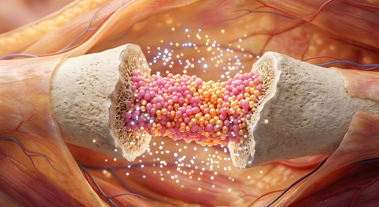
Medical illustration of bone regeneration.

For patients suffering from traumatic injuries that leave behind volumetric gaps – where significant bone and blood vessels are lost – the clock is always ticking. Without a nearby blood supply, cells in the centre of a large injury cannot survive, often leading to permanent tissue loss or failed grafts.

A team of eight scientists at The University of Texas at San Antonio has discovered a potential ‘perfect recipe’ to address this challenge. By blending two natural proteins found in the human body, the researchers created a specialised scaffold that allows bone and blood vessels to grow simultaneously at an accelerated rate.

The study, published in the journal Biomaterials Advances, identifies a 50:50 ratio of collagen and fibrin as the ideal environment for tissue repair.

The Lego blocks of healing

The technology relies on what scientists call interpenetrating polymer networks, or IPNs. In simpler terms, it is a microscopic support structure where different materials are entangled to create a stable foundation for new growth.

“An IPN network is two things that are entangled like a giant mess of Legos,” said Teja Guda, PhD, the Jacobson Distinguished Professor of Innovation and Entrepreneurship in the Department of Biomedical Engineering and Chemical Engineering at UT San Antonio and the study’s corresponding author. “We are leaving all the building blocks there and letting the cells build whatever Lego structure they like the most.”

In this biological ‘Lego’ set, one material is fibrin, the protein the body uses to form blood clots immediately after an injury. The other is collagen, the primary structural protein found in bones and other tissues.

Seeding the scaffold with MVFs and MSCs

To turn these protein gels into living tissue, the research team “seeded” the hydrogels with two critical types of biological starters: microvascular fragments (MVFs) and mesenchymal stem cells (MSCs). The MVFs have the capacity to grow into blood vessels, while the MSCs can, with the right environmental cues, grow into bone.

The researchers integrated these components by mixing the living MVFs and MSCs directly into the liquid protein solution before it underwent gelation. This 3D encapsulation ensured the cells were suspended throughout the entire depth of the scaffold rather than just sitting on the surface.

Balancing blood and bone

Standard medical treatments for severe bone loss typically involve autografts, where bone is harvested from another part of the patient’s body, or allografts, which use processed bone from a donor. These traditional grafts often fail to integrate because they lack an immediate blood supply to nourish the new tissue. Without rapid vascularisation, the transplanted bone can become necrotic, leading to a high rate of clinical failure in complex trauma cases.

The challenge for UT San Antonio researchers was finding the right balance between the two proteins to support both blood vessel and bone regeneration. Fibrin is excellent at recruiting the cells needed to form blood vessels, a process called angiogenesis. Collagen provides the mechanical strength needed to guide the development of bone, or osteogenesis.

“Whenever you have an injury where you are losing volume, you not only lose the tissue itself, but you’re also losing blood vasculature,” said Gennifer Chiou, a postdoctoral fellow at UT San Antonio and the study’s lead author. “We’re looking at how we can regenerate both the tissue and the vessel itself within specifically bone tissue.”

The team tested five different ratios of the two proteins. They found that while gels with more fibrin supported faster vessel sprouting, they lacked the stability needed for long-term bone growth. Conversely, high-collagen gels were too stiff for vessels to penetrate easily.

The 50:50 blend struck an ideal balance. The MVFs were able to sprout and branch out into a robust, interconnected network. Simultaneously, the MSCs developed in a stable environment, expressing the specific genetic markers needed to mature into bone-forming cells. This dual-growth approach ensures that as the new bone forms, it is continuously supplied with the blood and nutrients it needs to remain viable.

From the lab to the clinic

Because the materials used in the study – collagen, fibrin and the patient’s own blood vessels – are all naturally occurring in the body, the researchers believe the technology faces fewer regulatory hurdles than synthetic alternatives.

“There is almost nothing new in our material,” Guda said. “It’s your collagen, it’s your blood vessels, it’s your fibrin. The end goal is to provide evidence that will guide how clinicians think about healing wounds.”

The team hopes to proceed to preclinical trials in the near future, which will provide further support for the treatment to one day become standard practice.

By Audrey Gray

Source: UT San Antonio

Collagen’s Role in Breast Cancer Includes Triggering Metastasis

Breast cancer cells
Breast cancer cells. Image source: National Cancer Institute on Unsplash

Type XII collagen plays a key role in regulating the organisation of the tumour matrix, according to research published in the journal Nature Communications. The study investigators also discovered that high levels of collagen XII can trigger metastasis.

Cancer cells continually interact with the tumour microenvironment one component of which is the extracellular matrix. Collagen is an important part of this tumour microenvironment, but just how it influences tumours has not been understood.

“There’s still a lot we don’t know about the role of the extracellular matrix in cancer metastasis. Our study shows that collagen XII plays an important role in breast cancer progression and metastasis,” said Associate Professor Thomas Cox, senior author of the study.

“Imagine cancer cells as seeds, and the tumour microenvironment as the soil. By studying the soil – the extracellular matrix – we can begin to understand what makes some tumours more aggressive than others, and by extension, begin to develop new ways to treat cancer,” he explained.

The research also suggests that measuring the level of collagen XII in a patient’s tumour biopsy could potentially be used as an additional screening tool to identify aggressive breast cancers with higher rates of metastasis, such as in the triple-negative type of breast cancer. Furthermore, collagen XII might be a possible target for future treatments.

The extracellular matrix is a 3D meshwork of around 300–400 core molecules, including several collagen proteins. This matrix provides structural and functional support to cells and tissues in all parts of the body.

In this study, the researchers catalogued how the tumour matrix changes over time and have generated a comprehensive database of these changes, which has been made freely available to researchers.

The team focused on collagen XII, one of 28 types of collagen in the body. Collagen XII plays an important role in organising other collagens and can have profound effects on the 3D structure of the extracellular matrix.

The researchers studied tumours in mouse models from the earliest pre-clinical stages of cancer, right through to late-stage tumours. They found that as the tumours developed, many matrix molecules changed, and importantly the level of collagen XII was also increased.

“Collagen XII seems to be altering the properties of the tumour and makes it more aggressive,” said first author Michael Papanicolaou. “It changes how collagens are organised to support cancer cells escaping from the tumour and moving to other sites like the lungs.”

The team then genetically manipulated collagen XII production, looking at the effects of metastasis to other organs. They found that as levels of collagen XII increased, so did metastasis. These findings were then confirmed in human tumour biopsies, which showed that high levels of collagen XII are associated with higher metastasis and poorer overall survival rates.

Further research will focus on studying more human samples, and investigating possible therapeutic pathways.

Source: Garvan Institute of Medical Research

Why Cancer Cells Linger to Create Metastatic Cancer

Colon cancer cells. Source: National Cancer Institute on Unsplash

A major mystery in cancer research has been solved: How cancer cells remain dormant for years after leaving a tumour before awakening to create metastatic cancer.

According to findings by Mount Sinai researchers which were reported in Nature Cancer, the cells remain quiet by secreting a type of collagen, called type III collagen, in the environment around themselves, and only turn malignant once the level of collagen tapers off. The researchers found that by enriching the environment around the cells with this collagen, they could force the cells to remain in a dormant state and prevent tumour recurrence.

“Our findings have potential clinical implications and may lead to a novel biomarker to predict tumour recurrences, as well as a therapeutic intervention to reduce local and distant relapses,” said senior author Jose Javier Bravo-Cordero, PhD, Associate Professor of Medicine (Hematology and Medical Oncology) at The Tisch Cancer Institute at Mount Sinai. “This intervention aimed at preventing the awakening of dormant cells has been suggested as a therapeutic strategy to prevent metastatic outgrowth. As the biology of tumour dormancy gets uncovered and new specific drugs are developed, a combination of dormancy-inducing treatments with therapies that specifically target dormant cells will ultimately prevent local recurrence and metastasis and pave the way to cancer remission.”

Most cancer deaths result from metastases, which can occur several years after removal of a tumour. Previous work looked at how dispersed tumour cells awaken from dormancy; this new work showed how the cells remain dormant.

The study used high-resolution imaging techniques, including intravital two-photon microscopy, a technology that allows the visualisation of dormant cells in their environment in real time in a living animal. This technology allowed the researchers to track dormant tumour cells in mouse models using cancer cell lines. By using this technology, the researchers were able to visualise the changes in the architecture of the extracellular matrix as tumour cells became dormant and how it changed when these cells awoke.

The researchers demonstrated that an abundance of the collagen could potentially be used as a predictor of tumour recurrence and metastasis. In the mouse models, when type III collagen was increased around cancer cells that had left a tumour, cancer progression was interrupted and the disseminated cells were forced into a dormant state. Similar to wound treatment, in which collagen scaffolds have been proposed to treat complex skin wounds, this study suggests that by enriching the tumour microenvironment in type III collagen, metastasis may be prevented by sending tumour cells into a dormant state.

Source: The Mount Sinai Hospital / Mount Sinai School of Medicine