Category: Cancer

Stress-tolerant Cells may Drive Pancreatic Cancer’s Extreme Tenacity

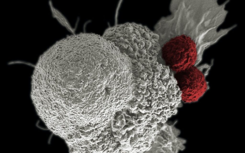
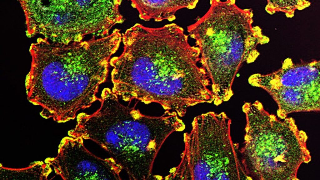
This image shows pancreatic cancer cells (nuclei in blue) growing as a sphere encased in membranes (red). Credit: NIH

Researchers at have discovered a molecular pathway critical to the initiation of pancreatic tumours, which could partly explain the disease’s high resistance to chemotherapy and its propensity for metastasis.

The study, published in Nature Cell Biology, found that pancreatic tumour-initiating cells must first overcome local ‘isolation stress’ by creating their own tumour-promoting microenvironment, and then recruit surrounding cells into this network. By targeting this tumour-initiating pathway, new therapeutics could limit the progression, relapse and spread of pancreatic cancer.

Pancreatic cancer is one of the most lethal cancers, notoriously resistant to treatment. Almost all patients experience cancer recurrence or metastasis.

In the early stages of tumour formation, cancer cells (those with cancerous mutations, called oncogenes) experience a loss of adhesion to other cells and the extracellular matrix. This isolation leads to a local lack of oxygen and nutrients. Most cells do not survive such isolation stress, but a certain group of cells can.

Tumour-initiating cells (TIC) play a major role in the formation, recurrence and metastatic spread of tumours. What sets them apart from other cancer cells is their resilience to these early substandard conditions. Like cacti in a desert, they can adapt to the harsh environment and set the scene for further tumour progression.

“Our goal was to understand what special properties these tumour-initiating cells have and whether we can control the growth and spread of cancer by disrupting them,” said senior study author David Cheresh, PhD.

To answer these questions, first author Chengsheng Wu, PhD, a postdoctoral fellow in Cheresh’s lab, subjected pancreatic cell lines to various forms of stress, including low oxygen and sugar levels. He then identified which cells could adapt to the harsh conditions and observed which genes and molecules were modified in these cells.

The stress-tolerant tumour-initiating cells showed reduced levels of a tumour-suppressive microRNA, miR-139-5p. This in turn led to the upregulation of lysophosphatidic acid receptor 4 (LPAR4), a G-protein-coupled receptor on the cell surface.

“LPAR4 is not normally found on happy cells, but it gets turned on in stressful environments to help the cells survive, which is particularly advantageous for tumor-initiating cells,” said Cheresh.

The researchers found that LPAR4 expression promoted the production of new extracellular matrix proteins, allowing the solitary cancer cells to start building their own tumour-supporting microenvironment.

The new extracellular matrix was particularly rich in fibronectin, a protein that binds to transmembrane receptors called integrins on surrounding cells. Once the integrins on these cells sensed the fibronectin, they began signalling the cells to express their own tumour-initiating genes. Eventually, these other cells were recruited into the fibronectin matrix laid by the tumour-initiating cells and a tumour started to form.

“Our findings establish a critical role for LPAR4 in pancreatic tumour initiation, and a likely role in other epithelial cancers, such as lung cancer,” said Cheresh. “It is central to tumour-initiating cells’ ability to overcome isolation stress and build their own niche in which tumours can form.”

Chemotherapy drugs are also designed to put cancer cells under stress, and may use this pathway. Indeed, Cheresh’s team found that treating cultured tumour cells and pancreatic tumours in mice with standard-of-care chemotherapeutics also led to the upregulation of LPAR4. The researchers said this might explain how such tumour cells could develop a stress tolerance and resistance to the drugs.

Further experiments also showed that using integrin antagonists to block cells’ ability to utilise the fibronectin matrix reversed the stress tolerance benefit of LPAR4 expression. Thus, the authors suggest targeting the LPAR4 pathway or disrupting the fibronectin/integrin interaction could be effective in preventing the growth, spread and drug resistance of pancreatic tumours.

“We can think of tumour-initiating cells as being in a transient state that can be induced by different stressors, so our clinical goal would be to prevent oncogenic cells from ever entering this state,” said Cheresh. “Now that we’ve identified the pathway, we can assess all the different ways we can intervene.”

The researchers suggested a new drug targeting this pathway could be used as a prophylactic in patients at high risk of developing the disease, or to prevent new tumours from forming in cancer cases with a high likelihood of metastasis.

Pairing the new drug with existing chemotherapeutics that put stress on mature tumour cells could also mitigate the effects of drug resistance and make cancer treatments more effective, authors said.

“Treating cancer can feel a little like whack-a-mole,” said Cheresh, “but if we have two or three hammers and we know where the moles are going to pop up next, we can beat the game.”

Source: University of California – San Diego

A Metabolic Switch for Childhood Obesity and Cancer

Researchers have unlocked a means to modify the function of an enzyme crucial to fat production, a finding could lead to more effective treatments for childhood obesity and cancer.

While the research, published in the Proceedings of the National Academy of Sciences, was in fruit fly larvae, the ability to alter the rates of lipid metabolism could have significant implications for human health, said Hua Bai, an associate professor of genetics, development and cell biology at Iowa State University.

“We’ve identified what’s basically a metabolic switch. It’s like the accelerator on a car,” he said.

The initial aim was investigating how ageing was affected by fatty acid synthase, an enzyme that plays a role in de novo lipogenesis, which is the process of turning excess dietary carbohydrates into fat. Typically, levels of fatty acid synthase rise and fall based on an animal’s cellular needs and diet.

Surprisingly, the researchers noticed that early in a fruit fly’s development, de novo lipogenesis increases without an accompanying boost in the expression of fatty acid synthase. That suggested there must be some other factor at play, Bai said.

After proteins such as fatty acid synthase are created based on genetic code, their function can be altered by one of several different types of post-translational modification. Bai’s team found one of those processes, acetylation, affected one of the 2540 amino acids that combine to make fatty acid synthase, changing how effective it was at producing fat.

In addition to its role in obesity, elevated levels of de novo lipogenesis are linked to cancer, so controlling it through a single amino acid could lead to highly targeted treatments, Bai said.

“Fine tuning the acetylation levels of fatty acid synthase would be a much more precise treatment than blocking the entire protein,” he said.

Though the findings may be applicable to humans, any medical application in humans is years away, he said.

“The potential is high, but further testing is needed in other animals,” he said.

Source: Iowa State University

Vitamin D Supplements may Reduce Skin Cancer Risk

Melanoma cells. Source: National Cancer Institute.

Fewer cases of melanoma were observed among regular users of vitamin D supplements than among non-users, researchers in Finland found. People taking vitamin D supplements regularly also had a considerably lower risk of skin cancer, according to a study of nearly 500 people with increased skin cancer risk, which was published in Melanoma Research.

A key micronutrient, vitamin D may play a role in many diseases. Previous studies investigating the link between vitamin D and skin cancers, have been inconclusive or contradictory, but they mainly focussed on serum levels of calcidiol, which is a metabolite of vitamin D. Serum calcidiol levels have been associated with both a slightly higher or lower risk of different skin cancers. This may be partly due to the fact that serum calcidiol analyses do not provide information on vitamin D metabolism in the human skin, which can express enzymes that generate biologically active vitamin D metabolites or inactivate them.

The new study took a different approach: 498 adult patients with an increased risk of a skin cancer, such as basal cell carcinoma, squamous cell carcinoma or melanoma, were recruited and classified into low risk, moderate risk and high risk. Based on their use of oral vitamin D supplements, the patients were divided into three groups: non-users, occasional users and regular users. Serum calcidiol levels were analysed in half of the patients and found to correspond to their self-reported use of vitamin D.

A key finding of the study is that there were considerably fewer cases of melanoma among regular users of vitamin D than among non-users, and that the skin cancer risk classification of regular users was considerably better than non-users’. Logistic regression analysis showed that melanoma risk among regular users was more than halved compared to non-users.

The findings suggest that even occasional users of vitamin D may have a lower risk for melanoma than non-users. However, there was no statistically significant association between the use of vitamin D and the severity of photoaging, facial photoaging, actinic keratoses, nevus count, basal cell carcinoma and squamous cell carcinoma. Serum calcidiol levels were not significantly associated with these skin changes, either. Since the research design was cross-sectional, the researchers were unable to demonstrate a causal relationship.

Other relatively recent studies, too, have provided evidence of the benefits of vitamin D in melanoma, such as of the association of vitamin D with a less aggressive melanoma.

“These earlier studies back our new findings from the North Savo region here in Finland. However, the question about the optimal dose of oral vitamin D in order to for it to have beneficial effects remains to be answered. Until we know more, national intake recommendations should be followed,” Professor of Dermatology and Allergology Ilkka Harvima of the University of Eastern Finland notes.

Source: University of Eastern Finland

How Well Do Acupuncture and Acupressure Help Cancer Patients to Sleep?

Source: Katherine Hanlon on Unsplash

Acupuncture and acupressure (which stimulates pressure points without the invasive needles) have been suggested as ways to help reduce sleep disturbance in patients with cancer. A recent analysis published in Worldviews on Evidence-Based Nursing finds moderate evidence that acupressure and acupuncture may be effective, with acupressure having a greater effect.

Researchers included 24 randomised controlled trials involving 2002 patients in the analysis. Compared with enhanced supportive care, acupressure had the largest effect size for reducing self-reported sleep disturbance (standardised mean difference [SMD] = −2.67, 95% CrI: −3.46 to −1.90; GRADE = moderate), followed by acupuncture (SMD = −1.87, 95% CrI: −2.94 to −0.81, GRADE = moderate) and electroacupuncture (SMD = −1.60, 95% CrI: −3 to −0.21; GRADE = low).

Based on these findings, the researchers conclude that acupressure can be recommended as the optimal treatment for reducing sleep disturbance in cancer patients. They also suggest further trials to investigate the effects of different acupuncture or acupressure techniques have on sleep in cancer patients, especially alongside other therapies.

“Based on available evidence, acupressure may be a more promising approach than acupuncture for reducing sleep disturbance in patients with cancer,” said corresponding author Denise Shuk Ting Cheung, BNurs, PhD, RN, of the University of Hong Kong. “Future studies should focus on the differential mechanisms of action of acupressure and acupuncture and link them to the multifactorial causes of sleep disturbance in patients with cancer.”

Source: Wiley

Extended Chemotherapy Slashes Childhood Leukaemia Recurrence

Photo by National Cancer Institute on Unsplash

Giving three years of chemotherapy to children with acute lymphoblastic leukaemia (ALL) instead of two years lowers the risk of their disease coming back after treatment by three times. The survival rate of all children with ALL, the most common form of childhood cancer, together has further increased to 94%. Less intensive therapy proved safe for three groups of children, resulting in a better quality of life. These findings on a large Dutch study into ALL were reported at the annual conference of the American Society of Hematology (ASH).

Many children with ALL have good outcomes. After two years of chemotherapy treatment, nine out of ten children are cured. But some children have a more aggressive disease, such as having the Ikaros mutation in their leukaemia cells, have a greater risk of recurrence after treatment. In order to improve the chances of survival and quality of life of all children with leukaemia, the treatment protocol has been continuously adapted over the years, based on the latest scientific insights.

Prof Rob Pieters, medical director and paediatric oncologist at the Princess Máxima Center for paediatric oncology in the Netherlands, presented the outcomes of the ALL-11 treatment protocol. The Dutch researchers tested the benefit of an adapted treatment in specific groups of children with leukaemia, including children with the Ikaros mutation. More than 800 children in the Netherlands were treated with this protocol between April 2012 and July 2020.

Threefold lower risk of recurrence

Children with Ikaros leukaemia received an extra year of chemotherapy in the ‘maintenance phase’ on top of the first two years of treatment. This change lowered the risk of their cancer coming back by threefold: this happened in only 9% of them, compared to 26% of the children in the previous treatment protocol.

87% of children with Ikaros leukaemia survived their disease for five years without their cancer coming back, an improvement on the 72% in the previous protocol. Because of the extra year of chemotherapy, this group of children had a slightly higher risk of infection, but these were treatable. The extended therapy did not lead to any additional side effects.

Analysis of data from all children with ALL, regardless of subtype, showed that the five-year survival rate has improved stepwise over the past 30 years from 80% to 94% under the ALL-11 protocol.

Safe reduction of treatment

In the ALL-11 protocol, doctors and researchers also looked at the benefit of a less intensive treatment plan for three groups of children. This included children with a leukaemia mutation linked to a very high chance of recovery, and children with Down syndrome who experience more severe side effects. These children received treatment without or with a lower dose of anthracyclines, a type of leukaemia drug that increases the risk of heart damage and infections. The reduced treatment proved successful: children had the same or even a better chance of survival, while their quality of life improved due to a lower risk of infections and damage to the heart.

Global interest

Globally, there is much interest in the Dutch research as it has been unclear how to improve therapy for children with Ikaros leukaemia. The results have now been presented for the first time at the largest blood cancer conference, and could lead to changes in treatment protocols for these children worldwide.

In the Netherlands, there are about 15 children with ALL each year for whom existing treatments stop working. Since 2019, they have been eligible for treatment with CAR T-cell therapy, a promising form of immunotherapy that now leads to a cure in 40% of these children.

Making a difference

Prof Monique den Boer, medical biologist and group leader at the Princess Máxima Center, played an important role in the adapted therapy for children with the Ikaros gene change. She says: ‘The Ikaros mutation was first discovered about 15 years ago in children with leukaemia who had a poor prognosis, partly thanks to the emergence of new DNA technologies. We saw that the cancer came back in many of these children shortly after the end of the two-year treatment plan. I am very proud that our lab findings have now found their way into the clinic and can make such a big difference for children with leukaemia.”

More cure with fewer side effects

Prof Pieters concludes: The five-year survival rate for children with acute lymphoblastic leukaemia has increased enormously since the 1960s, from zero to 94%, but the last steps are the most difficult. We are now one step closer to curing all children with ALL. We have also largely been able to remove a drug that poses a risk of heart damage from the treatment of children with a less aggressive form of the disease. The latest results for children with leukaemia therefore fit in perfectly with our mission: curing more children with cancer, with fewer side effects.”

Source: Princess Máxima Center

Study Identifies Risk Factors for CVD among Young Cancer Survivors

Source: Pixabay CC0

Adolescent and young adult (AYA) cancer survivors are at risk of experiencing treatment-related effects later in life, including cardiac damage. New research published in in CANCER has identified a number of sociodemographic and modifiable risk factors linked to these patients’ risk of developing cardiovascular disease (CVD).

The study by investigators at Duke University and The University of Texas MD Anderson Cancer Center relied on 2009–2018 data from the National Health Interview Survey, which collects information on a broad range of health topics through personal interviews of US households. Responses from 4766 AYA cancer survivors and 47 660 controls (without a history of cancer) were included. 

The risk of CVD was significantly higher in survivors than controls by sex, race/ethnicity, income, education, smoking status, and physical activity.

In the AYA survivor population, male sex, Black race, household income under $50 000 a year, and current or former smoking were all associated with higher odds of CVD. Household income < $50 000 a year disproportionately increased the odds of CVD in survivors compared with controls.  Performing any moderate to vigorous intensity physical activity was associated with lower CVD odds.  

“These results highlight the importance of long-term surveillance of AYAs after cancer treatment to ensure that appropriate screenings are initiated to reduce the risk of CVD and to promote healthy behavioural changes, such as physical activity, which impact long-term CVD outcomes,” said lead author Amy Berkman, MD, of the Duke University School of Medicine. 

Source: Wiley

New Immunotherapy Sees 73% Success Rate in Multiple Myeloma Patients

Woman using lab equipment
Source: NCI on Unsplash

A new immunotherapy to kill bone marrow cancer cells was successful in as many as 73% of patients in two clinical trials, according to Mount Sinai researchers. The new bispecific antibody therapy binds to both T cells and multiple myeloma cells and directs the T cells to kill multiple myeloma cells. The researchers described this strategy as “bringing your army right to the enemy.”

The success of the off-the-shelf immunotherapy, called talquetamab, was even seen in patients whose cancer resisted previous therapies. It uses a different target than other approved therapies: a receptor expressed on the surface of cancer cells known as GPRC5D.

Talquetamab was tested in phase 1 and phase 2 trials. The phase 1 trial, reported in NEJM, established two recommended doses that were tested in the phase 2 trial. The results of the phase 2 trial were reported at the American Society of Hematology annual meeting. The study participants had all received at least three different therapies without achieving lasting remission, suggesting talquetamab could offer new hope for patients with hard-to-treat multiple myeloma.

“This means that almost three-quarters of these patients are looking at a new lease on life,” said lead author of the studies, Ajai Chari, MD, Director of Clinical Research in the Multiple Myeloma Program at The Tisch Cancer Institute. “Talquetamab induced a substantial response among patients with heavily pretreated, relapsed, or refractory multiple myeloma, the second-most-common blood cancer. It is the first bispecific agent targeting the protein GPRC5d in multiple myeloma patients.”

Nearly all patients with myeloma who receive standard therapies continually relapse. Patients who relapse or become resistant to all approved multiple myeloma therapies have a poor prognosis, so additional treatments are urgently needed. This study, while an early-phase trial designed to detect tolerability and find a safe dose, is an important step in meeting that need.

This international Phase 1 clinical trial enrolled 232 patients between January 2018 and November 2021. Patients received a variety of doses of the therapy either intravenously or subcutaneously; future studies will focus on doses only administered subcutaneously either weekly or every other week

The efficacy and safety findings in the phase 1 study were validated in the phase 2 trial presented at ASH. The phase 2 trial included 143 patients treated on a weekly dose and 145 patients treated at a higher biweekly dose.

The overall response rate in these two groups was about 73%, Dr Chari said. The response rate was maintained throughout various subgroups examined, with the exception of patients with a rare form of multiple myeloma that also extends to organs and soft tissues. More than 30% of patients in both groups had a complete response (no detection of myeloma-specific markers) or better, and nearly 60% had a “very good partial response” or better (indicating the cancer was substantially reduced but not necessarily down to zero).

The median time to a measurable response was approximately 1.2 months in both dosing groups and the median duration of response to date is 9.3 months with weekly dosing. Researchers are continuing to collect data on the duration of response in the group receiving 0.8 mg/kg every other week and for patients in both dosing groups who had a complete response or better.

Side effects were relatively frequent, but typically mild. About three-quarters of patients experienced a common side effect of immunotherapy – cytokine release syndrome, a constellation of symptoms including fever. About 60% experienced skin-related side effects such as rash, about half reported taste changes, and about half reported nail disorders. Only 5–6% of patients stopped treatment due to side effects.

The response rate observed in the study, which Dr Chari explained is higher than that for most currently accessible therapies, suggests talquetamab could offer a viable option for patients whose myeloma has stopped responding to most available therapies, offering a chance to extend life and benefit from other new and future therapies as they are developed.

Source: Mount Sinai Hospital

‘Landmark’ Drug Doubles Progression Time of Advanced Breast Cancer

Source: National Cancer Institute

A new type of targeted medicine has shown ‘remarkable’ benefits for patients with advanced breast cancer in a major phase III clinical trial. Combined with hormone therapy, the drug capivasertib doubled the time it took for cancer to progress in people with advanced forms of the most common type of breast cancer.

Capivasertib is a potential first-in-class drug that blocks activity of the cancer-driving protein molecule AKT.

The findings, presented at the San Antonio Breast Cancer Symposium, show that capivasertib is a potential new treatment for people with oestrogen receptor (ER) positive, human epidermal growth factor receptor 2 (HER-2) negative breast cancer.

‘Effective across all patients treated in the trial’

The drug was found to be effective across all patients treated in the trial, including a group who had tumours with mutations in the AKT signalling pathway.

The phase III CAPItello-291 trial enrolled 708 women and men with an advanced form of ER-positive, HER-2 low or negative locally advanced or metastatic breast cancer.

Participants on the trial had seen their cancer recur or progress on standard hormone treatments, and the majority had also previously been treated with CDK4/6 inhibitors – drugs that block cancer cells from multiplying.

In current clinical practice, patients continue to receive fulvestrant hormone therapy, but this is often not effective, and many are left only with the option of chemotherapy, a treatment which can lead to debilitating side effects.

In the trial, adding capivasertib to fulvestrant hormone therapy doubled the median time to disease progression, from 3.6 months to 7.2 months. The treatment shrank tumours in 23% of patients, compared with 12% of patients who received fulvestrant hormone treatment plus a placebo.

Targeting the AKT pathway

The targeted drug was more effective for patients whose cancers had alterations to the AKT signalling pathway. Genetic alterations to the AKT pathway can drive both cancer’s development and treatment resistance.

Genetic alterations of the AKT pathway – revealed by tumour profiling – were present for 41% of patients on the trial. In this group treated with capivasertib and hormone therapy, it took an average of 7.3 months for the cancer to worsen compared with 3.1 months for those who received hormone therapy alone. Some 29% of patients with AKT pathway alterations who received capivasertib with hormone therapy saw their tumours shrink following treatment, compared with only 9.7% who received a placebo and hormone therapy.

Side effects from capivasertib with hormone therapy were manageable and consistent with previous studies.

More than two thirds of people with advanced breast cancer have ER-positive, HER-2 negative disease. ER-positive, HER-2 negative breast cancers have higher levels of the oestrogen receptor – allowing them to grow in the presence of oestrogen – but do not over-produce copies of the human epidermal growth factor.

Developing capivasertib

The development of capivasertib followed years of fundamental research at the Institute for Cancer Research (ICR), aimed at understanding how the AKT protein is regulated. In 2002, ICR scientists published the 3D structure of AKT and showed how the protein is activated.

Researchers then collaborated with Astex Pharmaceuticals to design small-inhibitors which would target AKT, based on its 3D structure.

In 2005, a series of prototype drug compounds discovered by the ICR and Astex was shown to have very promising activity against a range of human tumours grown in mice and was licensed to AstraZeneca. Then, in 2010, AstraZeneca announced its discovery of capivasertib, and began to develop the drug as a potential treatment for various forms of cancer.

The initial clinical development of capivasertib was centred on an early-stage trial which was led by the ICR and its partner hospital The Royal Marsden. Subsequently, phase II studies were completed in the UK in collaboration with the UK Cancer Research Network.

The trials used biomarkers that were developed at ICR to show proof of concept that the AKT protein was inhibited by capivasertib.

ICR researchers continue to study AKT biology to open up new ways of using treatments targeting the pathway.

Living well and longer with breast cancer

Trial leader Professor Nicholas Turner, Professor of Molecular Oncology at ICR London, and Consultant Medical Oncologist at The Royal Marsden NHS Foundation Trust, said:

“We are hopeful that capivasertib combined with hormone therapy will now become a new treatment option for patients whose cancer has progressed on hormone therapy plus a CDK4/6 inhibitor. We believe this new treatment could allow more women and men to live well and live longer with breast cancer.”

‘A landmark moment’

Professor Kristian Helin, Chief Executive at The Institute of Cancer Research, London, said:

“This is a landmark moment for the treatment of advanced forms of the most common type of breast cancer. It’s incredibly exciting to see a drug that was discovered following research conducted at the ICR now show remarkable benefits for patients in a phase III trial. Capivasertib could offer a completely new treatment option for these patients.

“I look forward to seeing further results from the CAPitello-291 trial, and I am hopeful that longer follow-up will show that capivasertib also extends the length of time that people survive with advanced breast cancer. But the existing findings are already strong enough for capivasertib to be submitted to regulators to be considered for approval as a new breast cancer treatment.”

Source: The Institute for Cancer Research

The Immune System Attacks Tumours at Dawn

Shown here is a pseudo-colored scanning electron micrograph of an oral squamous cancer cell (white) being attacked by two cytotoxic T cells (red), part of a natural immune response. Photo by National Cancer Institute on Unsplash

Researchers have demonstrated that the daily rhythms of the immune system – and in particular that of dendritic cells, its key sentinels – have a hitherto unsuspected impact on tumour growth. These results, published in Nature, indicate that simply changing the time of administration of a treatment could boost its effectiveness.

Like pathogens, cancer cells can be identified and targeted by a specific immune response, which can be enhanced by immunotherapy treatments.

In previous studies, the team from the University of Geneva (UNIGE) and the Ludwig-Maximilians University of Munich (LMU) had shown that the activation of the immune system is modulated according to the time of day, indicating a peak of efficiency early in the morning in humans.

The immune system is no exception to the circadian rhythm. ‘‘By studying the migration of dendritic cells in the lymphatic system, one of the most essential elements of the immune response, we had highlighted the fact that immune activation oscillates throughout the day, with a peak in the late behavioural resting phase,” summarises study leader Christoph Scheiermann, professor at the University of Geneva. In the current study, the group focussed on cancer to assess how this temporal modulation affected tumours.

Temporal profiling of dendritic cells

The scientists injected groups of mice with melanoma cells at six different times of the day and then monitored tumour growth for a fortnight. ‘‘By varying only the time of injection, we observed very surprising results: tumours implanted in the afternoon grew little, while those implanted at night grew much faster, in accordance with the rhythm of activation of the mice’s immune system’’, said Chen Wang, a researcher in Christoph Scheiermann’s lab and first author of this study. The research team then reproduced the experiment with mice that had no immune system. ‘‘There was no longer any difference related to the time of day, thus confirming that the latter is indeed induced by the immune response: the first immune cells activated are the dendritic cells of the skin, which are found 24 hours later in the lymph node. The T cells are then activated and attack the tumour.” Suppressing the internal clocks of the dendritic cells causes the rhythm of activation of the immune system to disappear, confirming their key role.

Lastly, the researchers administered an immunotherapy treatment at different times of the day to mice whose tumour implantation had taken place at the same time. ‘‘This therapeutic vaccine consisted of a tumour-specific antigen, very similar to what is used to treat patients. When administered in the afternoon, the beneficial effect was again increased.’’

Findings in humans

In order to find out whether these results were repeated in humans, the scientists re-examined the data of patients treated with cancer vaccines for melanoma. Melanoma-specific T cells in these patients responded better to treatments administered early in the morning, which corresponds to the human circadian profile, reversed in comparison with mice, which are nocturnal animals. ‘‘This is very encouraging, but it is only a retrospective study of a small cohort of ten people,’’ Christoph Scheiermann points out.

The researchers now want to confirm and refine these initial findings through clinical studies. However, the very idea that a treatment can become more potent depending on the time of day opens up some surprising possibilities.

Source: University of Geneva

Oestrogen Blockers Cut Mortality for Women with High Breast Cancer Risk

Photo by National Cancer Institute on Unsplash

Drugs that block the cancer-promoting activity of oestrogen are known to reduce the risk of developing new breast cancers. A new computer modelling study in the Journal of Clinical Oncology has shown that these treatments could also reduce the risk of dying from the disease in women who are at high risk.

“Recent studies have shown that women diagnosed with oestrogen receptor (ER) positive tumours continue to experience breast cancer recurrence and death for as long as 30 years after their primary diagnosis,” says Claudine Issacs, MD, of the Georgetown Lombardi Comprehensive Cancer Center, one of the study’s two senior authors.

She says this new evidence prompted researchers to revisit the lifetime benefits and harms of risk-reducing medications developed for the primary prevention of breast cancer to see if the drugs could reduce the rate of death from the disease in the long term.

“Based on the available data, recommendations for preventing ER-positive breast cancer with tamoxifen or aromatase inhibitors presumed that women at elevated risk who took the drugs simply reduced their chances of developing the disease, but our modelling study found that, over the long run, there could also be a significant impact on mortality” Isaacs says. “Giving an oestrogen blocker to a woman in her 30s who is at high risk could potentially forestall death due to breast cancer for 20 years or more, which would be significant.”

Over the past several decades, a number of large, federally-funded randomised clinical trials have shown that risk-reducing antioestrogen medications such as tamoxifen and aromatase inhibitors could decrease the incidence of ER-positive breast cancer by 30 to 50% in women who are at high-risk of developing the disease. Despite evidence from these trials, the drugs have remained underutilised, perhaps due to the risk, albeit low, of endometrial cancer conferred by the drugs as well as other factors.

“What has been missing from our conversation until now is our ability to say to women that these drugs can not only prevent them from getting breast cancer but they can ultimately prevent them from dying of the disease,” Isaacs says.

Studies have shown that chemoprevention drugs are most effective if taken for five years and not longer. This latest study shows that the impact on mortality could confer a lasting benefit for a decade or more.

The study used computer models developed by the Cancer Intervention and Surveillance Modeling Network (CISNET), a National Cancer Institute sponsored consortium, to determine the lifetime benefits and harms of oestrogen blockers for women with a five-year risk of developing breast cancer equal to or greater than three percent. The researchers evaluated the effects of oestrogen blockers, along with annual screening with mammograms and MRI if necessary, to calculate the risk of invasive breast cancer, breast cancer death, side-effects, false positives and chances of overdiagnosis.

Tamoxifen, and the use of annual screening, reduced the risk of developing new invasive breast cancers by 40% and reduced the risk of breast cancer deaths by 57%. This translates to 95 fewer invasive breast cancers and 42 fewer breast cancer deaths per 1000 women compared to women who didn’t get screening or risk-reducing drugs. Tamoxifen was not without downsides, potentially increasing new endometrial cancers by up to 11 per 1000. Furthermore, a randomised clinical trial would be too large and take too long to generate results, Isaacs noted.

Source: Georgetown University Medical Center