Category: Cancer

Removing Tumours – Without the Scalpel

A novel technology described in the journal Nanoscale enables targeted destruction of cancerous tumours, via a combination of ultrasound and the injection of nanobubbles into the bloodstream. Unlike invasive treatment methods or the injection of microbubbles into the tumour itself, this latest technology enables the destruction of the tumour in a non-invasive manner.

Dr Tali Ilovitsh at Tel Aviv University said: “Our new technology makes it possible, in a relatively simple way, to inject nanobubbles into the bloodstream, which then congregate around ​​the cancerous tumour. After that, using a low-frequency ultrasound, we explode the nanobubbles, and thereby the tumour.”

At present, the usual cancer treatment is surgical removal of the tumour, in combination with complementary treatments such as chemotherapy and immunotherapy.

Therapeutic ultrasound to destroy the cancerous tumour is a non-invasive alternative to surgery, a method which comes with advantages and disadvantages. On the one hand, it allows for localised and focused treatment; the use of high-intensity ultrasound can produce thermal or mechanical effects by delivering powerful acoustic energy to a focal point with high spatial-temporal precision. This method has been used to effectively treat solid tumours deep within in the body. Moreover, it makes it possible to treat patients who are unfit for tumour resection surgery. The disadvantage is that the heat and high intensity of the ultrasound waves could cause damage to neighbouring healthy tissues.

Reducing off-target damage

In the current study, Dr Ilovitsh and her team sought to overcome this problem. In the experiment, which used an animal model, the researchers were able to destroy the tumour by injecting nanobubbles into the bloodstream (as opposed to what has been until now, which is the local injection of microbubbles into the tumour itself), in combination with low-frequency ultrasound waves, with minimal off-target effects.

“The combination of nanobubbles and low frequency ultrasound waves provides a more specific targeting of the area of the tumour, and reduces off-target toxicity,” explains Dr Ilovitsh.

“Applying the low frequency to the nanobubbles causes their extreme swelling and explosion, even at low pressures. This makes it possible to perform the mechanical destruction of the tumours at low-pressure thresholds.”

“Our method has the advantages of ultrasound, in that it is safe, cost-effective, and clinically available, and in addition, the use of nanobubbles facilitates the targeting of tumours because they can be observed with the help of ultrasound imaging.”

Dr Ilovitsh adds that the use of low-frequency ultrasound also increases the depth of penetration, minimises distortion and attenuation, and enlarges the focal point. “This can help in the treatment of tumours that are located deep with the body, and in addition facilitate the treatment of larger tumour volumes. The experiment was conducted in a breast cancer tumour lab model, but it is likely that the treatment will also be effective with other types of tumours, and in the future, also in humans.”

Source: Tel Aviv University

Aerobic Exercise Creates a Metabolic Shield against Metastatic Cancer

Old man jogging
Photo by Barbra Olsen on Pexels

A new study at Tel Aviv University found that aerobic exercise can reduce the risk of metastatic cancer by 72%. According to the researchers, intensity aerobic exercise increases the glucose consumption of internal organs, thereby reducing the availability of energy to the tumour.  The paper was published in Cancer Research.

Previous studies have demonstrated that physical exercise reduces the risk for some types of cancer by up to 35%. This is similar to the positive impact of exercise on other conditions, such as heart disease and diabetes.

In this study, Prof Levy and Dr Gepner added new insight, showing that high-intensity aerobic exercise, which derives its energy from sugar, can reduce the risk of metastatic cancer by as much as 72%. “If the general message to the public so far has been ‘be active, be healthy’,” they say, “now we can explain how aerobic activity can maximise the prevention of the most aggressive and metastatic types of cancer.”

The study combined lab models trained under a strict exercise regimen, with data from healthy human volunteers examined before and after running. The human data, obtained from an epidemiological study that monitored 3000 individuals for about 20 years, indicated 72% less metastatic cancer in participants who reported regular aerobic activity at high intensity, compared to those who did not engage in physical exercise.

The animal model exhibited a similar outcome, enabling the researchers to identify its underlying mechanism. They found that aerobic activity significantly reduced the development of metastatic tumours in the lab models’ lymph nodes, lungs, and liver. The researchers hypothesised that in both humans and model animals, this favourable outcome is related to the enhanced rate of glucose consumption induced by exercise.

‘Exercise changes the whole body’

“Our study is the first to investigate the impact of exercise on the internal organs in which metastases usually develop, like the lungs, liver, and lymph nodes,” explains Prof Levy.

“Examining the cells of these organs, we found a rise in the number of glucose receptors during high-intensity aerobic activity – increasing glucose intake and turning the organs into effective energy-consumption machines, very much like the muscles. We assume that this happens because the organs must compete for sugar resources with the muscles, known to burn large quantities of glucose during physical exercise. Consequently, if cancer develops, the fierce competition over glucose reduces the availability of energy that is critical to metastasis.”

“Moreover,” she offers, “when a person exercises regularly, this condition becomes permanent: the tissues of internal organs change and become similar to muscle tissue. We all know that sports and physical exercise are good for our health. Our study, examining the internal organs, discovered that exercise changes the whole body, so that the cancer cannot spread, and the primary tumour also shrinks in size.”  

“Our results indicate that unlike fat-burning exercise, which is relatively moderate, it is a high-intensity aerobic activity that helps in cancer prevention,” adds Dr Gepner. “If the optimal intensity range for burning fat is 65–70% of the maximum pulse rate, sugar burning requires 80–85% – even if only for brief intervals.”

“For example: a one-minute sprint followed by walking, then another sprint. In the past, such intervals were mostly typical of athletes’ training regimens, but today we also see them in other exercise routines, such as heart and lung rehabilitation. Our results suggest that healthy individuals should also include high-intensity components in their fitness programs. We believe that future studies will enable personalized medicine for preventing specific cancers, with physicians reviewing family histories to recommend the right kind of physical activity. It must be emphasized that physical exercise, with its unique metabolic and physiological effects, exhibits a higher level of cancer prevention than any medication or medical intervention to date.”  

Source: Tel Aviv University

Chemo Drug Ifosfamide could Increase Disease Risk for Survivors’ Descendants

The common chemotherapy drug ifosfamide could leave a lasting toxic legacy for children and grandchildren of adolescent cancer survivors, suggests a study published online in iScience.

Researchers from Washington State University found that male rats given ifosfamide during adolescence had offspring and grand-offspring with increased incidence of disease. Other studies have shown that cancer treatments can increase patients’ chance of developing disease later in life, this is one of the first-known studies showing that susceptibility can be passed down to a third generation of unexposed offspring.

“The findings suggest that if a patient receives chemotherapy, and then later has children, that their grandchildren, and even great-grandchildren, may have an increased disease susceptibility due to their ancestors’ chemotherapy exposure,” said corresponding author Michael Skinner, a WSU biologist and corresponding author on the study.

Skinner emphasised that the findings should not dissuade cancer patients from undertaking chemotherapy since it can be a very effective treatment. Chemotherapy drugs kill cancerous cells and stop multiplication, but have many side effects, including on reproductive systems.

The researchers therefore recommend that cancer patients who plan to have children later take precautions, such as using cryopreservation to freeze sperm or ova before having chemotherapy.

In the study, researchers exposed a set of young male rats to ifosfamide over three days, mimicking a course of treatment an adolescent human cancer patient might receive. Those rats were later bred with unexposed female rats. The resulting offspring were bred again with another set of unexposed rats.

The first-generation offspring had some exposure to the chemotherapy drug since their fathers’ sperm was exposed, but researchers found greater incidence of disease in not only the first- but also the second-generation, who had no direct exposure to the drug. While there were some differences by generation and sex, the associated problems included greater incidence of kidney and testis diseases as well as delayed onset of puberty and abnormally low anxiety, indicating a lowered ability to assess risk.

The researchers also looked for epigenetic changes. Previous research has shown that exposure to toxicants, particularly during development, can create epigenetic changes that can be passed down through sperm and ova.

The results of the researchers’ analysis showed epigenetic changes in two generations linked to the chemotherapy exposure of the originally exposed rats. The fact that these changes could be seen in the grand-offspring, who had no direct exposure to the chemotherapy drug, indicates that the negative effects were passed down through epigenetic inheritance.

Skinner and colleagues at Seattle Children’s Research Institute are currently working on a human study with former adolescent cancer patients to learn more about the effects chemotherapy exposure has on fertility and disease susceptibility later in life.

A better knowledge of chemotherapy’s epigenetic shifts could also help inform patients of their likelihood of developing certain diseases, creating the possibility of earlier prevention and treatment strategies, Skinner said.

“We could potentially determine if a person’s exposure had these epigenetic shifts that could direct what diseases they’re going to develop, and what they’re going to potentially pass on to their grandchildren,” he said. “We could use epigenetics to help diagnose whether they’re going to have a susceptibility to disease.”

Source: Washington State University

Adverse Effects of Antibody Drug Conjugates for Cancer Treatment

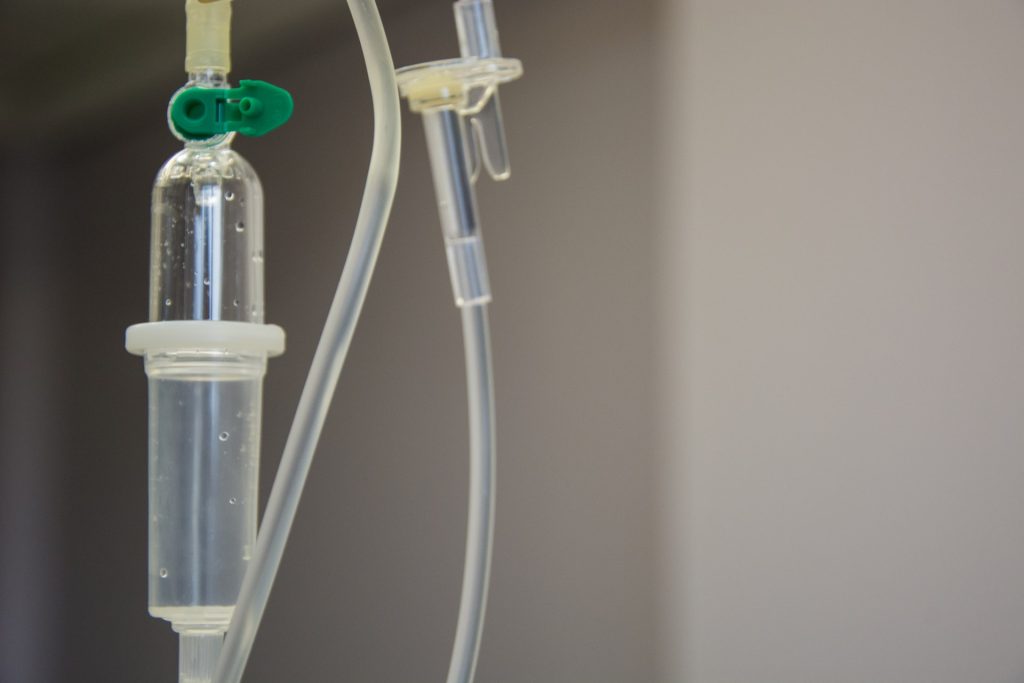
Intravenous IV drip
Source: Marcelo Leal on Unsplash

Over the past two decades, numerous complex cancer therapies called antibody drug conjugates (ADCs) have been tested in clinical trials and approved for use in patients. An analysis of multiple scientific databases has found that some ADCs have higher rates of adverse effects. The findings were reported in the journal CANCER.

An ADC has a complex structure comprised of a monoclonal antibody (MoAb) that targets a protein expressed on cancer cells, a toxic compound to kill the targeted cells (also called a payload or warhead), and a linker to join the two. The clinical efficacy and toxicity of ADCs are affected by each component. Data have established that ADCs as anticancer therapeutics have a unique mechanism of action, which includes the targeted delivery and release of its payload at the tumours site through MoAb antibody components, thus exerting simultaneous roles as both targeted therapy and chemotherapy

In 2000, gemtuzumab ozogamicin was the first ADC approved by the US Food and Drug Administration, and more than a dozen ADCs have been approved worldwide to date. To investigate the side effects associated with different ADCs, a team led by Prof Hong Zhu of Xiangya Hospital, Central South University, in China, conducted a systematic review and meta-analysis of published clinical trials of ADCs that reported treatment-related toxicities.

The researchers found 2511 records of 169 relevant trials involving 22 492 patients. The incidence of treatment-related adverse events was 91.2% for all events and 46.1% for serious adverse events (grade 3 or higher). The most common adverse events overall were lymphopenia, nausea, neutropenia, vision blurriness, and peripheral neuropathy. The most common serious adverse events were neutropenia, hypaesthesia, thrombocytopenia, neutropenia with fever, and lymphopenia. Certain ADCs were linked with higher average incidences of adverse events.

“Different ADCs appear to vary in their treatment-related adverse events. Our results provide an important reference for clinicians and patients on how to address ADCs’ toxicity in clinical practice,” said Prof Zhu.

Source: Wiley

Irradiating only Select Lymph Nodes Reduces Cancer Recurrences

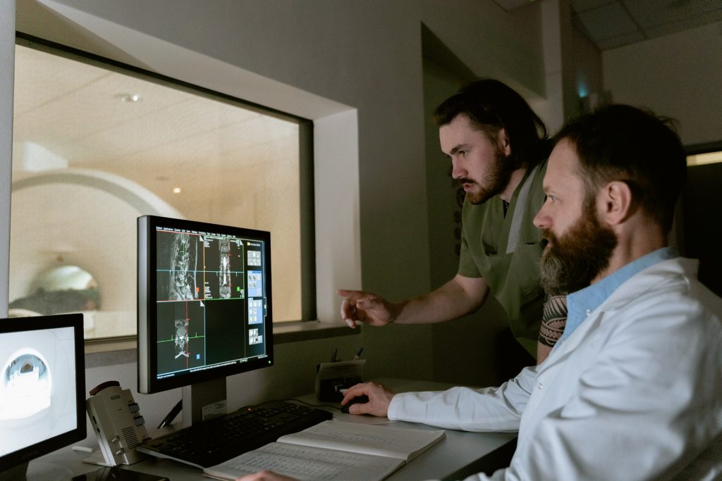
MRI or CT machine
Photo by Mart Production on Pexels

A new study published in Nature Communications suggests that the recurrence of certain cancers can be significantly decreased by only irradiating a select set of lymph nodes near a tumour instead of all of them. By doing so, some immune cells that can recognise the cancer are able to survive.

“Doctors have traditionally irradiated all the lymph nodes surrounding a tumour in a process called ‘elective nodal irradiation,'” said senior author Sana Karam, MD, PhD, associate professor of radiation oncology at the University of Colorado School of Medicine. “But the problem with this scorched-earth approach in the era of immunotherapy is that doing so also eliminates the source of immune cells for the immunotherapy to work on. Lymph nodes are the hub for priming and expanding the major immune cells that can go and fight the cancer.”

Karam and her team learned that irradiating all of the lymph nodes around a tumour significantly decreased immune memory and antigen spread, creating a much greater risk of metastasis. “That was an unexpected finding,” says Karam.

The team tested this theory pre-clinically on several different tumour models of head and neck, along with a breast cancer and melanoma model. The results remained the same: when radiation killed immune cells that recognised the cancer, the patient’s immune system lost the ability to fight the cancer systemically.

On the other hand, the researchers found that irradiating certain lymph nodes known as sentinel lymph nodes was absolutely critical. Failure to irradiate those resulted in recurrence nearby.

“This study confirms why elective nodal irradiation has long been the clinical standard of care, as it does decrease regional recurrence,” said Laurel B. Darragh, the paper’s first author and an MD/PhD student in Dr. Karam’s lab. “But it also shows that to combine radiation with immunotherapy effectively, we need to re-evaluate our strategies and restrict nodal irradiation to the sentinel lymph nodes. This eliminates regional neck recurrence, and far away spread, while preserving long-term, systemic immune response.”

The findings were corroborated by data from a recent human patient clinical trial where pre-surgical radiation was limited to the gross tumour and the sentinel lymph nodes while avoiding all other lymph nodes so as to sustain immune memory. Researchers found that the immune system in these patients was highly activated, which has been shown to correlate with a better prognosis.

“We’re hopeful that this data will set the stage for future clinical trial design, not only reducing patients’ side effects but also improving long-term outcomes,” said Karam.

Source: University of Colorado Anschutz Medical Campus

Exercise Improves Quality of Life in Breast Cancer Radiotherapy

Tired woman after exercise
Photo by Ketut Subiyanto on Pexels

Radiotherapy is an important part of breast cancer treatment but can lead to cancer-related fatigue and negatively impact patients’ health-related quality of life. Fortunately, latest research by has revealed exercise may make radiotherapy more tolerable for patients, offering benefits for their emotional, physical and social wellbeing.

Researchers at Edith Cowan University (ECU) included 89 women in the study, with 43 completing a home-based 12-week program, consisting of a weekly exercise regime of one to two resistance training sessions and an accumulated 30–40 minutes of aerobic exercise. The 46 controls did not participate in the exercise regime.

The results, published in Breast Cancer, showed that patients who exercised recovered from cancer-related fatigue quicker during and after radiotherapy compared to the control group and saw a significant increase in health-related quality of life post radiotherapy with no reported adverse effects.

Study supervisor Professor Rob Newton said this showed home-based resistance and aerobic exercise during radiotherapy is safe, feasible and effective in accelerating recovery from cancer-related fatigue and improving health-related quality of life.

“A home-based protocol might be preferable for patients, as it is low-cost, does not require travel or in-person supervision and can be performed at a time and location of the patient’s choosing,” he said.

“These benefits may provide substantial comfort to patients.”

Important changes observed

Australia’s current national guidelines for cancer patients recommend moderately intense aerobic exercise for 30 minutes per day, five days a week, or vigorously intense aerobic exercise for 20 minutes a day for three days a week.

They also call for 8–10 strength-training exercises with 8–12 repetitions per exercise, for two-to-three days per week.

However, study lead Dr Georgios Mavropalias said benefits were still observed with less exercise.

“The amount of exercise was aimed to increase progressively, with the ultimate target of participants meeting the national guideline for recommended exercise levels,” he said.

“However, the exercise programmes were relative to the participants’ fitness capacity, and we found even much smaller dosages of exercise than those recommended in the national guidelines can have significant effects on cancer-related fatigue and health-related quality of living during and after radiotherapy.”

The study also found participants good adherence to exercise programmes once they started. The exercise group reported significant improvements in mild, moderate and vigorous physical activity up to 12 months after the supervised exercise programme finished.

“The exercise programme in this study seems to have induced changes in the participants’ behaviour around physical activity,” Dr Mavropalias said.

“Thus, apart from the direct beneficial effects on reduction in cancer-related fatigue and improving health-related quality of life during radiotherapy, home-based exercise protocols might result in changes in the physical activity of participants that persist well after the end of the program.”

Source: Edith Cowan University

Temporary Low-protein Diet could Enhance Colon Cancer Treatment

Photo by Ella Olsson on Pexels

A brief switch to a low-protein diet could be a key to enhancing colon cancer treatment, say researchers investigating cancer metabolism. They reported their findings in the journal Gastroenterology.

Like all cells, cancer cells need nutrients to survive and grow. One of the most important nutrient sensing molecules in a cell is called mTORC1. Often called a master regulator of cell growth, it lets cells sense different nutrients, thereby growing and proliferating. When nutrients are limited, cells dial down nutrient sensing cascade and turn off mTORC1.

While mTORC1 is known to be hyperactive in colon cancer, the key question is whether colon tumours hijack nutrient sensing pathways to fire up the master regulator.

“In colon cancer, when you decrease the nutrients available in the tumours, the cells don’t know what to do. Without the nutrients to grow, they undergo a kind of crisis, which leads to massive cell death,” said senior author Yatrik M. Shah, PhD, professor at Michigan Medicine.

Researchers found in cells and in mice that a low-protein diet blocked the nutrient signalling pathway that fires up a master regulator of cancer growth.

The regulator, mTORC1, controls how cells use nutritional signals to grow and multiply. It’s highly active in cancers with certain mutations and is known to cause cancer to become resistant to standard treatments. A low-protein diet, and specifically a reduction in two key amino acids, changed the nutritional signals through a complex called GATOR.

GATOR1 and GATOR2 work together to keep mTORC1 in business. When a cell has plenty of nutrients, GATOR2 activates mTORC1. When nutrients are low, GATOR1 deactivates mTORC1. Limiting certain amino acids blocks this nutrient signalling.

Previous efforts to block mTORC have focused on inhibiting its cancer-causing signals. But these inhibitors cause significant side effects — and when patients stop taking it, the cancer comes back. The study suggests that blocking the nutrient pathway by limiting amino acids through a low-protein diet offers an alternative way to shut down mTORC.

“We knew that nutrients were important in mTORC regulation but we didn’t know how they directly signal to mTORC. We discovered the nutrient signalling pathway is just as important to regulate mTORC as the oncogenic signalling pathway,” said study first author Sumeet Solanki, Ph.D., a research investigator at the Rogel Cancer Center.

Researchers confirmed their findings in cells and mice, where they saw that limiting amino acids stopped the cancer from growing and led to increased cell death. They also looked at tissue biopsies from patients with colon cancer, which confirmed high markers of mTORC correlated with more resistance to chemotherapy and worse outcomes. Solanki said this could provide an opportunity to direct treatment for patients with this marker.

“A low-protein diet won’t be standalone treatment. It has to be combined with something else, such as chemotherapy,” Solanki said.

The risk with a low-protein diet is that people with cancer often experience muscle weakness and weight loss, which limiting protein could exacerbate.

“Putting cancer patients on a protein-deficient diet long-term is not ideal. But if you can find key windows – like at the start of chemotherapy or radiation – when patients could go on a low protein diet for a week or two, we could potentially increase the efficacy of those treatments,” Shah said.

Further research will refine this concept of a therapeutic window to limit amino acids. Researchers will also seek to understand how these pathways are creating resistance to treatment and whether an inhibitor could block the GATOR complexes.

Source: Michigan Medicine – University of Michigan

Metastases Survive by Adapting to Different Tissues

Source: National Cancer Institute on Unsplash

In a breakthrough for understanding metastases, researchers have found that, as metastatic cancers spread to different parts of the body, they adapt their metabolism to the tissue in which they grow. The findings, which help further break down the puzzle of metastasis, are published in PNAS.

Metabolism in the body is an important target for cancer treatments, where the focus is on stopping the progress of cancer cells.

“Obviously, the local environment affects the cancer cells more than previously known. The metastatic tumours should show the same metabolic properties no matter where in the body they are located, but we discovered that the cancer cells largely adapted their metabolism to the new tissue in order to continue to develop and grow. This is important knowledge, which shows that we cannot consider the metastases as their original tumours,” says Fariba Roshanzamir, PhD in Systems and Synthetic Biology at Chalmers and the study’s lead author.

Cutting off cancer metabolism

Fariba Roshanzamir works in Professor Jens Nielsen’s research group at Chalmers and has, together with Swedish and international colleagues, been able to establish the groundbreaking results. The study focused primarily on triple-negative breast cancer but the conclusions can, according to the researchers, be applied to all types of metastatic cancer. This opens new doors to develop more effective treatments.

“If we manage to shut down the metabolism in a tumour, it will stop working and this study provides important keys to better understand what to target. Selecting metabolic inhibitors that specifically target the metastases in the organs to which the tumour has spread, rather than treating them as their original tumours, is of great importance to be able to find good strategies for treatments in the future,” she says.

Source: Chalmers University of Technology

X Chromosome is Shut Down in Some Male Cancers

Source: NIH

Researchers report in Cell Systems that they have discovered another difference between cancer cells and normal cells besides mutations: the X chromosome, typically only inactivated in XX female cells, can also be inactivated across different male-derived cancers.

“To balance the expression of genes between the sexes, in normal development, one copy of the female X chromosome is inactivated at random across the human body. We wanted to know if this process that occurs in normal development goes awry in genetically unstable male or female cancer cells,” says senior author Srinivas Viswanathan, a cancer geneticist and medical oncologist at the Dana-Farber Cancer Institute.

By using publicly available datasets comprising of thousands of DNA samples from cancer patients around the world, the team of researchers stumbled upon the high expression of XIST – the gene responsible for shutting down gene expression on the X chromosome – in about 4% of the male cancer samples analysed.

While XIST may be expressed in very early development in all sexes, X inactivation is thought to be a female-specific process later in development. It was previously shown that some female cancer cells may lose the ability to turn off one of the X chromosomes, leading to increased X-linked gene expression, but this ability of X inactivation had still only been studied primarily in female cells.

Within the 4% of anomalous male cancer samples identified, 74% were from reproductive cancers already shown to inactivate the X chromosome, but that left 26% of samples from other cancer types. These included liver, brain, skin, heart, lung, and thyroid cancers.

“We were very surprised by this result since XIST is a transcript typically used to classify female cancers, and so we wanted to ensure that this was not merely a result of mis-annotation. Yet, we do in fact see that some male cancers of diverse subtypes activate XIST and display features of X inactivation,” says Viswanathan.

“We have to be aware of the caveats of working with these types of datasets. These samples have been in many people’s hands, and there is more room for human error,” said co-corresponding author Cheng-Zhong Zhang, cancer biologist at the Dana-Farber Cancer Institute. “This is the biggest source of uncertainty for us; we have to be creative in how we look at the data and find controls.”

One possible explanation for why this phenomenon is occurring is genetic instability. Cancers often have multiple copies of chromosomes, and if two X chromosomes happen to be in one cell, then it may be necessary to inactivate one of them by activating XIST, regardless of whether that cell is in a female or male individual.

“Another possibility is there are some important genes on the X chromosome that, when silenced, enable the cancer to grow. We will investigate this in future studies,” says Viswanathan.

“In some ways, sex is the ultimate biomarker in that it subdivides the human population, but we often don’t think about how genetic differences between the sexes may inform cancer prognosis or response to therapy,” says Viswanathan.

Source: Cell Press

Could More Fruit & Veg Help Male Sexual Health Issues?

Banana
Photo by Mike Dorner on Unsplash

A systematic review has revealed that plant-based or plant-heavy diets may offer a level of protection against prostate cancer and other male sexual health issues according.

The analysis included 23 studies, 12 of which included prostate cancer, and suggested a link between a plant-based diet and reduced prostate cancer risk. Some evidence also suggested benefits for erectile dysfunction and benign prostate hyperplasia. The findings were reported at the Sexual Medicine Society of North America (SMSNA) annual meeting.

“Medicine has moved to a more holistic approach overall, and with that, more researchers have started to look into [the question of] ‘Can we use these plant-based diets to help manage and prevent conditions like prostate cancer, erectile dysfunction [ED], and benign prostate hyperplasia [BPH]?’ Nathan Feiertag, MD, a medical student at Albert Einstein College of Medicine in New York City, told MedPage Today. “There were relatively few studies that we were able to find for this literature review, but that’s the current state.”

With the growing popularity of plant-based diets, studies have shown their benefits for patients with hypertension or diabetes. Dr Feirtag said that less is known about their effect on prostate cancer, ED and BPH.

Dr Feiertag told MedPage Today that “Urologists can maybe consider our review as an opportunity to incorporate or modify existing diet counselling for their patients, especially the ones who are eager to implement lifestyle changes, particularly as it pertains to prostate hyperplasia, ED, and prostate cancer.”

The review mostly consisted of cohort studies, along with cross-sectional studies, and a handful of randomised controlled trials. Studies included those on vegan diets, vegetarian diets, and plant-heavy diets, such as the Mediterranean diet. In a number small cohort studies, there was a significant decrease in prostate cancer velocity, though not sustained at six months, Dr Feiertag said.

Two of the five ED studies found a link between plant-based diets and improved International Index of Erectile Function scores, though one reported worsening scores. The two studies included on ED reported a reduced relative risk of ED for patients on plant-based diets. For BPH, five of six studies reported an inverse relationship between plant-based diets and developing BPH.

Limitations including not being generalisable due to the number of observational and cohort studies that relied on patient-reported evaluations of diet. Additional high-quality studies are needed to confirm the link between diet and urological conditions.

Fortunately, the studies all reported no non-association or no harmful effects of following a plant-based or plant-forward diet. “For the patients who want to change their diet, this is useful for them. It definitely won’t hurt,” Dr Feiertag told MedPage Today.

Source: MedPage Today