North Korean defectors who resettled in South Korea share genetics but markedly contrasting early-life exposures with South Korean residents. Research published in the Journal of Internal Medicine compared overall and site-specific cancer incidence rates between North Korean defectors and native South Koreans.
Breast cancer cells. Image by National Cancer Institute
Using the Korean National Health Insurance database, researchers matched 25 798 North Korean defectors and 1 276 601 South Korean residents. Defectors had higher risks of infection-related cancers (such as liver and cervical cancers) and lower risks of breast, colon, and prostate cancers (which are more prevalent in developed countries). Over time, though, their cancer profile changed, suggesting adaptation to South Korean society.
“The study provides a model for understanding how cancer epidemiology evolves in such transitions, offering lessons that may help guide prevention and health planning for other vulnerable groups in transition worldwide,” said corresponding author Sin Gon Kim, MD, PhD, of the Korea University College of Medicine.
Credit: Darryl Leja National Human Genome Research Institute National Institutes Of Health
A major UK clinical trial co-led by researchers at UCL will test whether lower doses of common hormone therapies can treat advanced prostate cancer while reducing the severe side effects many patients experience.
The ENHANCE trial, jointly funded by Cancer Research UK and Prostate Cancer UK, will recruit 1,500 men with advanced prostate cancer from hospitals across the UK to compare standard and half-dose treatment using four commonly prescribed hormone drugs. UCL is sponsoring the trial and Professor Allan Hackshaw (Director of CRUK & UCL Cancer Trials Centre, UCL Cancer Institute) is joint-lead investigator, alongside Professors Ananya Choudhury and Peter Hoskin at the University of Manchester.
Men diagnosed with advanced prostate cancer, where the disease has spread beyond the prostate, are typically treated with powerful hormone therapies that slow tumour growth. While these drugs can extend life, they can also cause debilitating side effects such as extreme fatigue, hot flushes and high blood pressure, which can make it difficult for some patients to remain on treatment.
Researchers hope the £3.2 million ENHANCE trial will show that lower doses of these drugs can be just as effective while improving patients’ quality of life. The trial has not yet been launched and is awaiting the relevant regulatory and ethics approvals.
Professor Allan Hackshaw said: “Although focus is often on cancer trials to improve survival, we also need to find more tolerable ways of treating cancer without compromising survival. Side effects of cancer therapies matter a lot to patients, especially when they are frequent.
“We believe that several modern cancer drugs can be given at a much lower dose than what they were licensed for. Not only would this improve patient’s quality of life, but healthcare costs would be lower allowing more access especially in countries that cannot afford these drugs at their current high dose. The ENHANCE study can therefore influence clinical practice worldwide.
“Furthermore, Black men are more likely to get prostate cancer, and they too suffer from the same side effects of hormone treatment. They are often under-represented in clinical trials, so the ENHANCE study is a good opportunity to show that they get the same benefits from lowering the treatment dose as other ethnic groups.”
ENHANCE will compare full and half-dose treatment across four hormone therapies widely used for advanced prostate cancer: abiraterone, enzalutamide, darolutamide and apalutamide. If successful, the findings could influence prostate cancer treatment guidelines in the UK and internationally as early as 2030, improving care for thousands of men and reducing costs for the NHS.
Dr Ian Walker, Executive Director of Policy at Cancer Research UK, said: “Thanks to research, there’s been huge progress in prostate cancer treatments. Today, more than 8 in 10 men diagnosed with the disease in the UK will survive for 10 years or more. There’s more that can be done to save even more lives though, and in addition to finding more effective treatments, we need to find kinder ones too. The ENHANCE trial is looking to do just that and could mean that men affected by prostate cancer live not just longer lives but have a better quality of life.”
Dr Matthew Hobbs, Director of Research at Prostate Cancer UK, said: “No man should be forced to compromise between survival and their day-to-day wellbeing. This is a crucial issue for men with prostate cancer. That’s why Prostate Cancer UK is thrilled to be working alongside Cancer Research UK, pooling our resources and expertise to deliver the impact men need by funding this bold new trial that puts men’s wellbeing at its centre.”
Prostate cancer is now the most common cancer in UK men. Around 55 900* men are diagnosed each year. While survival rates have tripled since the 1970s, many men still face difficult side effects from treatment.
Retired solicitor Jonathan Edwards, 80, from Cheshire, experienced severe side effects after starting the hormone-blocking drug enzalutamide following his prostate cancer diagnosis in 2024, but when his nurse reduced the dose, his cancer remained under control and his quality of life improved dramatically.
Jonathan said: “It was such a shock when I was diagnosed. I had several health issues and after many tests was eventually told that I was suffering from prostate cancer and that it had spread beyond the prostate wall to my bones. I was referred to The Christie Hospital for treatment and was prescribed hormone blockers. The side effects made me extremely tired; I was sleeping through the day on and off and I had frequent hot flushes and generally felt weak.
“When the nurse suggested lowering the dose I was not sure what to expect. The difference soon became apparent and I felt normal again. I know that I will stay on the medication for as long as it is effective but, in the meantime, I am able to live a pretty normal life. I now exercise more and do not usually need an afternoon sleep. Happily, my PSA level started to go down until, after a few months, it was undetectable and has so far remained undetectable.
“My life has been transformed by the medication, my energy levels are higher, and I can socialise as normal. Traveling was a problem but now I can plan trips as long as I work around the 12-week cycle of injections and consultations. I am delighted that this trial has the potential to help other men going through the same thing in the future by enabling them to be treated for prostate cancer with their quality of life still largely intact.”
At least 10 per cent of trial participants will be Black men. Historically under-represented in clinical trials, Black men are often treated based on data that may be less applicable to them. Although data shows Black men are more likely to develop prostate cancer, more evidence is needed to understand their risk of aggressive disease and the role of overdiagnosis.
Alongside testing lower doses, the trial will collect tissue, blood and urine samples to identify biomarkers that could help determine which men are most suitable for reduced-dose treatment, shaping more personalised care in the future.
*Based on the average annual number of new cases of prostate cancer (ICD10 C61) diagnosed in the UK in the years 2018-2019, 2021.
Patients with leptomeningeal metastasis (LM) have historically had few treatment options. Now, researchers from The University of Texas MD Anderson Cancer Center have found a combination of targeted therapies, tucatinib and trastuzumab, plus the chemotherapy drug, capecitabine, may improve symptoms and extend survival in some breast cancer patients with LM.
The Phase II study, published in Nature Cancer, included 17 female patients with newly diagnosed LM and HER2+ breast cancer. Median overall survival (OS) in those treated with the combination therapy increased from a historical average of 4.4 months to 10 months. At the 18-month mark, 41% of patients were still alive. Under the combination treatment, disease progression also stalled, with a median of seven months before central nervous system progression, and seven of 12 evaluable patients also had improved neurologic deficits.
“The combination achieved a clinically meaningful improvement in overall survival compared to historical controls,” said lead author Rashmi Murthy, MD, associate professor of Breast Medical Oncology. “For these patients, who often face limited treatment options, our results represent a step forward, offering new hope in how we treat and manage leptomeningeal metastasis.”
Why are there limited treatments for patients with leptomeningeal metastasis?
Leptomeningeal metastasis is difficult to treat primarily because the blood-brain barrier may block drugs from reaching the spinal fluid, where the metastatic cells are found. Additionally, LM is not a solid tumor but is made up of metastatic cells living in fluid, making them more difficult to target. Historically, there also are few studies about this specific disease.
“In addition to encouraging survival outcomes, throughout this study we observed improvements in neurologic symptoms,” said co-lead author Barbara O’Brien, MD, associate professor of Neuro-Oncology. “Treatments for breast cancer leptomeningeal metastasis have historically focused on stabilising disease rather than improving symptoms, making these findings particularly meaningful and encouraging.”
How do the treatments in this combination therapy work?
Tucatinib is a targeted therapy pill that blocks the HER2 protein, which helps some breast cancers grow. Trastuzumab is a targeted antibody that attaches to the HER2 protein on cancer cells and helps the immune system destroy them. Finally, capecitabine is a chemotherapy pill that turns into 5-fluorouracil (5-FU) in the body to eliminate fast-growing cancer cells.
The single arm, non-randomised, multi-phase study enrolled patients at four sites in the U.S., including UT MD Anderson. Eligible patients were at least 18 years old with histologically proven metastatic HER2+ breast carcinoma. These patients were treated with 21-day cycles of oral tucatinib (300 mg) twice daily, plus oral capecitabine (1000 mg/m2) twice daily on days 1-14 and intravenous trastuzumab (6 mg/kg) on day 21.
What are other key findings of the study?
Side effects included diarrhoea, nausea, vomiting, hand-foot syndrome, and liver function test elevation. Most adverse effects improved or resolved with appropriate care and dose modifications. One patient saw alanine aminotransferase elevation after one cycle, which led to discontinuation of the combination, and symptoms resolved after one month.
Study limitations include early termination due to slow accrual following Food & Drug Administration (FDA) approval of the combination therapy. Additionally, LM from HER2+ metastatic breast cancer is rare, resulting in limited published data. As a result, the study design was informed by the small amount of available retrospective evidence.
Credit: Darryl Leja National Human Genome Research Institute National Institutes Of Health
Prostate cancer screening compares favourably to screening for breast cancer in identifying significant cancers, reducing mortality and avoiding unnecessary harms, according to new research. The findings are presented on Sunday 15 March 2026 at the European Association of Urology Congress (EAU26) in London. The research is also accepted for publication in European Urology.
The researchers maintain that the similarities between the two forms of screening mean it is no longer rational to reject prostate cancer screening on one hand while endorsing screening for breast cancer on the other. Nevertheless, they recommend some caution given their research compares a trial with a population-based screening programme and across two different cancers.
Although breast and prostate cancer are the most commonly diagnosed cancers in Europe amongst men and women respectively, screening for the diseases is vastly different. Organised breast cancer screening programmes have been established across Europe for more than three decades. Prostate cancer screening has lagged behind, primarily due to concerns around the effectiveness of the PSA blood test and the risks of overdiagnosis and overtreatment. Nevertheless, many men undergo variable, ‘opportunistic’ screening for the disease, mostly based on self-referral.
Several prostate cancer screening trials in Europe have now reported long-term outcomes, showing a reduced risk of death from prostate cancer [1]. This risk reduction is similar to that seen in breast screening programmes.
The new analysis compares the two types of cancer screening in terms of the effectiveness of the diagnostic tests and levels of overdiagnosis. The researchers, from the German Cancer Research Centre in Heidelberg, Germany, drew on data from the PROBASE prostate cancer screening trial in Germany and the country’s breast cancer screening programme.
They used data from 39,392 men who underwent an initial PSA blood test as part of the PROBASE trial at age 45 or 50. They compared this with data from just over 2.8 million women, aged 50–69, who had a mammography as part of Germany’s organised breast cancer screening programme. They found:
PSA blood testing followed by an MRI scan leads to a higher number of false positives than mammography (37-42% vs 10%).
A similar proportion of men and women were referred for biopsy (0.8-2.4% for men and 1.1% for women) as men in the PROBASE trial were triaged before referral using various factors to determine the likelihood of significant cancer (known as risk stratification)
Biopsies were far more likely to identify significant cancer in prostate screening than in breast screening (50-68% vs 10%), indicating that fewer men were referred for biopsy unnecessarily.
The percentages of invasive cancers identified were similar across both prostate and breast cancer screening (60-74% vs 73%).
Prostate cancer screening was more likely to identify non-aggressive cancers than breast cancer screening (26-31% vs. 22%). However, in prostate cancer the option of active surveillance is well-established, and the researchers maintain this would limit the risk of overtreatment. Active surveillance involves monitoring lower grade cancers and only starting treatment (radiotherapy or surgery) if they progress.
Dr Sigrid Carlsson, who leads Clinical Epidemiology of Early Cancer Detection at the German Cancer Research Centre (DKFZ) in Heidelberg, is lead author of the research. She said: “Until we have a population-based screening programme for prostate cancer, we can’t make an exact like-for-like comparison with breast cancer. But we can make some informed assumptions based on the data from our trial, which shows that if prostate cancer screening were extended to the wider population, then the outcomes are likely to be very similar to breast cancer. Although our study used German data, the findings are applicable to other countries. The final question we now need to answer is: what will this cost compared to what we are already paying for opportunistic screening? And that work is already underway.”
Tobias Nordström is a clinical urologist and Associate Professor at the Karolinska Institute, Sweden and a member of the EAU Scientific Congress Office. He said: “There is much that prostate cancer screening can learn from breast cancer screening and that is why this analysis is an important addition to our knowledge base. As these kinds of comparisons are very challenging, the results do need to be taken with a level of caution. That said, the clear overall similarities between the outcomes for breast and prostate cancer screening show that we are moving in the right direction, ensuring prostate cancer screening offers more benefits than harm.”
New research from a University of Cincinnati Cancer Center study found external beam radiation therapy (EBRT) is safe to administer to patients with liver cancer even after they undergo a targeted internal radiation therapy with Yttrium-90 (Y90).
Led by first author Sarah Feldkamp, MD, and senior author Jordan Kharofa, MD, the research was published in the American Journal of Clinical Oncology. Feldkamp explained that while traditional EBRT delivers radiation from a machine outside the body, Y90 provides carefully aimed treatment for liver cancer through microscopic beads injected into the blood supply.
The location and size of each tumour help clinicians decide which radiation approach to take with each patient. But following Y90, doctors questioned whether the treatment exhausted the liver’s radiation tolerance, meaning additional EBRT could lead to toxicity.
In the study, the team reviewed 94 patients with liver cancer treated with EBRT from 2016 to 2024, including 15 who were additionally treated with Y90.
“EBRT can be delivered after Y90 without an increase in toxicity,” said Feldkamp, a resident in the Department of Radiation Oncology in UC’s College of Medicine. “These results, though not particularly surprising to our team given collective personal experience, do contradict commonly held assumptions by others in the field.”
Kharofa said the study offers “meaningful reassurance” that EBRT can be offered after Y90 when treatment is carefully individualized to each patient.
“That expands the options we can offer patients with residual or recurrent disease, and I think it will change how some clinicians approach this sequencing question,” said Kharofa, senior advisor and chair of the Protocol Review and Monitoring System at the Cancer Center and professor in the Department of Radiation Oncology in UC’s College of Medicine.
“This research suggests that having had Y90 in the past doesn’t automatically close the door on radiation as a next step,” he continued. “The key is working with a team experienced in individualizing treatment plans, and that’s exactly the kind of care we aim to provide.”
Feldkamp noted that the relatively small size of this patient population makes a follow-up study or larger clinical trial more difficult. However, other institutions conducting similar single- or multisite reviews could provide more clarity on liver toxicity following radiation treatment for liver cancer.
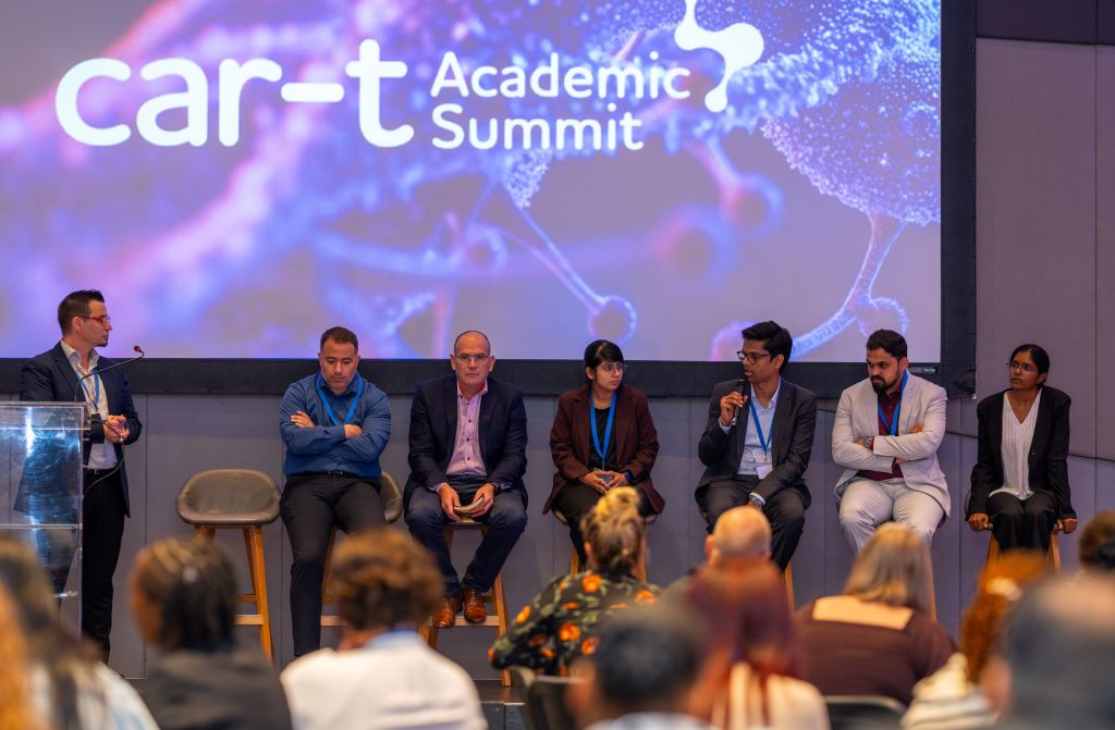
Cipla recently brought together doctors and blood cancer experts for an academic summit to talk about an advanced cancer treatment called CAR‑T cell therapy, and what it could mean for people in Africa in the future.
CAR‑T cell therapy is a form of personalised medicine in which a person’s own immune cells are collected and modified in a specialised laboratory so they can better recognise and attack certain blood cancers. It is used in some countries for patients with specific types of lymphoma and leukaemia when other treatments have not worked. It is only available in a few highly specialised hospitals around the world.
The cost challenge
In the same way that quality, affordable antiretrovirals changed HIV from a fatal disease to a chronic condition in the early 2000s, one of the biggest challenges now is to make CAR-T cell therapy more widely accessible as costs are prohibitively expensive.
CAR‑T cell therapy remains complex and expensive to deliver, and the cost of treatment is a major barrier to access worldwide. In many high‑income countries, the cost of a single CAR‑T treatment can reach the equivalent of hundreds of thousands of US dollars per patient. In South Africa, high‑complexity cellular and stem cell procedures can cost in the order of millions of rand per patient, which means such therapies are beyond the reach of most people in both public and private sectors.
Paul Miller, CEO of Cipla Africa, said: “Treatment costs are a major hurdle for patients. Efforts to develop scientifically rigorous, clinically validated CAR‑T therapies at more sustainable costs could, in future, be very important for patients across Africa.”
Miller added: “Globally, there is increasing focus on making cutting‑edge therapies more accessible. By developing local expertise and manufacturing capabilities, countries can reduce reliance on expensive imports and work toward lowering costs over time.”
How CAR-T cell therapy works
If a patient is eligible, CAR‑T treatment usually starts with collecting some of their white blood cells through a process similar to donating blood. In a special laboratory, these cells are genetically modified so that they can better recognise and target cancer cells. The cells are then multiplied and later given back to the patient in a single infusion.
Studies in other countries have shown that CAR‑T therapy can help some patients with difficult‑to‑treat blood cancers achieve long‑lasting remissions. However, it does not work for everyone and can cause serious side effects, so patients must be treated and monitored in experienced centres.
CAR‑T cell therapy has evolved over several decades, and current research focuses on improving precision, safety, scalability and global accessibility, with the aim of making these treatments available to more patients across more cancer types in future.
Equitable access
Africa carries a heavy burden of both infections and cancer. South Africa, for example, has one of the largest populations of people living with HIV in the world, and these patients have a higher risk of certain blood cancers. This makes access to good‑quality, proven cancer care especially important.
People living with HIV face an increased risk of B‑cell malignancies, including aggressive lymphomas, making the need for effective and equitable cancer care all the more pressing.
Even though cancer treatment has improved a lot in Europe, North America and Asia, most patients in low‑ and middle‑income countries still do not have access to the newest therapies. The main barriers are high cost, the need for advanced laboratories and equipment.
Medical experts with deep clinical experience in environments from South Africa, Morocco and India contributed to the academic programme, bringing a global perspective to an African challenge and sharing important lessons learned.
The promise of CAR-T cell therapy
CAR‑T cell therapy has shown encouraging results in certain relapsed or refractory blood cancers, with some patients achieving deep and durable responses. Internationally, thousands of patients have now received CAR‑T treatment in approved centres.
Gene and cell therapies are subject to strict regulations and rigorous quality standards in many countries. In addition to cost, logistics and the “vein‑to‑vein” traceability chain are important factors that health systems must be equipped to manage.
“Cipla is committed to partnering with healthcare professionals, policymakers and institutions to chart a clear and equitable path for CAR-T therapy access across Africa, ensuring that the most vulnerable patients are not left behind in the next chapter of cancer care,” said Miller.
An analysis of biomarkers in patient blood samples could help with early detection of cachexia, or cancer wasting syndrome, according to new research published in Cancers. The study by Cedars-Sinai Health Sciences University investigators, explores biologic signals detectable in the blood that could be used to design future strategies for assessing patient risk and developing therapies aimed at mitigating fatigue and muscle and fat loss experienced by many patients with cancer.
“We found that in patients with advanced non-small cell lung cancer, cachexia biomarkers change over time,” said Kamya Sankar, MD, co-medical director of the Thoracic Disease Research Group at Cedars-Sinai Cancer and corresponding author of the study. “And treatments targeting one of the early cachexia biomarkers we identified, an inflammatory protein called GDF-15, are already under evaluation in clinical trials.”
Investigators measured the blood of 27 patients with non-small cell lung cancer at two different time points. In patients with early cachexia, they found higher levels of inflammatory proteins such as GDF-15. In patients with later-stage cachexia, they found increased mitochondrial DNA, which comes from the parts of cells that convert food into energy.
Larger, prospective studies are required to validate the clinical benefit of these biomarkers, but they could serve as the basis for risk assessment of patients and may inform design of future clinical trials of therapies for cancer-associated cachexia, Sankar said.
Additional Cedars-Sinai authors include Elham Kazemian, Nicole Lorona, Carlos D. Cruz-Hernández, Mitra Mastali, Akil A. Merchant, Jennifer Van Eyk, Karen L. Reckamp, Neil A. Bhowmick, and Jane C. Figueiredo.
Other authors include Alex K. Bryant and Puneeth Iyenga
Small cell lung cancer cells (green and blue) that metastasised to the brain in a laboratory mouse recruit brain cells called astrocytes (red) for their protection. Credit: Fangfei Qu
Artificial intelligence tools are increasingly being developed to predict cancer biology directly from microscope images, promising faster diagnoses and cheaper testing. But new research from the University of Warwick, published in Nature Biomedical Engineering, suggests that many of these systems may be using visual shortcuts rather than true biology – raising concerns that some AI pathology tools are currently too unreliable for real-world patient care.
“It’s a bit like judging a restaurant’s quality by the queue of people waiting to get in: it’s a useful shortcut, but it’s not a direct measure of what’s happening in the kitchen,” says Dr Fayyaz Minhas, Associate Professor and principal investigator of the Predictive Systems in Biomedicine (PRISM) Lab in the Department of Computer Science, University of Warwick, and lead author of the study.
“Many AI pathology models are doing the same thing, relying on correlations between biomarkers or on obvious tissue features, rather than isolating biomarker-specific signals. And when conditions change, these shortcuts often fall apart.”
To reach this conclusion, the researchers analysed more than 8000 patient samples across four major cancer types – breast, colorectal, lung and endometrial – and compared the performance of leading machine learning approaches. While the models often achieved high headline accuracy, the team found this frequently came from statistical “shortcuts.”
For example, instead of detecting mutations in the cancer-associated BRAF gene, a model might learn that BRAF mutations often occur alongside another clinical feature such as microsatellite instability (MSI). The system then learns to use this combination of cues to predict BRAF status rather than learning the causal BRAF signal itself – meaning accurate cancer predictions work only when these biomarkers co-occur and become unreliable when they do not.
Kim Branson, SVP Global Head of Artificial Intelligence and Machine Learning, GSK and co-author says, “We’ve found that predicting a BRAF mutation by looking at correlated features like MSI is often like predicting rain by looking at umbrellas – it works, but it doesn’t mean you understand meteorology.
“Crucially, if a model cannot demonstrate information gain above a simple pathologist-assigned grade, we haven’t advanced the field; we’ve just automated a shortcut. The roadmap for the next generation of pathology AI isn’t necessarily bigger models; it’s stricter evaluation protocols that force algorithms to stop cheating and learn the hard biology.”
When performance of AI models was assessed within stratified patient subgroups, such as only high-grade breast cancers or only MSI-positive tumours, accuracy fell substantially, revealing that the models were dependent on shortcut signals that disappear once confounding factors are controlled.
For certain prediction tasks, the performance advantage of deep learning over human-derived clinical information was modest. AI systems achieved accuracy scores of just over 80% when predicting biomarkers, compared with around 75% using tumour grade alone – a measure already assessed by pathologists.
Machine learning methods can still prove valuable for research, drug development candidate screening and for clinical triaging, screening, or supplementary decision support. However, the researchers argue that future AI tools must move beyond correlation-based learning and adopt approaches that explicitly model biological relationships and causal structure.
They also call for stronger evaluation standards, including subgroup testing and comparison against simple clinical baselines, before looking at deployment in routine care.
Dr Minhas concludes, “This research is not a condemnation of AI in pathology. It is a wake-up call. Current models may perform well in controlled settings but rely on statistical shortcuts rather than genuine biological understanding. Until more robust evaluation standards are in place, these tools should not be seen as replacements for molecular testing, and it is essential that clinicians and researchers understand their limitations and use them with appropriate caution.”
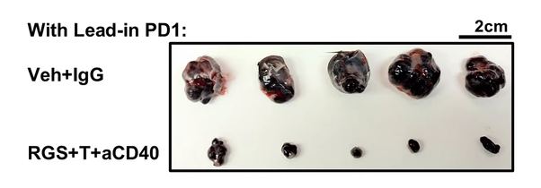
For patients with advanced melanoma without BRAF mutation who no longer respond to immune checkpoint inhibitors, treatment options remain frustratingly limited. A new study from Vanderbilt researchers led by Professor Emerita of Pharmacology Ann Richmond outlines a promising therapeutic strategy that may re-sensitise these resistant tumours to immunotherapy.
Figure 5b from the paper shows the difference between tumours treated with a just an antibody and vehicle (solution) or with trametinib and rigosertib and a CD40 agonist. Image cropped and shared from the paper by Yan et al. published in Nature Communications in 2026 in accordance with a CC BY-NC-ND 4.0 license.
The research introduces a three-drug combination that enhances immune activity and suppresses tumour-promoting immune cells by leveraging a low dose of the MEK inhibitor trametinib and multi-kinase inhibitor rigosertib alongside a CD40 agonist to shift the tumour microenvironment toward immune activation. Notably, all three agents have been either approved by the U.S. Food and Drug Administration or are currently in clinical trials, which may speed their path to patient testing.
“While agonist CD40 therapy can be helpful for treatment of melanoma, this therapy also induces the CD11b+ B regulatory cells that suppress the T cell response to tumours,” Richmond said. “We showed that combining CD40 therapy with trametinib and rigosertib prevents the induction of these B regulatory cells.”
Immune checkpoint inhibitors have become a mainstay of melanoma treatment, working by releasing the molecular “brakes” that prevent T cells from attacking cancer. But resistance to ICI is common in metastatic melanoma, especially in tumours that evolve immune-suppressive microenvironments. While CD40 agonists can activate immune cells, this therapy also unexpectedly expands CD11b+ regulatory B cells.
By combining CD40 activation with MEK and PI3K inhibition, the researchers blocked the expansion of suppressive B cells while retaining the benefits of CD40 stimulation. In preclinical mouse models of melanoma, the triple combination not only suppressed tumour growth but also restored responsiveness to checkpoint blockade.
Key findings
B cells as a resistance mechanism: CD40 therapy alone induced regulatory B cells that dampen T cell–mediated tumor immunity.
Triple combination prevents immune suppression: Co-treatment with trametinib and rigosertib blocked the agonist CD40 induction of regulatory B cells, allowing immune responses to proceed.
ICIs regain effectiveness: The drug cocktail slowed tumor progression and re-sensitized resistant melanomas to anti-PD-1 therapy.
Translational promise
Because trametinib, rigosertib, and CD40 agonists are already in human trials or approved for other indications, this therapeutic strategy may advance more quickly than approaches requiring new drug development. Richmond’s team sees potential for testing the triple therapy in clinical trials for melanoma patients who have progressed on ICI.
“This approach provides a new route to enhance antitumor immunity in patients with tumors that no longer respond to immunotherapy,” Richmond said.
Long-term data suggests an overall cure rate of 42%
Photo by National Cancer Institute on Unsplash
Unlike some other forms of lymphoma, advanced stage follicular lymphoma is considered incurable. But a new analysis of long-term data on patients treated for the disease years ago with standard regimens of immunotherapy and a chemotherapy combination known as CHOP suggests that many of those patients can now be considered cured.
The analysis is just published in the journal JAMA Oncology.
“A subset of advanced-stage follicular lymphoma patients can achieve cure with CHOP-based chemoimmunotherapy, as relapse rates decline over time,” said Wilmot Cancer Institute Director Jonathan W. Friedberg, MD, MMSc, at the University of Rochester Medical Center, who is senior and corresponding author on the paper.
“This finding represents a paradigm shift in our understanding and approach to follicular lymphoma, with broad implications for initial patient discussions and future research strategies.”
Historically, follicular lymphoma has been considered an incurable disease, with most patients experiencing relapses even years after initial treatment.
The JAMA Oncology paper reports on an analysis of follow-up data from patients with advanced follicular lymphoma who had been treated with a standard first-line chemoimmunotherapy regimen on a large clinical trial.
Roughly 70 percent of the patients remained alive 15 years after beginning treatment, and a statistical method known as cure modelling estimated that 42% of treated patients had been cured.
Cure modelling incorporates background mortality rates in an analysis of patient survival data to estimate what fraction of a group of patients can be considered cured of a disease. Such modelling accounts for the fact that over time some patient deaths will occur that are unrelated to the given disease.
The researchers applied a cure model to 15-year follow-up data from the S0016 clinical trial conducted by the SWOG Cancer Research Network, a clinical trials group funded by the National Cancer Institute (NCI), part of the National Institutes of Health (NIH), with the participation of other groups within the NCI-funded National Clinical Trials Network (NCTN).
This phase 3 trial, which opened in 2001, enrolled patients with untreated advanced-stage CD20-positive follicular lymphoma and randomised 531 of them to one of two treatments, both of which were built around a core chemotherapy regimen known as CHOP (cyclophosphamide, hydroxydaunorubicin, vincristine, and prednisone). One arm treated patients with rituximab plus the CHOP combination (R-CHOP), while the other arm used CHOP followed by radioimmunotherapy (CHOP-RIT). Primary results of the S0016 trial were published in 2013 (Press, OW. J Clin Oncol. 2013).
The S0016 modeling, including cure analysis, was carried out by Michael LeBlanc, PhD, a biostatistician at Fred Hutch Cancer Center and director of SWOG’s Statistics and Data Management Center (SDMC), and Hongli Li, MS, also at Fred Hutch and the SWOG SDMC.
It showed that, with a median follow-up time of 15.5 years after a patient had begun treatment, the rate of disease relapse dropped substantially over time, falling from 6.8% of patients relapsing in the first 5 years to only 0.6% relapsing between years 15 and 20.
Fifteen years after starting treatment, about 70%of patients remained alive. The analysis also showed no statistically significant difference between the two treatment arms in the rates of 15-year overall survival.
Based on their work, the authors conclude that a substantial subset of patients with advanced-stage follicular lymphoma can, when treated with a standard regimen that includes immunotherapy and chemotherapy, achieve a functional cure – defined as having no chance of lymphoma recurring during the patient’s expected lifespan.
“These results reinforce that front-line chemoimmunotherapy remains an important option – particularly for appropriate patients – because it can deliver long-term disease control after a time-limited course of treatment, ” said first author Mazyar Shadman, MD, MPH, of Fred Hutch Cancer Center. Shadman is medical director of cellular immunotherapy at Fred Hutch, where he holds the Innovators Network Endowed Chair.
“As we bring novel agents into the first-line setting, the durability seen here sets a high benchmark; new strategies should aim not only to improve short-term response rates but to match or exceed long-term remission and cure potential.”
The idea that many of these patients can be cured could change how newly diagnosed patients are counseled and could eliminate the need for indefinite oncology and radiologic follow-up visits after treatment, with patients eventually transitioning from oncology care back to a primary care team.