Author: ModernMedia

Oxford Researchers Develop Uniquely Shaped Microstent to Combat Glaucoma

A schematic of the eye’s anterior segment, demonstrating the anatomical placement of the microstent. The stent diverts aqueous humour from the anterior chamber to the suprachoroidal space through the flexible tube, creating a subconjunctival bleb supported by the expanding element. Credit: Yunlan Zhang, Zhong You, Jared Ching.

A team of researchers at the University of Oxford have unveiled a pioneering ‘microstent’ which could revolutionise treatment for glaucoma, a common but debilitating condition. The study has been published in The Innovation, Cell Press.

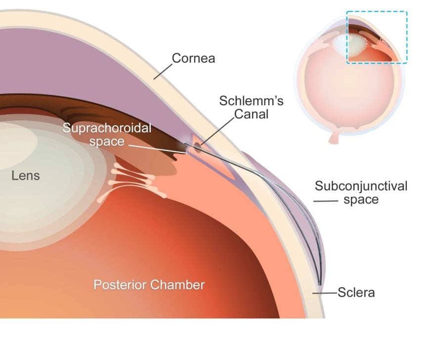
Glaucoma is a leading cause of vision loss, second only to cataracts. Globally, 7.7 million people were blind or visually impaired due to glaucoma in 2020. The condition can cause irreversible damage to the optic nerve, due to increased pressure within the eyeball. Current treatment options – principally surgery to create openings in the eye or insert tubes to drain fluid – are highly invasive, carry risk of complications, and have limited durability.

‘Our deployable microstent represents a significant advancement in glaucoma treatment,’ said lead author Dr Yunlan Zhang (University of Oxford at the time of the study/University of Texas). ‘Current surgical implants for this type of glaucoma have been shown to have limited long-term effectiveness, being susceptible to failure due to fibrosis (scarring) in the eye.’ 

The new microstent features a unique structural shape that allows it to expand once in the eye. At 200µm, less than a quarter of a millimetre, the stent’s tiny diameter enables it to fit within the needle of a standard hypodermic syringe, for minimally-invasive insertion. Once in place and expanded, the microstent spans the fluid-filled space between the white of the eye and the membrane that covers it.

By supporting this space, the stent reduces the excessive fluid buildup and resulting intraocular pressure in the eye which is responsible for the most common type of glaucoma, primary open-angle glaucoma. Initial trials carried out in rabbits found that the microstents lowered eye pressure in less than a month with minimal inflammation and scarring. Furthermore, the microstent achieved a greater reduction of eye pressure than a standard tubular implant.

This development has the potential to transform the landscape of glaucoma therapy. By offering an enhanced solution in the minimally invasive glaucoma surgery field that combines mechanical innovation with biocompatibility, we hope to improve patient outcomes and quality of life.

Senior co-author Dr Jared Ching (Department of Engineering Science, University of Oxford).

Senior co-author, Professor Zhong You (Department of Engineering Science, University of Oxford) said: ‘Our microstent is made from a durable and super-flexible nickel-titanium alloy called nitinol, renowned for its proven long-term safety for ocular use. Its unique material and structural properties help prevent subsequent movement, improve durability, and ensure long-term efficacy.’

The research team used advanced modelling techniques to guide the microstent’s design and ensure compatibility with the anatomy of the eye. The device’s superelastic properties enable it to accommodate how the eye changes and stretches over time without permanent deformation, enhancing its durability and functionality.

Over half a million people in the UK have glaucoma – 2% of everyone over the age of 40 – and it is one of the most common causes of blindness worldwide. The introduction of this microstent could mark a pivotal step in enhancing treatment efficacy and accessibility.

The study ‘A Novel Deployable Microstent for the Treatment of Glaucoma has been published in The Innovation, Cell Press.

Source: Oxford University

Gene Therapy Shot at Birth May Shield Children from HIV for Years

Colourised transmission electron micrograph of an HIV-1 virus particle (yellow/gold) budding from the plasma membrane of an infected H9 T cell (purple/green).

A new study in Nature shows that delivering a single injection of gene therapy at birth may offer years-long protection against HIV, taking advantage of a critical window in early life that could reshape the fight against paediatric infections in high-risk regions.

This study is among the first to show that the first weeks of life, when the immune system is naturally more tolerant, may be the optimal window for delivering gene therapies that would otherwise be rejected at older ages.

“Nearly 300 children are infected with HIV each day,” said first author Amir Ardeshir, associate professor of microbiology and immunology at the Tulane National Primate Research Center, who conducted the study alongside fellow researchers at the California National Primate Research Center. “This approach could help protect newborns in high-risk areas during the most vulnerable period of their lives.” 

“This is a one-and-done treatment that fits the critical time when these mothers with HIV in resource-limited areas are most likely to see a doctor.”

Amir Ardeshir

In the study, nonhuman primates received a gene therapy that programs cells to continuously produce HIV-fighting antibodies. Timing proved critical to the one-time treatment offering long-term protection.

Those that received the treatment within their first month of life were protected from infection for at least three years with no need for a booster, potentially signifying coverage into adolescence in humans. In contrast, those treated at 8–12 weeks showed a more developed, less tolerant immune system that did not accept the treatment as effectively.

“This is a one-and-done treatment that fits the critical time when these mothers with HIV in resource-limited areas are most likely to see a doctor,” Ardeshir said. “As long as the treatment is delivered close to birth, the baby’s immune system will accept it and believe it’s part of itself.”

More than 100 000 children acquire HIV annually, primarily through mother-to-child transmission after birth from breastfeeding. Antiretroviral treatments have shown success in suppressing the virus and limiting transmission. However, adherence to treatment and access to doctors both decline after childbirth, particularly in areas with limited access to healthcare.

To deliver the treatment, researchers used an adeno-associated virus (AAV), a harmless virus that can act as a cargo truck to deliver genetic code to cells. The virus was sent to muscle cells, unique in their longevity, and delivered instructions to produce broadly neutralising antibodies, or bNAbs, which are capable of neutralising multiple strains of HIV.

This approach solved a longstanding problem with bNAbs. Previous studies found them effective at fighting HIV, but they required repeated infusions, which are costly and pose logistical challenges in low-resource settings.

“Instead, we turn these muscle cells – which are long-lived – into micro factories that just keep producing these antibodies,” Ardeshir said.

Newborns showed greater tolerance and expressed high levels of bNAbs, which successfully prevented infection, while older infants and juveniles were more likely to have produced anti-drug antibodies that shut down the treatment.

Researchers also found that exposing fetuses to the antibodies before birth helped older infants accept the gene therapy later, avoiding the immune rejection that often occurs with age.

Still, Ardeshir said a one-time injection at birth offered a more cost-effective and feasible real-world solution, while putting less burden on the mother for a follow-up visit.

Questions remain as to how the results translate to human infants and children, who may be less susceptible to AAV-delivered treatments. The study also used one strain of simian–human immunodeficiency virus, which doesn’t reflect the variety of HIV strains.

If successful, however, this treatment could dramatically reduce mother-to-child HIV transmission rates in high-risk regions such as sub-Saharan Africa, where 90% of paediatric HIV cases can be found. It may also be adapted to protect against other infectious diseases like malaria, which disproportionately affects young children in low-income countries.

“Nothing like this was possible to achieve even 10 years ago,” Ardeshir said. “This was a huge result, and now we have all the ingredients to take on HIV.”

Source: Tulane University

Unusual Allies: Vagus Nerve Cells Emerge as Defenders Against Flu Damage

Study finds sensory cells that detect tissue damage, irritants also rein in harmful immune responses to protect the lungs

Clusters of mouse vagus nerve sensory cells reveal the presence of TRPV1, a molecular sensor that detects irritants, heat, and inflammation. A new HMS study reveals nerve cells with this sensor play a central role in taming inflammation and tissue damage in the lung during flu infection. Image: Chiu Lab.

A group of nerve cells known for their role in detecting chemical irritation, tissue damage, heat, and pressure now emerge as critical defenders against the worst ravages of the flu caused by an overactive immune response, according to new research by scientists at Harvard Medical School and the Harvard T.H. Chan School of Public Health.

The cells, called TRPV1 vagal nociceptors, live in the vagus nerve, which sends signals from internal organs – including the heart, lungs, and gut – to the brain to help regulate heart rate, breathing, digestion, and other functions. In the lungs, these cells trigger the protective cough reflex that forces the airways to expel foreign particles, mucus, and other irritants.

But the new research, published in Science Immunology and conducted in mice, shows that in the setting of flu, these cells do much more – they rein in the immune system and avert the smoldering inflammation that often occurs in the aftermath of a viral infection and can injure healthy tissue.

Each year, the flu sickens millions and kills between 290 000 and 650 000 people worldwide, according to the World Health Organization. While the immune system helps fight off the virus, an excessive inflammatory response can inflict tissue damage and worsen illness. The findings are especially relevant in the wake of the COVID-19 pandemic, which revealed how an aberrant immune response following viral infection can sometimes lead to serious organ damage and even organ failure.

“Our research shows that the infected lung is a battleground where nerves and immune cells engage in a delicate dance to safeguard our health,” said co-senior study author Isaac Chiu, professor of immunology in the Blavatnik Institute at HMS. “Understanding this powerful neuro-immune signaling axis will be increasingly important as we design better ways to prevent and treat immune-mediated damage in viral infections, which can sometimes be worse than the direct damage caused by the virus itself.”

The findings, he added, raise the possibility that vagus nerve function may be one variable that explains why some people with the flu go on to develop long-lasting and devastating immune-driven damage in their lungs while others recover once the initial infection is resolved.

“Flu infections are highly variable in severity and there is a need to understand why certain groups of people, such as the elderly, experience worse disease,” said study first author Nicole Almanzar, a doctoral student in the Chiu Lab. “Our study demonstrates that the nervous system is actively involved in regulating the response of the lungs to infection, offering a new perspective on viral infections that could help explain why particular factors increase risk of severe infections.”

The new study illuminates the complex interaction between body and brain and adds to a growing body of research showing that the nervous and immune systems engage in highly orchestrated crosstalk during infection to modulate body defenses.

Previous research led by Chiu has revealed the intricate interplay between the two systems in bacterial pneumoniaflesh-eating diseasemeningitispain, and itch.

One of Chiu’s earlier studies found that during bacterial infections of the lung, the same set of vagal nerve neurons suppressed the immune defences. In the new study, however, the immune-taming function of these cells worked to shield the lung from excessive damage during viral infection.

“Context clearly matters,” Chiu said.

Disabling the neurons worsened flu damage in the lungs

In a set of experiments, researchers exposed a group of mice with genetically disabled or chemically silenced TRPV1 neurons to influenza A virus. Mice without these nerve cells fared notably worse than mice with functioning TRPV1 cells. Even though the overall amount of virus in the lungs was the same in both groups, mice lacking TRPV1 neurons suffered more severe lung pathology, higher levels of harmful inflammation, and lower survival rates. Interestingly, Almanzar noted, even though the overall viral load remained the same, the spread of the virus within the lobes of the lungs was more pronounced in mice without these protective neurons.

The researchers also found that the absence of TRPV1 neurons altered the lung’s immune landscape. The lungs of mice lacking these neurons had an overabundance of neutrophils and macrophages – two types of immune cells that, in excess, can worsen tissue damage. At the same time, interferon signalling – one of the body’s most important viral-defence pathways – was seriously impaired in these immune cells.

In another experiment, the researchers used an antibody to deplete inflammation-driving cells in flu-infected mice lacking TRPV1 neurons. These animals had notably better survival than untreated mice lacking these protective neurons. The observation further underscores how nerve cells help prevent harmful immune reactions that can sometimes be more dangerous than the virus itself.

The researchers noted that they do not yet know precisely how TRPV1 neurons restrain the march of inflammatory cells at the molecular level – a question they plan to explore in subsequent work.

“The vagus nerve is powerfully controlling inflammation, but how it does so remains a mystery to be solved,” Chiu said. “But we’re excited that it plays such a strong role in viral infections.”

Harnessing the immune brake for therapy

Moving forward, this insight opens the door to exciting new avenues for therapy. Instead of only targeting the flu virus or dampening immune activity, the research team said, future treatments could mimic the function of nerve cells to ensure the delicate balance between protective and damaging immune responses is not thrown off.

The idea is not that far-fetched, the researchers said, noting that the FDA recently approved a therapy for rheumatoid arthritis by vagus nerve stimulation.

“Imagine if you could harness this brake to control inflammation in the lungs and beyond,” Chiu said. “By stimulating related circuits where the vagus nerve shuts down immune cells, one could envision treating immune-mediated dysfunction of many kinds, including that caused by viral infections.”

Source: Harvard Medical School

People with High Sensitivity Have Greater Mental Health Risk

Photo by Andrew Neel on Unsplash

New research, led by Queen Mary University of London and published in Clinical Psychological Science, has revealed that highly sensitive people (HSP) are more likely to experience mental health problems compared to individuals who are less sensitive. 

The meta-analysis of 33 studies, the first of its kind, looked at the relationship between sensitivity and common mental health problems such as depression and anxiety. Researchers found there was a significant, positive relationship between the two, concluding that highly sensitive people are more likely to experience depression and anxiety compared to those who are less sensitive.

In the study, sensitivity was defined as a personality trait that reflects people’s capacity to perceive and process environmental stimuli such as bright lights, subtle changes in the environment and other peoples’ moods. Often overlooked in mental health studies and clinical practice, which tend to focus on neuroticism and its association with mental health conditions, this research shows that understanding a person’s sensitivity level is important and can have therapeutic implications. 

People with sensitive personality traits may benefit from different treatment plans

For example, people with more sensitive personality traits may be more likely to benefit from treatment plans which involve techniques such as applied relaxation and mindfulness, which can also prevent relapse. 

Tom Falkenstein, a psychotherapist and a PhD student at Queen Mary University of London, said: “This is the most extensive systematic review on sensitivity and mental health in adolescents and adults to date, and is the first ever meta-analysis on the topic to estimate the impact of this relationship. We found positive and moderate correlations between sensitivity and various mental health problems such as depression, anxiety, post-traumatic stress disorder, agoraphobia and avoidant personality disorder. Our findings suggest that sensitivity should be considered more in clinical practice which could be used to improve diagnosis of conditions.”

“In addition, our findings could help improve treatment for these individuals. Around 31% of the general population are considered highly sensitive, and, as our findings show, are more likely to respond better to some psychological interventions than less sensitive individuals. Therefore, sensitivity should be considered when thinking about treatment plans for mental health conditions. Our work shows it is crucial that the awareness of sensitivity is improved among mental health care professionals, so clinicians and practitioners can recognise the trait in their patients, and tailor treatment to their sensitivity.”

Michael Pluess, Professor in Developmental Psychology at University of Surrey and Visiting Professor at Queen Mary University of London said: “This is the first meta-analysis providing robust evidence that highly sensitive people are more prone to common mental health problems. However, it is important to remember that highly sensitive people are also more responsive to positive experiences, including psychological treatment. Our results provide further evidence that sensitive people are more affected by both negative and positive experiences and that the quality of their environment is particularly important for their well-being.”

The systematic review and meta analysis of 33 studies was carried out by an academic team from several universities including Queen Mary University and the University of Surrey. 

Source: Queen Mary University London

Off-label Use of Ketamine for Chronic Pain is Unsupported by Evidence

Results show no clear evidence of benefit for ketamine in chronic pain and identified an increased risk of adverse effects such as delusions, delirium, paranoia, nausea, and vomiting

Photo by Towfiqu Barbhuiya on Unsplash

The off-label use of ketamine to treat chronic pain is not supported by scientific evidence, a new Cochrane review has found.

Ketamine is an anaesthetic commonly used for procedural sedation and short-term pain relief. Ketamine is also frequently prescribed off-label to manage chronic pain conditions such as nerve pain, fibromyalgia and complex regional pain syndrome. It is one of several NMDA receptor antagonists – a group of drugs thought to reduce pain by blocking certain brain receptors involved in pain signalling.

The review, conducted by researchers from UNSW Sydney, Neuroscience Research Australia (NeuRA), and Brunel University of London, examined 67 trials involving over 2300 adult participants. It assessed five NMDA receptor antagonists: ketamine, memantine, dextromethorphan, amantadine, and magnesium.

Results show no clear evidence of benefit for ketamine in chronic pain and identified an increased risk of adverse effects such as delusions, delirium, paranoia, nausea, and vomiting. Evidence was rated low to very low certainty, due to small study sizes and poor methodological quality.

“We want to be clear – we’re not saying ketamine is ineffective, but there’s a lot of uncertainty,” said Michael Ferraro, Doctoral Candidate at UNSW and NeuRA, first author of the review. “The data could point to a benefit or no effect at all. Right now, we just don’t know.”

Researchers looked at the effects across various chronic pain conditions and dosing strategies but found no clear evidence of benefit in any specific condition or dose. Side effects were a major concern, particularly with intravenous use.

“The most common adverse events we saw were psychotomimetic effects such as delusions, delirium and paranoia, as well as nausea and vomiting.” said Ferraro. “These effects are distressing for many patients. Clinicians often try to balance the dose for pain relief without triggering those symptoms, but this isn’t always achieved.”

The review also found no studies that reported on two key outcomes: whether ketamine reduced depressive symptoms or opioid use. This is notable, as ketamine is often proposed for patients with depressive symptoms or opioid tolerance.

“This group of drugs, and ketamine in particular, are in relatively common use for chronic pain around the world. Yet we have no convincing evidence that they are delivering meaningful benefits for people with pain, even in the short term,” said Neil O’Connell, Professor at Brunel University of London, co-senior author of the review. “That seems a good reason to be cautious in the clinic and clearly indicates an urgent need to undertake high quality trials.”

The authors hope the review will help inform patients and clinicians weighing up potential benefits and harms, and guide future research. While more evidence is needed, this review highlights the importance of high-quality trials to understand whether ketamine has a role in chronic pain care.

“We’ve seen the harm that can come from taking medicines developed for acute pain and applying them to chronic pain, opioids are a prime example. Now we’re seeing a similar pattern with ketamine,” said co-senior author James McAuley, Professor at UNSW and senior researcher at NeuRA . “As opioid prescribing is slowly reduced, there’s a growing demand for alternatives, but we need to be careful not to rush into widespread use without strong evidence.”

Source: Cochrane

Discovery Offers Hope for Breathing Recovery After Spinal Cord Injuries

Innovative research paves way for more effective treatment for ALS and other neurodegenerative diseases

View of the spinal cord. Credit: Scientific Animations CC4.0

Respiratory complications are the most common cause of illness and death for the 300 000 Americans living with spinal cord injury, according to the Christopher & Dana Reeve Foundation.  

But the results of a new study, led by researchers at Case Western Reserve University’s School of Medicine, show promise that a group of nerve cells in the brain and spinal cord, called interneurons, can boost breathing when the body faces certain physiological challenges, such as exercise and environmental conditions associated with altitude.

The researchers believe their discovery could lead to therapeutic treatments for patients with spinal cord injuries who struggle to breathe on their own. Their findings were recently published in the journal Cell Reports.

“While we know the brainstem sets the rhythm for breathing,” said Polyxeni Philippidou, an associate professor in the Department of Neurosciences at Case Western Reserve University School of Medicine and lead researcher, “the exact pathways that increase respiratory motor neuron output, have been unclear – until now.”

The research team included collaborators from the University of St. Andrews in the United Kingdom, the University of Calgary in Canada and the Biomedical Research Foundation Academy of Athens in Greece.

The study

By identifying a subset of interneurons as a new and potentially easy-to-reach point for treatment in spinal cord injuries and breathing-related diseases, the researchers believe doctors may be able to develop therapies to help improve breathing in people with such conditions.

The study showed that blocking signals from these spinal cord cells made it harder for the body to breathe properly when there was too much CO2 in the blood, a condition known as hypercapnia.

“These spinal cord cells are important for helping the body adjust its breathing in response to changes like high CO2 levels,” Philippidou said.

In this study, the team used genetically modified mouse models to explore the pathways involved in breathing. The researchers mapped neuron connections, measured neuron electrical activity, observed the models’ behaviour and used microscopy to visualise neuron structure and function – all focused on spinal cord nerve cells involved in breathing.

“We were able to define the genetic identity, activity patterns and role of a specialized subset of spinal cord neurons involved in controlling breathing,” Philippidou said.

The team is now testing whether targeting these neurons in neurodegenerative diseases like amyotrophic lateral sclerosis (ALS), also known as Lou Gehrig’s disease, and Alzheimer’s disease can help restore breathing.

Source: Case Western Reserve University

Ischaemia Speeds Tumour Growth by Aging the Immune System

Immune cells (red) accumulating within a tumour (blue) and multiplying (green). When blood flow is blocked either in the heart or legs, these immune cells change in a way that enables tumour growth. Credit: NYU Langone

Cutting off blood flow can prematurely age the bone marrow, weakening the immune system’s ability to fight cancer, according to a new study from NYU Langone Health.

Published online August 19 in JACC: CardioOncology, the study showed that peripheral ischaemia, restricted blood flow in the arteries in the legs, caused breast tumours in mice to grow at double the rate seen in mice without restricted flow. These findings build on a 2020 study from the same team that found ischemia during a heart attack to have the same effect.

Ischaemia occurs when fatty deposits, such as cholesterol, accumulate in artery walls, leading to inflammation and clotting that restrict the flow of oxygen-rich blood. When this happens in the legs, it causes peripheral artery disease, which can increase the risk of heart attack or stroke.

“Our study shows that impaired blood flow drives cancer growth regardless of where it happens in the body,” says corresponding author Kathryn J. Moore, PhD, tProfessor of Cardiology at NYU Grossman School of Medicine. “This link between peripheral artery disease and breast cancer growth underscores the critical importance of addressing metabolic and vascular risk factors as part of a comprehensive cancer treatment strategy.”

Importantly, the research team found that restricted blood flow triggers a shift toward immune cell populations that cannot efficiently fight infections and cancer, mirroring changes seen with aging.

Systemic Skewing

To examine the mechanisms behind the link between cardiovascular disease and cancer growth, the study authors developed a mouse model with breast tumours and induced temporary ischaemia in one hind limb. The team then compared cancer growth in mice with and without impaired blood flow.

Their findings build on the nature of the immune system, which evolved to attack invading bacteria and viruses, and under normal conditions detects and eliminates cancer cells. These protective functions rely on stem cell reserves in the bone marrow, which can be activated as needed to produce key white blood cell populations throughout life.

Normally, the immune system responds to injury or infection by ramping up inflammation to eliminate threats, then scaling back to avoid harm to healthy tissue. This balance is maintained by a mix of immune cells that either activate or suppress inflammation. The researchers found that reduced blood flow disrupts this equilibrium. It reprograms stem cells in the bone marrow to favour the production of “myeloid” immune cells (monocytes, macrophages, neutrophils) that dampen immune responses, while reducing output of lymphocytes like T cells that help to mount strong antitumour responses.

The local environment within tumours showed a similar shift, accumulating more immune-suppressive cells, including Ly6Chi monocytes, M2-like F4/80+ MHCIIlo macrophages, and regulatory T cells, that shield cancer from immune attack.

Further experiments showed that these immune changes were long-lasting. Ischaemia not only altered the expression of hundreds of genes, shifting immune cells into a more cancer-tolerant state, but also reorganised the structure of chromatin, the protein scaffolding that controls access to DNA. This made it harder for immune cells to activate genes involved in fighting cancer.

“Our results reveal a direct mechanism by which ischemia drives cancer growth, reprogramming stem cells in ways that resemble aging and promote immune tolerance,” says first author Alexandra Newman, PhD, a postdoctoral scholar in Dr Moore’s lab. “These findings open the door to new strategies in cancer prevention and treatment, like earlier cancer screening for patients with peripheral artery disease and using inflammation-modulating therapies, to counter these effects.”

Moving forward, the research team hopes to help design clinical studies that evaluate whether existing inflammation-targeted therapies can counter post-ischaemic changes driving tumour growth.

Source:

Minimally Invasive Procedure Relieves Painful Symptoms of Knee Osteoarthritis

Photo by Towfiqu barbhuiya

A procedure that can be performed under mild sedation in less than two hours by an interventional radiologist relieves chronic knee pain caused by osteoarthritis, an NYU Langone Health study shows.

As they gradually break down, knee joints in people with osteoarthritis are known to become inflamed, which triggers the growth of small blood vessels (angiogenesis) and increased blood flow to joints. The study procedure, called genicular artery embolisation, kept this abnormal blood flow from ferrying in immune cells that cause the inflammation and related pain. 

For the new study, the researchers delivered chemical beads (biocompatible hydrogels) through an image-guided plastic tube to block blood flow in any of a half dozen arteries feeding the synovium lining in the knee. More than 60% of the 25 men and women who had the procedure at its facilities in Manhattan experienced significant improvements one year later.

Results of the study appear online in the Journal of Vascular Interventional Radiology.

“Our study shows that genicular artery embolisation is a safe and effective, minimally invasive treatment for those with moderate to severe symptomatic knee pain tied to osteoarthritis,” said study co-investigator and interventional radiologist Ryan M. Hickey, MD. “This work also provides evidence that genicular artery embolisation is offering more than just pain relief and could be modifying the disease process itself.”

“This procedure is particularly suited to those patients who are either not yet ready for knee replacement surgery or for whom surgery is not an option because of age or other risk factors, such as obesity, uncontrolled diabetes or heart disease, or smoking,” added Dr Hickey.

He says there is urgent need for alternative, less-invasive treatments for osteoarthritis. An estimated 24 million cases of osteoarthritis in a knee are diagnosed each year in the United States, a number he expects only to grow with the aging population.

Among the study’s other key results: significant, one-year postsurgical declines (on average 12%) in blood levels of vascular endothelial growth factor (VEGF), a protein that is needed to stimulate the formation of new blood vessels. Past research has also linked VEGF to other structural changes in the knee from osteoarthritis. Another protein biomarker, interleukin 1 receptor agonist (IL-1Ra), showed a similar decrease (15%). IL-1Ra is known for its role in countering inflammation. Tests of a half dozen other immune molecules involved in inflammation were inconclusive.

“Our research suggests that declines in vascular endothelial growth factor could serve as a valuable biomarker or chemical trace for determining success with genicular artery embolisation, offering a much-needed objective benchmark by which to measure its effectiveness,” said study principal investigator and lead author Bedros Taslakian, MD, MA.

“Our study findings also indicate that genicular artery embolisation may, if further larger clinical trials prove successful, slow down the progression of osteoarthritis by observing significant decreases in signalling proteins, specifically vascular endothelial growth factor and interleukin 1 receptor agonist, one year after the procedure,” said Dr Taslakian.

The improvements seen in the 25 patients in the current study were captured by standard patient survey scores for knee pain, stiffness, and the ability to bend, stand up, or walk up and down stairs freely. While subjective surveys are useful in monitoring disease progression, Dr Hickey says, independent blood tests like the NGF measure are more accurate and convenient for patients for tracking small declines or improvements over time.

The NYU Langone team has plans to further investigate precisely how embolisation alleviates inflammation and leads to pain relief.

Dr Hickey also says that more research is needed to establish how long the procedure’s benefits last and which osteoarthritis patients are most likely to benefit.

All study volunteers were diagnosed with moderate to severe osteoarthritis in the knee that had been unresponsive to first-line therapy. This includes knee injections of corticosteroids to reduce inflammation; fluid aspiration (arthrocentesis) to remove excess fluid from the joint; and injections of platelet-rich plasma to repair damaged tissue, as well as physiotherapy. Study participant ages ranged from 50 years old to 78 years old, with all having their embolisation procedures performed between January 2021 and January 2023.

As part of the procedure, interventional radiologists accessed each patient’s arteries through a small incision in the thigh, using video X-ray to guide the catheter to the precise knee artery selected earlier for embolisation. Study participants were then monitored during routine checkups for at least one year and across two dozen measures of pain, knee stiffness, and their ability to move about. Patients needed to achieve a four-point difference on a scale of 20 points to establish a clinically significant reduction in pain. Side effects from the procedure, the researchers say, were minimal and limited to dark skin blemishes on the knee and mild pain near the incision site.

Source: NYU Langone Health

Kids of Obese Parents More Likely to Develop Obesity due to Inheriting Related Genes

Mom’s genes play a larger role than dad’s in determining whether kids will be obese

A new study finds that kids with obesity are more likely to have obese parents because they inherit obesity-related genes, and to a smaller extent, are impacted indirectly by genes carried by the mother – even when those genes aren’t passed down. A new study led by Liam Wright of the University College London, UK, and colleagues, reports these findings August 5th in the open-access journal PLOS Genetics.

Studies commonly show that children with obesity often have parents with obesity, but the cause of this trend has been poorly understood. Children may inherit genes from their parents that increase their risk of obesity, or they could be shaped by conditions in the womb, or by the food and lifestyle choices their parents make.

In the new study, researchers investigated the effects of the parents’ genetics on the weight and diet of their children. They looked at a measure of obesity called the body mass index (BMI), along with the diet and genetic data from more than 2500 mother-father-child trios. They focused on obesity related genes in the parents – both the ones that were directly passed down to their children, and the genes that weren’t, but that may indirectly impact weight by shaping the child’s environment, which are called genetic nurture effects. They found that, though mothers’ and fathers’ BMIs were consistently correlated with the child’s BMI, this trend could be mostly explained through the genes that children directly inherit. Genetic nurture effects from obesity-related genes in the mother that were not inherited had a smaller impact, only during the child’s adolescence.

The results suggest that a mother’s BMI may be particularly important for determining a child’s BMI, both due to the effects of genes that children directly inherit, and through indirect nurture effects from genes that weren’t passed down. Meanwhile, fathers had little impact on their child’s BMI, apart from the genes that were directly inherited. The study’s authors suggest that analyses that don’t consider the inherited genes are likely to give misleading estimates of the parents’ influence on a child’s weight.

The authors add, “Our results suggest mother’s weight could affect their children’s weight; policies to reduce obesity could have intergenerational benefits.”

Provided by PLOS

Bouts of Exercise Could Help in the Fight Against Cancer

Photo by John Arano on Unsplash

A single bout of either resistance or high intensity interval training could help in the cancer battle, new research from Edith Cowan University (ECU) has found.

ECU PhD student Mr Francesco Bettariga found that a single bout of exercise increased the levels of myokines, a protein produced by muscles which has anti-cancer effects, and which could reduce the proliferation of cancer growth by 20 to 30 per cent.

“Exercise has emerged as a therapeutic intervention in the management of cancer, and a large body of evidence exists that shows the safety and effectiveness of exercise as medicine, either during or post cancer treatment,” Mr Bettariga said, first author of the study which appears in Breast Cancer Research and Treatment.

His research with survivors of breast cancer measured myokine levels before, immediately after and 30 minutes post a single bout of either resistance of high intensity interval training and found that both sets of exercise had a resultant increase in myokine levels.

While higher levels of myokines were expected in a healthy population, post a vigorous workout, Mr Bettariga investigated whether breast cancer survivors would see the same results, given the impact that cancer treatments and cancer itself often has on the body.

“The results from the study show that both types of exercise really work to produce these anti-cancer myokines in breast cancer survivors. The results from this study are excellent motivators to add exercise as standard care in the treatment of cancer,” Mr Bettariga said.

He added that the long-term implications of elevated myokine levels should be further investigated, particularly in relation to cancer recurrence.

Further research by Mr Bettariga investigated how changes in body composition, following consistent exercise, could impact inflammation, which plays a key role in breast cancer recurrence and mortality by promoting tumour progression.

Persistent inflammation not only promotes tumour progression by influencing cell proliferation, survival, invasiveness, and metastasis, but also inhibits immune function. Given that the cancer itself and the side-effects of treatments can elevate levels of inflammatory biomarkers, survivors of breast cancer are at increased risk of cancer progression, recurrence and mortality.

“Strategies are needed to reduce inflammation which may provide a less supportive environment for cancer progression, leading to a lower risk of recurrence and mortality in survivors of breast cancer,” Mr Bettariga said.

The new research found that by reducing fat mass and increasing lean mass, through consistent and persistent exercise, cancer survivors had a better chance at reducing inflammation.

“If we are able to improve body composition, we have a better chance of decreasing inflammation because we are improving lean mass and reducing fat mass, which is responsible for releasing anti and pro-inflammatory markers,” Mr Bettariga said.

Unfortunately, quick fixes to reduce fat mass would not have the same beneficial effects, Mt Bettariga stressed.

“You never want to reduce your weight without exercising, because you need to build or preserve muscle mass and produce these chemicals that you can’t do through just diet alone.”

Source: Edith Cowan University