
Tag: glaucoma

Statins May Protect People from Developing Glaucoma
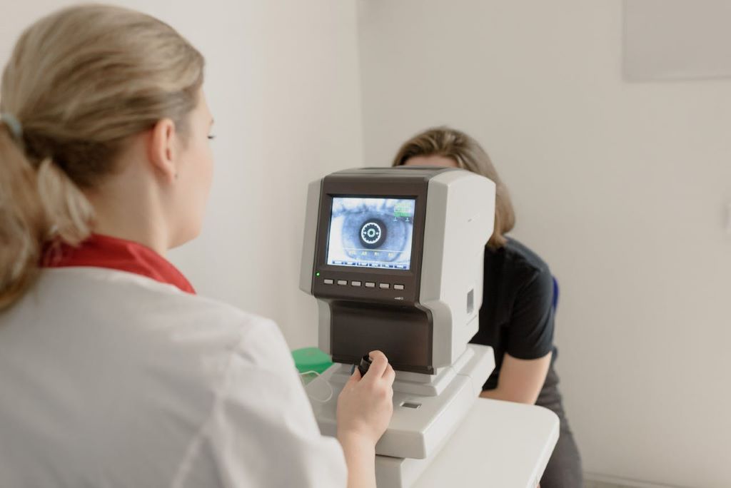
Photo by Ksenia Chernaya

New research from the University of California San Diego School of Medicine has found that taking statins may be linked to a lower risk of developing glaucoma and a reduced chance of it getting worse. The study found that people with existing glaucoma who are taking statins appear less likely to need stronger treatments like extra eye drops or surgery to keep it under control. The findings are published online in the British Journal of Ophthalmology.

“Glaucoma is one of the leading causes of irreversible blindness, and statins are among the most common medications used to lower cholesterol and reduce the risk of heart disease,” said senior study author Robert Weinreb, MD, Distinguished Professor in the Viterbi Family Department of Ophthalmology at UC San Diego School of Medicine and ophthalmologist at UC San Diego Health. Weinreb is also director of the Hamilton Glaucoma Center and the Gleiberman Center for Glaucoma Research at the Shiley Eye Institute.

“If this association is confirmed in future studies, it could have implications for a very large number of people.”

Glaucoma is caused by permanent damage to the optic nerve and is typically treated with prescription eye drops, oral medications, laser treatment or a combination of these approaches. Statins are commonly prescribed to lower cholesterol levels, but studies have indicated that they may also prevent or slow nerve cell death. However, their potential role in preventing or slowing glaucoma is poorly understood.

To strengthen the evidence base addressing this gap in knowledge, researchers searched a database covering 165 million individuals across the world.

They compared more than 210 000 people who had a record of statin use between August 2006 and August 2026 with a matched control group of 210 000 people who had no record of using statins. Individuals in both groups had to have a recorded ophthalmological follow-up.

Statin use was associated with a 15% lower risk of developing open-angle glaucoma, the researchers found.

At five years, there were 4771 new diagnoses of glaucoma in the statin group (2.3%) and 5,491 in the control group (2.6%).

In a separate analysis, more than 30,000 individuals with glaucoma who were taking statins were compared with almost 250,000 individuals with glaucoma who were not taking statins.

Statins were linked to a lower risk of progressing to need glaucoma surgery or other treatment.

At five years, statin use was associated with a 36% lower risk of incisional glaucoma surgery and a 55% reduced risk of minimally invasive glaucoma surgery (MIGS) – a keyhole procedure to lower the intraocular pressure in the eye.

“Statins are known to have pleiotropic effects beyond cholesterol lowering, including anti-inflammatory and potential neuroprotective properties. This study suggests those effects could be relevant to optic nerve health, and future studies will be able to tell us if this is a causal, clinically actionable relationship.”

— Robert Weinreb

Statins were also associated with a 60% lower risk of selective laser trabeculoplasty (SLT) — a procedure that lowers the pressure in the eye by using laser pulses. There was also a 12% reduced risk of eye drops to reduce the pressure in the eye linked to statin use.

After 10 years, statin use continued to be associated with a lower risk of incisional glaucoma surgery, MIGS, SLT, and use of eye drops, the researchers found.

The researchers caution that this is an observational study and as such, no firm conclusions can be drawn about cause and effect. Another limitation of the study is that the researchers did not have access to individual patient data, including intraocular pressure measurements or surgical details. This means they were unable to determine whether surgery was performed due to increased disease severity or because the patient was not adhering to treatment.

Nevertheless, they say their findings add to the growing literature about the neuroprotective effect of statins. Prospective randomized trials are now needed to better understand the effects of statin use on glaucoma incidence and treatment.

“Statins are known to have pleiotropic effects beyond cholesterol lowering, including anti-inflammatory and potential neuroprotective properties,” said Weinreb. “This study suggests those effects could be relevant to optic nerve health, and future studies will be able to tell us if this is a causal, clinically actionable relationship.”

This story was adapted from a press release issued by the British Journal of Ophthalmology.

Read the full study: “Statin Use is Associated with Lower Risk of Open-Angle Glaucoma Incidence and Treatment Escalation”

Source: University of California – San Francisco

New Technology Advances Understanding of Eye Pressure and Glaucoma

For millions of people at risk of glaucoma, healthy eye pressure can mean the difference between preserving vision and losing it. Yet despite decades of research, scientists still do not fully understand what regulates daily changes in eye pressure or how those fluctuations may contribute to glaucoma. A series of three studies led by Christopher Passaglia, USF Health professor and associate chair in the Department of Medical Engineering, is helping fill in those gaps.

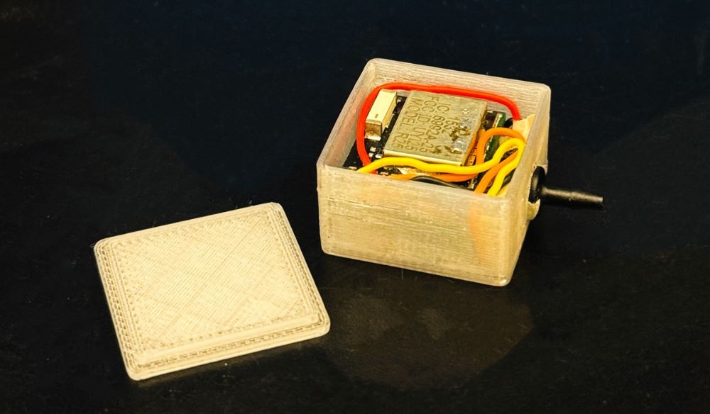
Recently published in Investigative Ophthalmology & Visual Science, Passaglia’s latest research, with USF alumna Alexandra Zamitalo serving as lead author, centres on a first-of-its-kind wireless eye-pressure monitoring system developed in his lab. The technology collects continuous measurements in rats, which share daily eye-pressure rhythms similar to those observed in humans.

Using the technology, researchers investigated how the body’s circadian rhythm may help regulate pressure inside the eye. This internal clock helps manage sleep, hormone production and other daily functions, with light serving as an important signal that keeps it synchronized.

No one was continuously measuring eye pressure until we figured out a way. It was then that we started seeing patterns that had largely been invisible before, providing one of the most detailed looks yet at the biological mechanisms that control eye pressure. This could eventually help researchers develop new approaches for understanding and treating glaucoma.

Christopher Passaglia
USF Professor

Eye Pressure and the Body’s Circadian Rhythm

Most people think of eye pressure as a single number measured during an annual eye exam. But Passaglia notes that eye pressure constantly changes and follows a daily pattern linked to the body’s circadian rhythm.

“Researchers have known for years that eye pressure rises and falls over 24 hours,” Passaglia said. “The problem is that most measurements are taken when patients are awake and sitting in a doctor’s office. What happens while a person is sleeping is rarely captured.”

Passaglia’s monitoring device uses a miniature pressure sensor connected to the rat’s eye through a tiny tube, collecting data while the animals go about their normal activities. Unlike a traditional eye exam, which provides only a snapshot in time, the technology captures pressure changes throughout the day and night.

The measurements showed that eye pressure follows a daily rhythm, rising at night to levels that, if sustained continuously, are associated with glaucoma. Yet the animals do not develop the disease for reasons researchers do not yet fully understand. Researchers also found that the nighttime increase is driven by neural signals traveling from the brain to the eye. That suggests pressure is connected to the biological timing system that coordinates functions throughout the body, rather than being regulated solely within the eye.

“If you think about what the brain does, it’s constantly coordinating activities throughout the body,” Passaglia said. “What we’re showing is that eye pressure appears to be another one of those processes under that kind of control.”

Because elevated eye pressure is a major risk factor for glaucoma, understanding what drives those fluctuations could help researchers determine why some people develop the disease and how it progresses over time.

The Surprising Influence of Light

To test whether circadian rhythms influence eye pressure, researchers disrupted the animals’ normal light-dark cycle by exposing them to constant light.

We were actually surprised by how strong the effect was,” Passaglia said. “When we exposed them to constant light, the rhythm disappeared and mean pressure started climbing. That was a much stronger effect than we expected.”

The results suggest light may play a larger role in regulating eye pressure than researchers previously understood. While the studies were conducted in rats and do not suggest that everyday exposure to artificial light causes glaucoma, they provide further evidence that the eye is closely connected to the body’s internal clock and raises new questions about how disrupted light cycles may affect long-term eye health.

Pathway for Future Glaucoma Research

While the findings are not expected to immediately change how glaucoma is treated, identifying the neural pathways, chemical messengers and eye tissues involved in regulating eye pressure gives researchers a clearer picture of how the system works from the brain to the eye.

By revealing when and how circadian signals influence eye pressure, the research may help explain why some therapies work differently at different times of day and whether treatment timing could influence effectiveness.

“What we’re really trying to understand is the fundamental biology,” Passaglia said. “Once you understand the mechanism, then you can start thinking about better ways to intervene.”

Original written by Joey Garcia, University Communications and Marketing 

Source: University of South Florida

Understanding the Night-time Rise in Intraocular Pressure Could Lead to Glaucoma Treatment

Norepinephrine-induced RHOB is a key regulator of circadian intraocular pressure rhythm

Photo by Cottonbro on Pexels

Glaucoma is an eye disease that causes progressive damage to the optic nerve, leading to loss of sight. One major risk factor is an increase in intraocular pressure (IOP), or the pressure inside the eye. This pressure fluctuates throughout the day and is known to rise at night. However, the detailed molecular mechanisms on why this happens have not been fully understood.

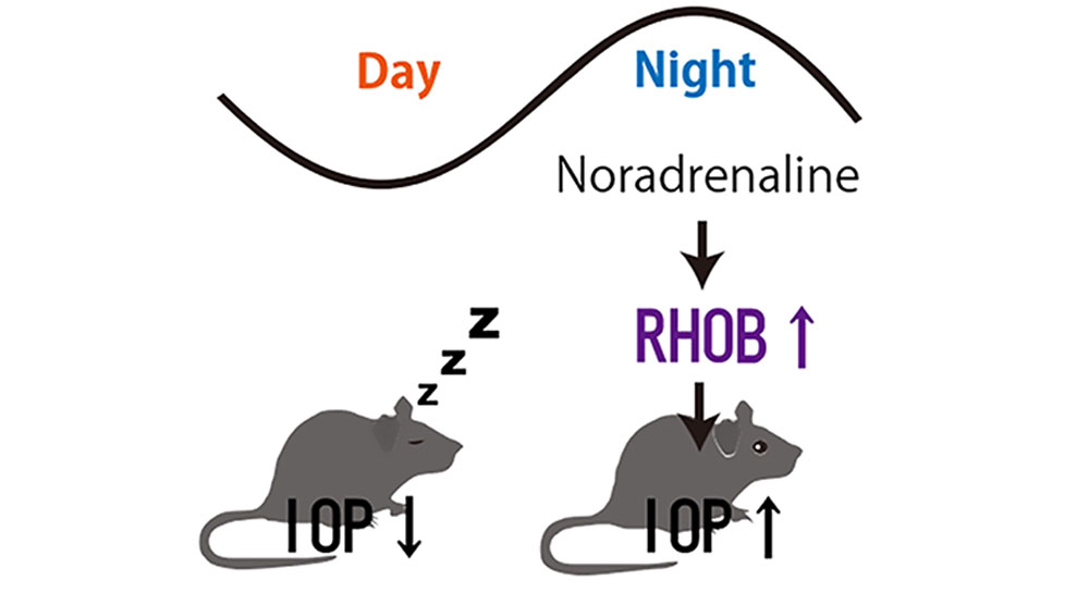
Using human cells and mouse models, researchers at Kyushu University have found a new molecular pathway that explains why IOP increases at night. The neurotransmitter norepinephrine (also known as noradrenaline) increases the levels of a molecule called RHOB in the eye’s drainage system called the trabecular meshwork. This weakens the eye’s ‘cleaning function’, resulting in increased IOP. The team expects these findings will lead to the development of new approaches for the early detection of glaucoma, as well as new treatments for controlling IOP by targeting RHOB. Their results were published in the journal Communications Biology.

IOP is maintained by balancing fluid production and fluid drainage within the eye. As with many bodily functions, it is regulated by the circadian clock. In the case of the eye, IOP increases at night. Elevated IOP is a key factor in identifying the onset of glaucoma, but because pressure tends to be lower during the day, regular checkups can potentially miss these warning signs.

Because the circadian clock regulates IOP, disruptions to a person’s internal clock can lead to increased risk of glaucoma. This is why there is a higher risk of glaucoma in the elderly whose body clocks are desynchronising.

Fig.1. Graphical abstract of the research results
Using mice, researchers found that norepinephrine released from the sympathetic nervous system increases RHOB levels and suppresses the eye’s drainage system, thereby contributing to the rise in intraocular pressure at night.

“Previous studies have found that signals from the sympathetic nervous system contribute to the nighttime rise in eye pressure, but that underlying process was not well understood,” explains first author of the study, Associate Professor Keisuke Ikegami from Kyushu University’s Faculty of Agriculture. “Most of the fluid in the eye is drained through a tissue called the trabecular meshwork. These cells also help keep the drainage pathway clear by taking up and removing small particles and waste. We also know that norepinephrine is a chemical that is released by the sympathetic nervous system. We decided to investigate how norepinephrine can change the function of fluid drainage in the eye and whether it can explain why eye pressure increases at night.”

The team began by exposing human and mouse trabecular meshwork cells to norepinephrine and compared changes in their genetic activity. They identified 18 genes that increased in both systems and focused on one called RHOB. RHOB is a molecule that is involved in controlling cell shape, movement, and intracellular transport.

Norepinephrine increased RHOB in the trabecular meshwork cells, and when RHOB was removed from human cells, their ability to take up and clear particles increased. In contrast, increasing RHOB reduced the cleaning activity and fluid movement in the eye. Testing in mice, the team used eye drops that inhibit a chemical pathway that controls RHOB activity, the RHO-ROCK pathway. The results showed that the eye drops reduced the nighttime rise in eye pressure.

While these results are not intended for immediate clinical application, they have identified the RHOB pathway as a new potential target for suppressing nocturnal increase in IOP. While ROCK inhibitors are used in glaucoma treatments today, further verification is needed to determine the most effective time of day for administration and how they alter IOP rhythm.

“Loss of vision from glaucoma occurs slowly, so early detection is crucial. We hope our work will lead to new treatment regimens and strategies for administering medicines to achieve the greatest effect,” concludes Ikegami.

Source: Kyushu University

Common Eye Ointment can Damage Glaucoma Implants, Study Warns

Research shows that petrolatum-based eye ointments can cause the device to swell and potentially rupture, prompting an urgent update to clinical guidance.

Photo by Tima Miroshnichenko


Widely-used eye ointments can cause glaucoma implants to swell and potentially rupture, according to new research from Nagoya University in Japan. This study is the first to show, using clinical and experimental evidence, that petrolatum-based eye ointments can compromise the PRESERFLO® MicroShunt, an implant used in over 60 countries to treat glaucoma.

Glaucoma is an eye disease that damages the optic nerve and can lead to vision loss. It often results from increased intraocular pressure caused by blocked drainage of eye fluid. A recent study estimated that 76 million people globally are affected by glaucoma.

Progression of visual field loss (from left to right) due to glaucoma
(Credit: Ryo Tomita)

MicroShunt is a small filtration device implanted in the eye to improve fluid drainage in glaucoma patients. Compared to traditional surgeries, it lowers post-operative complications and reduces reliance on additional medications.

MicroShunt is made from a styrenic thermoplastic elastomer based on a polystyrene-block-polyisobutylene-block-polystyrene (SIBS) block polymer, which is highly biocompatible, flexible, and less likely to cause inflammation or scarring. However, this material is vulnerable when it comes into contact with hydrocarbon- and oil-based materials. Due to its high oil affinity, exposure to petrolatum-based eye ointments may allow oil components to penetrate the device, causing swelling and potential changes in its shape and flexibility.

The MicroShunt manufacturer’s instructions state that “the MicroShunt should not be subjected to direct contact with petrolatum-based (ie, petrolatum jelly) materials, such as ointments and dispersions.” But this precaution is not widely recognised or consistently followed in clinical practice.

“Swollen MicroShunts can be structurally fragile,” said ophthalmologist and Assistant Professor Ryo Tomita of Nagoya University Graduate School of Medicine, the study’s first author. “During surgery, I observed a rupture in a swollen MicroShunt. If more clinicians are aware of this risk, they will be able to prevent similar problems.”

Tomita and colleagues, including Assistant Professor Taiga Inooka and Associate Professor Kenya Yuki from Nagoya University Hospital and the Graduate School of Medicine collaborated with Dr. Takato Kajita and Junior Associate Professor Atsushi Noro from the Graduate School of Engineering to examine changes in the MicroShunt after exposure to a petrolatum-based eye ointment.

The medical team reviewed clinical cases, while the engineering team conducted laboratory analyses. The findings were published in Graefe’s Archive for Clinical and Experimental Ophthalmology.

Clinical evidence

The clinical study examined seven glaucoma patients whose MicroShunt implants were later removed for different reasons. The results revealed a clear pattern. In three cases, the MicroShunt was exposed outside the conjunctiva, and patients received a petrolatum-based eye ointment. All three explanted devices showed significant swelling, and two of them ruptured.

In three other cases, the MicroShunt remained covered by the conjunctiva, and no ointment was administered. These devices retained their original structure. Crucially, in one additional case, the MicroShunt was exposed outside the conjunctiva, but no ointment was applied. The device did not swell. This indicates that direct contact with the ointment, rather than conjunctival rupture alone, is the primary cause of swelling.

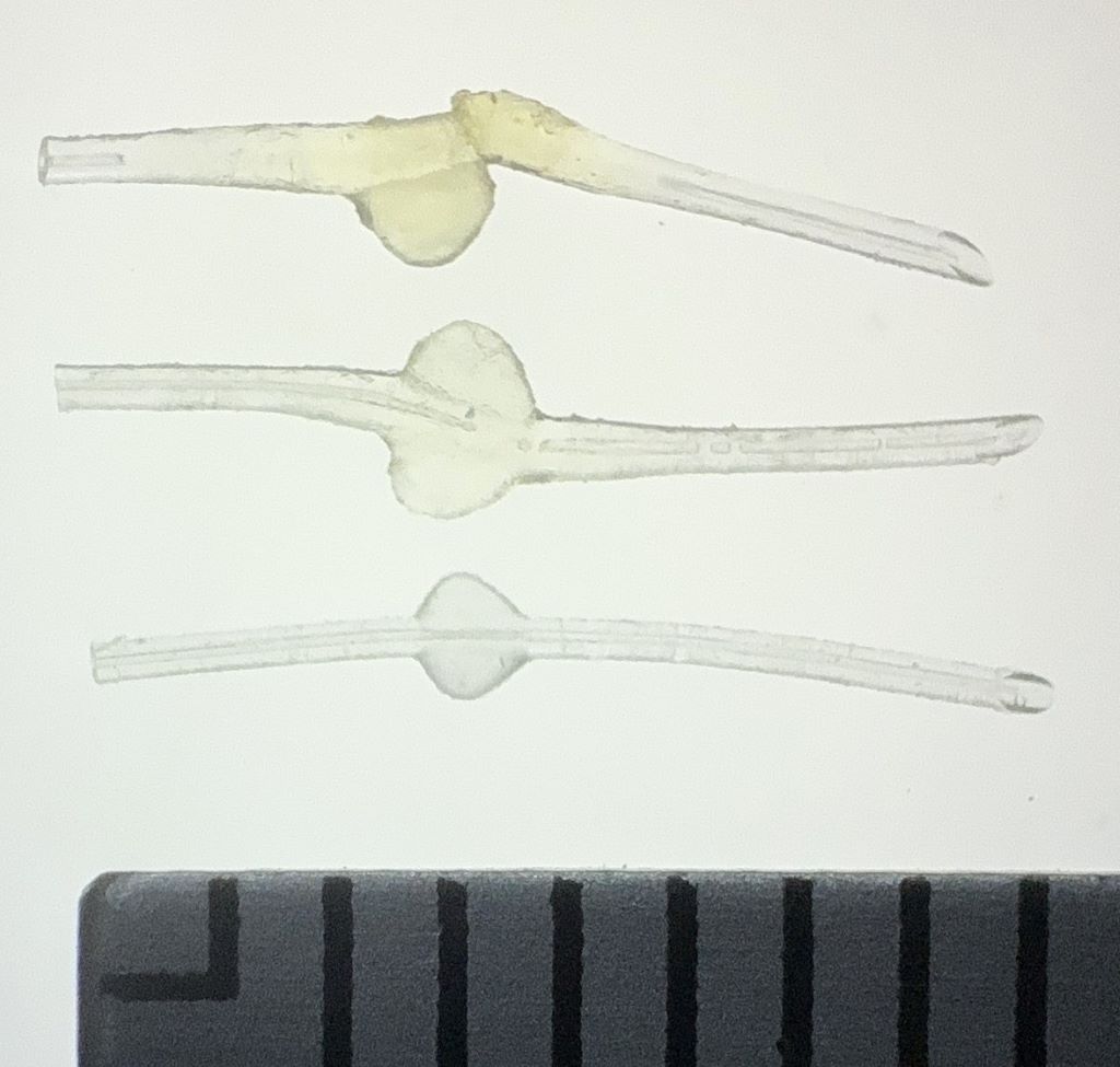
Photographic comparison of MicroShunt illustrating size changes
Top: MicroShunt explanted from a patient, exhibiting diffuse swelling with fracture and loss of one fin
Middle: MicroShunt explanted from another patient, showing localized swelling around the fin
Bottom: Unused MicroShunt (control)

Scale: 1 division = 1 mm   
(Credit: Ryo Tomita)

Laboratory confirmation

Laboratory experiments confirmed the clinical findings. The team immersed unused MicroShunts in petrolatum-based eye ointment to reproduce the swelling seen in clinical cases. Microscopic measurements showed significant changes. After 24 hours in the ointment, the MicroShunt’s outer diameter increased to 1.44 times its original size, and the fin-like portion widened to 1.29 times its initial value.

Chemical analysis identified the cause of this change. After 24 hours of immersion, oil components made up approximately 45% of the MicroShunt’s total weight, rising to 73% after three months. These results confirmed the primary cause of swelling to be the absorption of oil-based ointment constituents into the material.

Clinical implications

The research team emphasises that clinicians should avoid using petrolatum-based ointments on patients with MicroShunt implants, particularly when the device is exposed outside the conjunctiva. Alternative post-operative treatments should be considered, while further research is needed to assess whether swelling impacts MicroShunt performance even when rupture does not occur.

“Our study found that commonly used medical materials can cause unexpected complications if their chemical properties and usage environments are not fully understood,” Noro stated. “From both medical and engineering perspectives, we emphasise the importance of understanding the chemical properties of medical materials and appropriately managing their usage environments.”

Paper information:

Ryo Tomita, Taiga Inooka, Takato Kajita, Hideyuki Shimizu, Ayana Suzumura, Jun Takeuchi, Tsuyoshi Matsuno, Hidekazu Inami, Koji M. Nishiguchi, Atsushi Noro, and Kenya Yuki. (2026) Petrolatum-based ointment application induces swelling of the PRESERFLO MicroShunt. Graefe’s Archive for Clinical and Experimental Ophthalmology
DOI: 10.1007/s00417-025-07075-2

Oxford Researchers Develop Uniquely Shaped Microstent to Combat Glaucoma

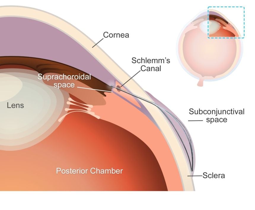
A schematic of the eye’s anterior segment, demonstrating the anatomical placement of the microstent. The stent diverts aqueous humour from the anterior chamber to the suprachoroidal space through the flexible tube, creating a subconjunctival bleb supported by the expanding element. Credit: Yunlan Zhang, Zhong You, Jared Ching.

A team of researchers at the University of Oxford have unveiled a pioneering ‘microstent’ which could revolutionise treatment for glaucoma, a common but debilitating condition. The study has been published in The Innovation, Cell Press.

Glaucoma is a leading cause of vision loss, second only to cataracts. Globally, 7.7 million people were blind or visually impaired due to glaucoma in 2020. The condition can cause irreversible damage to the optic nerve, due to increased pressure within the eyeball. Current treatment options – principally surgery to create openings in the eye or insert tubes to drain fluid – are highly invasive, carry risk of complications, and have limited durability.

‘Our deployable microstent represents a significant advancement in glaucoma treatment,’ said lead author Dr Yunlan Zhang (University of Oxford at the time of the study/University of Texas). ‘Current surgical implants for this type of glaucoma have been shown to have limited long-term effectiveness, being susceptible to failure due to fibrosis (scarring) in the eye.’ 

The new microstent features a unique structural shape that allows it to expand once in the eye. At 200µm, less than a quarter of a millimetre, the stent’s tiny diameter enables it to fit within the needle of a standard hypodermic syringe, for minimally-invasive insertion. Once in place and expanded, the microstent spans the fluid-filled space between the white of the eye and the membrane that covers it.

By supporting this space, the stent reduces the excessive fluid buildup and resulting intraocular pressure in the eye which is responsible for the most common type of glaucoma, primary open-angle glaucoma. Initial trials carried out in rabbits found that the microstents lowered eye pressure in less than a month with minimal inflammation and scarring. Furthermore, the microstent achieved a greater reduction of eye pressure than a standard tubular implant.

This development has the potential to transform the landscape of glaucoma therapy. By offering an enhanced solution in the minimally invasive glaucoma surgery field that combines mechanical innovation with biocompatibility, we hope to improve patient outcomes and quality of life.

Senior co-author Dr Jared Ching (Department of Engineering Science, University of Oxford).

Senior co-author, Professor Zhong You (Department of Engineering Science, University of Oxford) said: ‘Our microstent is made from a durable and super-flexible nickel-titanium alloy called nitinol, renowned for its proven long-term safety for ocular use. Its unique material and structural properties help prevent subsequent movement, improve durability, and ensure long-term efficacy.’

The research team used advanced modelling techniques to guide the microstent’s design and ensure compatibility with the anatomy of the eye. The device’s superelastic properties enable it to accommodate how the eye changes and stretches over time without permanent deformation, enhancing its durability and functionality.

Over half a million people in the UK have glaucoma – 2% of everyone over the age of 40 – and it is one of the most common causes of blindness worldwide. The introduction of this microstent could mark a pivotal step in enhancing treatment efficacy and accessibility.

The study ‘A Novel Deployable Microstent for the Treatment of Glaucoma has been published in The Innovation, Cell Press.

Source: Oxford University

Vitamin Supplements Slow Down the Progression of Glaucoma

Photo by Ksenia Chernaya

A vitamin supplement that improves metabolism in the eye appears to slow down damage to the optic nerve in glaucoma. Promising results have been published in the journal Cell Reports Medicine. The researchers behind the study have now started a clinical trial on patients.

In glaucoma, the optic nerve is gradually damaged, leading to vision loss and, in the worst cases, blindness. High pressure in the eye drives the disease, and eye drops, laser treatment or surgery are therefore used – with varying effect – to lower the pressure in the eye and thus slow down the disease.

Glaucoma researchers have long theorised that the substance homocysteine is somehow relevant to understanding the disease. Now, researchers at Karolinska Institutet have investigated the role of homocysteine in several ways. In the current study, the researchers discovered that when rats with glaucoma were given elevated levels of homocysteine, their disease did not worsen. 

Investigated metabolic pathways

The researchers also found that high levels of homocysteine in the blood of people with glaucoma did not correlate with how quickly the disease progressed, and that glaucoma was not more common in people with a genetic susceptibility to forming high levels of homocysteine. Based on these findings, the researchers concluded that homocysteine does not drive the disease but is a consequence of it.

Since homocysteine is a natural part of the body’s metabolism, the researchers wanted to investigate metabolic pathways involving homocysteine in both rodents and humans with glaucoma. They then saw several abnormalities, the most important of which were metabolic changes linked to the retina’s ability to use certain vitamins. This change meant that metabolism was slowed down locally in the retina – and this played a role in the development of the disease. 

“Our conclusion is that homocysteine is a bystander in the disease process, not a player. Altered homocysteine levels may reveal that the retina has lost its ability to use certain vitamins that are necessary to maintain healthy metabolism. That’s why we wanted to investigate whether supplements of these vitamins could protect the retina”, says co-lead on the paper James Tribble, researcher and assistant professor at the Department of Clinical Neuroscience at Karolinska Institutet.

Promising results lead to clinical trial

In experiments on mice and rats with glaucoma, the researchers gave supplements of the B vitamins B6, B9 and B12, as well as choline. This had a positive effect. In mice that had a slower developing glaucoma, the damage to the optic nerve was completely halted. In rats, which had a more aggressive form of the disease with faster progression, the disease was slowed down. 

In these experiments, eye pressure was left untreated, which the researchers highlight as particularly interesting – it suggests that the vitamin mix affects the disease in a different way than lowering eye pressure does. 

“The results are so promising that we have started a clinical trial, with patients already being recruited at S:t Eriks Eye Hospital in Stockholm”, says James Tribble. 

Both patients with primary open-angle glaucoma (slower progression) and pseudoexfoliation glaucoma (faster progression) are included. 

Read more about the clinical trial here. 

Source: Karolinska Institutet

The Search for an Effective Treatment for Glaucoma

Photo by Ksenia Chernaya

Pete Williams at Karolinska Institutet is one of the few researchers in Sweden concentrating on glaucoma. The goal is an effective treatment, something that stops the degenerative process in the nerve cells of the eye. He is the senior author of a new paper in Nature Communications on how deficiency of the enzyme NMNAT2 renders the nerve cells of the eye vulnerable to neurodegeneration and could be a key in the search for a treatment. 

Glaucoma is very common. Eighty million people worldwide have the eye disease. There is no cure, but there are treatments that lower the pressure in the eye and that can slow down the progression of the disease, which otherwise leads to irreversible blindness.

Not always treatable

“Most people who have heard of glaucoma believe that it can be treated with eye drops and surgery. Unfortunately, this is not entirely true. For many of the patients, the treatment lower the eye pressure but doesn’t prevent further vision loss,” says Pete Williams.

Knowledge about glaucoma has taken time to develop because the disease progresses slowly. This means that in the past, it took many years before researchers could see if a particular treatment had any effect. However, in the last decade or so, the availability of instruments that measure changes in the eye much earlier than the patient experiences them has given new impetus to research into the eye disease.

The importance of NAD

In the 1980s, research into neurodegenerative diseases discovered a link with NAD, a co-enzyme, ie a molecule that binds to an enzyme and makes it active.  Pete Williams’ group was the first to show that NAD levels were low in animal models of glaucoma.

“NAD has many important functions in the body. A lack of it is important for neuronal health and survival in many diseases, but we don’t yet know how to use this information to create a better treatment ” says Pete Williams.

When the body makes NAD, it uses an enzyme NMNAT1. However, in neurons, it needs another enzyme: NMNAT2 (which is only found in neurons). 

“In our recent paper in Nature Communications, we show that NMNAT2 is needed to protect neurons in the eye and that gene therapy can be used to increase levels,” says Pete Willams. “

The research team is now moving on to try to develop new substances that target NMNAT2 in nerve cells. 

Source: Karolinksa Institutet

Many Old People may be Unaware that They Have Glaucoma

Photo by Mari Lezhava on Unsplash

Research on 70-year-olds carried out at the University of Gothenburg, found that nearly 5% had glaucoma – with half of whom were unaware that they had the disease. The study also confirmed hereditary factors were involved in the disease and that intraocular pressure was normal in two-thirds of those newly diagnosed.

Glaucoma is a common eye disease that damages the optic nerve and thereby the field of vision and can lead to blindness. One of the most common risk factors for it is raised intraocular pressure, exceeding the normal range of 11–21mmHg.

The research, published in Acta Ophthalmologica, was carried out by Lena Havstam Johansson, a PhD student at the University of Gothenburg and a specialist nurse at Sahlgrenska University Hospital. The study shows that 4.8% of the 560 study participants examined by eye specialists had glaucoma.

“Of those who were diagnosed with glaucoma via the study, 15 people – or 2.7% of all participants – were unaware that they had the disease before being examined,” says Lena Havstam Johansson. “So half of those who turned out to have glaucoma were diagnosed because they took part in the study.”

For those who were newly diagnosed, the discovery of the disease meant they could start treatment with daily eye drops to reduce intraocular pressure, slowing the progression of optic nerve damage.

Glaucoma impacts some areas of life – but not others

People with glaucoma had similar levels of physical activity to those without the disease and did not smoke more, or drink more alcohol. They rated their overall quality of life as being just as good as others, they were not more tired or more depressed.

“This was a positive surprise, and was a finding that I hope can bring comfort to many people who have been diagnosed with glaucoma. It’s hard to live with a disease that gradually impairs vision, but life can still be good in many ways.”

By contrast, people with glaucoma reported that their vision-related quality of life was poorer.

“It’s harder to climb stairs, see curbs in the evening, and notice things in your peripheral vision. This means that people with glaucoma may avoid visiting others, or going to restaurants or parties, and instead stay at home. They lose their independence, and may feel frustrated about it.”

Ongoing study of 70-year-olds

The research was carried out as part of the H70 study, examining the health of older people, which has been conducted at the University of Gothenburg for fifty years. The H70 study continuously invites all 70-year-olds born in a certain year in Gothenburg to attend several comprehensive physical and cognitive examinations. The 1203 70-year-olds included in the glaucoma study were born in 1944. For these studies, 1182 participants answered written questions about their eye health and the presence of glaucoma in their family. Eye specialists at Sahlgrenska University Hospital also examined 560 of the participants.

The findings confirm that there are hereditary factors behind the disease, as those diagnosed with glaucoma were more likely to have a close relative with the same diagnosis. The results also confirm that glaucoma involves higher intraocular pressure, although they also show that the majority of those who were newly diagnosed (67%) still had normal eye pressure.

During the early stages of the disease, the healthy eye can compensate for the loss of vision, meaning that many people believe their vision is as good as before. These studies confirm that glaucoma often does not initially involve a loss of visual acuity, which may make it harder to detect the disease.

Source: University of Gothenburg

Early Diabetes, Hypertension Accelerates Development of Glaucoma

Credit: National Eye Institute

Developing Type 2 diabetes or hypertension earlier in life is linked to the earlier development of the most common form of glaucoma, according to a study published in Clinical Ophthalmology. These findings could lead to better screening protocols for primary open-angle glaucoma (POAG), the leading cause of irreversible blindness worldwide, which makes up nearly 90% of all cases of glaucoma.

“Currently, we lack the tools to cure glaucoma, but with enough advanced notice, we can preserve patients’ vision. Early detection of glaucoma is the key to better control of intraocular pressure and preventing blindness,” said study leader Karanjit Kooner, MD, PhD, MBA, Associate Professor of Ophthalmology at UTSW.

Tens of millions of people have POAG around the globe. Because this disease has few symptoms in its earliest stages, Dr. Kooner explained, patients are frequently diagnosed in its later stages when vision has already been permanently damaged. Although researchers have identified several risk factors for POAG – including Type 2 diabetes, hypertension, migraines, and obstructive sleep apnoea — how they might influence the onset of POAG is not well understood.

To answer this question, Dr Kooner and his colleagues collected data from the medical records of 389 of his patients with POAG. The researchers found no link between migraines and/or obstructive sleep apnoea and the age of POAG onset. However, the researchers found that the age of Type 2 diabetes and/or hypertension diagnosis was significantly linked with the onset of POAG – the earlier patients presented with either or both of these conditions, the earlier they tended to develop POAG.

Dr Kooner noted that both Type 2 diabetes and hypertension are diseases that affect blood vessels of both the optic nerve and retina, thus potentially causing changes that predispose patients to POAG, another condition with a vascular root. If these connections hold up in future research, he said, Type 2 diabetes and hypertension could be added to the list of factors that can trigger POAG screening — including a family history of POAG, elevated intraocular pressure, and Black race — and lead to earlier diagnosis of POAG, preserving patients’ vision and quality of life.

Source: UT Southwestern Medical Center

Little to No Link Between Glaucoma and Cognitive Function

Older woman smiling
Photo by Ravi Patel on Unsplasj

Previous studies looking for an association between the neurodegenerative disorder glaucoma and cognitive function have produced mixed results. Now, findings from a large study recently published in the Journal of the American Geriatrics Society suggest that any association, if it exists, will only be small.

Glaucoma, the leading cause of irreversible blindness, is a progressive optic neuropathy with incompletely understood pathogenesis that results in progressive vision loss, often beginning with peripheral visual field defects. As a neurodegenerative process, glaucoma is associated with trans-synaptic degeneration in the brain, specifically in the lateral geniculate nucleus and visual cortex. Some prior studies have suggested that the pathogenesis of primary open angle glaucoma (POAG) and normal tension glaucoma (NTG), specifically, may be part of a broad neurodegenerative mechanism with ocular and non-ocular manifestations. Evidence also suggests that impaired vision is associated with a significant increase in the risk of accelerated cognitive decline and incident dementia. Therefore, there is interest in measuring an association between glaucoma and dementia.

The study included 7073 US adults aged 51 years and older who were interviewed by phone every two years. Those who developed glaucoma tended to have higher cognitive function scores but steeper rates of cognitive score decline over a maximum follow-up time of 18 years. The observed associations between glaucoma and cognitive function were small and unlikely to be clinically meaningful. 

“In this large longitudinal study, a diagnosis of glaucoma was not associated with steeper rates of cognitive decline; however, this study did not have access to clinical data to determine whether glaucoma-related vision loss is a risk factor for cognitive decline and dementia,” said senior author Joshua R. Ehrlich MD, MP, of the University of Michigan Medical School. “This is an important question for future studies to consider.” 

Source: Wiley