Ethical patient record keeping is essential to modern healthcare, ensuring safe, effective, and trustworthy care. Accurate documentation supports continuity, clinical decision-making, and legal accountability, while also carrying ethical importance by safeguarding sensitive information with honesty, confidentiality, and respect. Poor record keeping can decrease patient outcomes, create confusion, impact multidisciplinary collaboration, and create legal risk.
Managing records ethically protects patient privacy and autonomy while allowing patients access and control over their health data. With the growth of digital health technologies, new challenges such as data security, electronic informed consent, and cybersecurity have emerged. Breaches or cyberattacks can erode trust, discourage information sharing, and disrupt care delivery, highlighting the need to balance accessibility with strong safeguards.
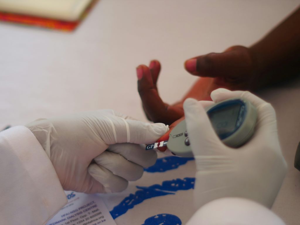
Join our expert speaker panel to explore how you can effectively manage your patient records to ensure integrity and continue to build trust vital to patient-centred healthcare.
During this webinar, attendees will review the HPCSA Booklet 9:Guidelines on the keeping of patient health records where the pertinent South Africa laws and basic principles around ethical practices for maintaining patient health records are laid out. The webinar will offer insights into both paper and electronic record-based practices.
The audience will have an opportunity to engage with faculty who offer insights from a clinical, legal, medical malpractice insurance, cybersecurity, and medical practice setup and management perspective through short lectures, interactive case studies, a series of multiple choices questions and Q&A sessions.
This webinar will focus on specialist practices. Administrative staff working in these practices are welcome to join the discussion.
In this month’s podcast, QuickNews looks at a new Lancet study, “Non-autoimmune, insulin-deficient diabetes in children and young adults in Africa.” In this study, researchers report that a significant subset of what has previously been classified as Type 1 diabetes in sub-Saharan Africa may in fact be a distinct, novel form of the disease.
The individuals in this subset did not exhibit the typical autoimmune markers (islet autoantibodies) usually found in classic Type 1 diabetes in other parts of the world. The researchers instead identified a novel, non-autoimmune, insulin-deficient subtype of diabetes that is also distinct from Type 2 diabetes.
Further evidence of this new subtype was found in Black individuals in the USA, albeit less frequently, but not in White individuals. The discovery throws a spotlight on the heterogeneity of diabetes diagnoses in sub-Saharan Africa, and points to the need to consider alternative causes and explore new prevention and treatment strategies for this distinct form of the disease.
The placenta has long been thought to produce serotonin during pregnancy. But in a new study, Yale researchers shatter the deep-rooted hypothesis – and show that the placenta doesn’t produce serotonin but instead regulates its delivery to the embryo and foetus. They found that serotonin comes from the pregnant parent, with the placenta acting as a “serotonin shield” that controls how much reaches the embryo and foetus.
The findings, published in the journal Endocrinology, could offer critical insights into how a parent’s serotonin levels might affect the development of their baby’s body and brain, the researchers say.
“The placenta is in essence the ‘serotonin shield’ that regulates how much serotonin is ultimately delivered to the embryo and foetus, not the source of serotonin,” said Harvey Kliman, a research scientist in the Department of Obstetrics, Gynecology, and Reproductive Sciences at Yale School of Medicine and corresponding author of the study. “Why does this matter? Because now we correctly know where this delivery is regulated.”
Often called a “happiness hormone,” serotonin regulates mood, so it’s often associated with the brain. In reality, less than 5% of serotonin is made in the brain, with 95% of it made in the gut. But serotonin does more than just regulate mood. It’s also a growth hormone. In the gut, it gets taken up by platelets and is delivered to parts of the body that need to grow, including in wound healing.
During pregnancy, serotonin also helps with growth: It travels into the placenta through a special protein known as the serotonin transporter (SERT) where it plays a critical role in the development of the embryo and foetus.
Serotonin from the mother is taken up by the foetal placenta, which then produces a myriad of hormones, growth factors, and regulators that are delivered to the foetus.
For the new study, researchers sought to better understand these relationships by using a pure source of placenta cells, unlike in previous studies that looked at either whole animals or isolated mouse placentas. To do so, they first purified human cytotrophoblasts, which are the stem cells that make all the cells of the placenta. They then added serotonin to those cells to see where it would go and discovered it concentrated in the nucleus. Next, they used a selective serotonin reuptake inhibitor (SSRI) that blocked SERT, escitalopram, to show that the normal growth, function, and differentiation of these cells was completely blocked.
They also used another inhibitor called cystamine to block serotonylation, or the process by which serotonin is added to proteins like histone 3, which turns genes “on” and “off.” Again, that completely blocked the normal growth of the cells.
Blocking either SERT or serotonylation led to significant changes in gene expression of RNAs in the cytotrophoblasts, they found. Some genes, including ones involved in making, moving, and growing cells, became downregulated, or less active, when serotonin couldn’t enter the cell. Other gene, including ones that help cells stay alive and protect them, became upregulated, or more active. According to the researchers, these findings show that serotonin is critical for the growth of the cytotrophoblasts, the placenta, and by extension, the foetus.
Additionally, researchers discovered that the cytotrophoblasts don’t contain tryptophan hydroxylase (TPH-1), or the enzyme that makes serotonin, indicating the cells within the placenta can’t produce serotonin on their own.
“This suggests that factors that either inhibit serotonin transport through the placenta, or increase it, may have a significant impact on the placenta, embryo, foetus, and ultimately, the newborn and its brain,” Kliman said.
For example, Kliman says this explains why taking SSRIs, which decrease the levels of serotonin into the placenta, leads to smaller babies, and why, conversely, increased levels of serotonin may lead to bigger babies, with bigger brains, who may be at increased risk for developmental disabilities like autism.
Kliman and his lab have long investigated the link between placentas and children with autism, specifically the number of trophoblast inclusions (TIs) in the placenta. TIs are like wrinkles or folds in the placenta, caused by cells multiplying more than they should, typically only seen in pregnancies where there are genetic problems with the foetus.
This new study is the culmination of research first published in 2006 that found significantly more TIs in the placentas from children with autism, and later in 2021, that the genetics of the foetus, and not the parent’s uterine environment, determine how many TIs are in the placenta.
“This puts a big nail into the theory that vaccines cause autism,” suggested Kliman. “Autism, in essence, starts in the womb, not after delivery, and is most likely due to the genetics of the placenta and to a lesser extent, the maternal environment the placenta finds itself in.”
Gut Microbiome. Credit Darryl Leja National Human Genome Research Institute National Institutes Of Health
About a dozen studies in the past five years have made claims linking nearly every type of human cancer with the presence of microbiomes, “communities” of bacteria, viruses and fungi that live in or on peoples’ bodies. Now, scientists at Johns Hopkins Medicine say a study that sequenced human cancers found far less microbial DNA sequences than earlier studies reported in the same cancer tissue samples.
“It’s the nature of science to validate, confirm and reproduce findings,” says Steven Salzberg, PhD, Professor of Biomedical Engineering, Computer Science, and Biostatistics at The Johns Hopkins University. “Over time, we see a more complete picture of new research, and in this case, we did not find any associations between microbiomes and many types of cancer.”
Salzberg says details of the new study, published Sept. 3 in Science Translational Medicine, surveyed the whole genome sequences generated from 5734 tissue samples collected from 25 cancer types and stored in a large National Cancer Institute-funded database, The Cancer Genome Atlas (TCGA). About half of the samples are from normal tissues and blood, the other half from solid tumours and blood-based cancers.
The TCGA’s whole genome sequencing data contains millions of chopped up pieces of DNA molecules, known as reads, from each tissue sample. The original goal of the TCGA studies was to identify mutations in the DNA sequence of genes that might be associated with various cancer types. Sometimes, though, the original tumors might have microbes in them, and the reads could be used to identify those microbes.
Because reads often contain contaminants from bits of DNA left behind in sequencing machinery or picked up from the air or surfaces, samples can acquire DNA from those sources, as well as from the original tumour tissues. Salzberg says extraordinary efforts were made to identify such contaminants, preventing their study from displaying false results.
To rule out contaminants, Salzberg and his team relied on extensive experience with genomic sequencing and careful analysis of control samples to identify reads belonging to sequences known or highly likely to have contaminated samples.
For the current study, a continuation of one that the Johns Hopkins team published in 2023, Salzberg and first author Yuchen “Peter” Ge, a graduate student in biomedical engineering at Johns Hopkins, removed human DNA sequences from the TCGA data sets by mapping each read against two human reference genomes – one from the Telomere-to-Telomere (T2T) project and another from the Genome Reference Consortium.
After removing human DNA, the research team was left with, on average, 2.4 million reads per sample, or about 0.35% of the total 6.5 billion tumour sample reads. Of these, the research team found 323 million human DNA reads that weren’t eliminated in the first pass and 986 million reads they classified as contaminants.
They next compared the remaining sequencing reads against a database containing 50 651 genomes representing 30,355 species of bacteria, viruses, fungi and archaea (single-celled organisms that aren’t bacteria or viruses).
After removing human DNA sequences and contaminants, the average proportion of microbial DNA reads in solid tumour samples was 0.57% and 0.73% in blood cancers.
The Johns Hopkins researchers then compared their new results to a study published five years ago in the journal Nature [since retracted, because of concerns about contaminants in the microbial data], and found the previous study identified 56 times as many microbial reads as the new study for half of the total microbial reads. And 5% of the time, the previous study found 9,000 times the number of microbial reads as the current Johns Hopkins study. Salzberg says the microbial reads in the retracted study were highly likely to be contaminants.
“This disparity in the number of microbial reads didn’t occur in just a few samples,” says Salzberg. “Over the whole study, the previous researchers found far more microbial reads than we did.”
In another comparison of a study published in Cell in 2022 and the current Johns Hopkins work, the 2022 study reported fungal DNA amounts that were hundreds of times more than what was found in the current Johns Hopkins study, largely due to contaminants.
Among the DNA samples in the current Johns Hopkins study, in which they did find microbiome DNA, the researchers found microbes that have long been linked with human cancer, such as HPV (linked with cervical and some head and neck cancers), Helicobacter pylori (linked to stomach cancer), and Fusobacterium nucleatum and Bacteroides fragilis (linked with GI cancers).
The current Johns Hopkins study and the previous ones published in Cell and Nature reported microbiomes of Saccharomyces cerevisiae, commonly known as baker’s yeast. “It’s one of the most common contaminants in sequencing labs,” says Salzberg. They also found a virus that infects plant fungi, Rosellinia necatrix partitivirus 8, which has no known link to human disease.
Salzberg said the need to carefully document claims about the links between cancer and microbiomes is “especially important” as efforts ramp up to diagnose cancers early using microbiome information.
The Johns Hopkins researchers have made their sequencing analysis data available online to other scientists in the supplementary materials in Science Translational Medicine and online.
Scientists are investigating a small region of the brain that plays a major role in memory, spatial navigation, and perhaps Alzheimer’s disease. One of the first parts of the brain affected by Alzheimer’s disease is the entorhinal cortex – a region that plays a big role in memory, spatial navigation, and the brain’s internal mapping system.
With support announced in September from the Commonwealth of Virginia’s Alzheimer’s and Related Diseases Research Award Fund, Virginia Tech scientists Sharon Swanger and Shannon Farris are working to understand why this area is especially vulnerable.
Swanger studies how brain cells communicate across synapses, while Farris focuses on how memory at the molecular level. Their overlapping expertise made the collaboration a natural fit.
“We’ve both been studying for a while,” said Swanger, assistant professor at the Fralin Biomedical Research Institute at VTC. “This new collaborative project brings together my work on synapses and Shannon’s on mitochondria in a way that addresses a big gap in the field.”
“This kind of state-level support is critical,” said Farris, also an assistant professor at the research institute. “It gives researchers in Virginia the chance to ask questions that may eventually make a difference for people living with Alzheimer’s. It’s meaningful to be part of research that could help people facing that journey.”
A key focus of their research is mitochondria – tiny structures inside brain cells that provide the energy needed for a variety of cellular functions in neurons including transmission. In Alzheimer’s disease, mitochondria stop working properly early in the course of the disease.
Farris and Swanger are investigating whether mitochondria in a vulnerable memory-related circuit may become overloaded with calcium, a key signaling chemical for multiple neuronal and synaptic processes. That overload could contribute to the early breakdown of memory.
“The connection between these cells is one of the first to fail in Alzheimer’s,” Farris said. “We found that this synapse has unusually strong calcium signals in nearby mitochondria – so strong we can see them clearly under a light microscope. Those kinds of signals are hard to ignore. It gives us a model where we can really watch what’s happening as things start to go wrong.”
To test their hypothesis, the researchers will study brain tissue from healthy mice and mice with Alzheimer’s. By comparing how mitochondria function and how brain cells communicate across synapses in each group, they hope to find early signs of stress or failure in the entorhinal cortex–hippocampus circuit.
People who take medication for ADHD have a lower risk of suicide attempts, substance abuse, traffic accidents, and criminality than people with ADHD who do not take medication. This is shown in a new study by researchers from Karolinska Institutet and University of Southampton, published in the journal BMJ.
ADHD, which affects about 5% of children and 2.5% of adults globally, is associated with an increased risk of suicide attempts, substance abuse, accidents, and crime, among other things.
The researchers behind the study wanted to investigate whether ADHD medication reduces the risk of these outcomes by analysing Swedish national registry data between 2007 and 2020.
A total of nearly 150 000 individuals between the ages of 6 and 64 with newly diagnosed ADHD were included. The average age in the group was 17, and 41% were women. Of these, 57% started medication, with methylphenidate being the most common drug.
The researchers compared people who had started medication within three months of diagnosis with those who had not, and assessed the outcomes over two years after diagnosis.
Reduction in the risk of serious outcomes
The results showed that ADHD medication was associated with a significant reduction in the risk of several serious outcomes: suicide attempts decreased by 17%, substance abuse by 15%, traffic accidents by 12%, and criminality by 13%. The effects were even more pronounced in certain subgroups – for example, a 25% reduction in substance abuse and criminality was noted in individuals who had had recurring problems with these issues.
One possible explanation is that the medication leads to reduced impulsivity, which can reduce the risk of crime by curbing aggressive behaviour, as well as improved attention, which can reduce the risk of traffic accidents by reducing distractions,” says the study’s last author, Zheng Chang, a researcher at the Department of Medical Epidemiology and Biostatistics, Karolinska Institutet. He continues:
“These results provide evidence that ADHD medication can affect important health and societal outcomes, which should be taken into account both in clinical practice and in the public debate on drug treatment.”
The study is a collaboration between Karolinska Institutet and the University of Southampton. The study was funded by the Swedish Research Council and the Swedish Research Council for Health, Working Life and Welfare, Forte, among others. Some researchers have received fees from pharmaceutical companies, but for work outside the current study.
Reaching the age of 100 does not necessarily mean a life fraught with illness. A new study from Karolinska Institutet shows that centenarians not only live longer, they also stay healthier than other older people, with fewer diseases that develop more slowly.
The study, published in eClinicalMedicine, compared people who reached the age of 100 with those who died earlier. The results show that centenarians not only suffer from fewer diseases, they also develop them more slowly.
While many older people accumulate several diagnoses quickly during their final years, the disease burden of centenarians seems to level off from around the age of 90. They more often have diseases that are limited to a single organ system and significantly fewer concurrent conditions.
The study also shows that cardiovascular disease is less common and occurs later in life among centenarians. Neuropsychiatric diseases are also less prevalent among those who live the longest.
”Our results challenge the widespread belief that a longer life inevitably means more diseases. We show that centenarians follow a distinct ageing curve, with slower disease progression and greater resistance to common age-related diseases,” says last author Karin Modig, associate professor at the Institute of Environmental Medicine, Karolinska Institutet.
Ages in a different ways
The study covered the entire Swedish birth cohort between 1920 and 1922, totalling over 270,000 individuals. The researchers followed the participants’ health from the age of 70 and up to three decades. The progression of disease in centenarians was compared with those who lived shorter lives using national health registers. The results show that centenarians not only delay disease – they seem to age in a fundamentally different way.
”We show that exceptional longevity is not just about delaying ill health. It reflects a unique pattern of ageing. The results suggest that centenarians have preserved homeostasis and resistance to disease despite ageing and physiological stresses – something that may be due to a favourable combination of genes, lifestyle and environment,” says Karin Modig.
The study was funded by Karolinska Institutet. No conflicts of interest have been reported.
Although metformin is the most widely prescribed diabetes drug in the world, its mechanism of action is still not clear. Kobe University endocrinologist OGAWA Wataru has now made significant progress, finding that it changes blood metal levels in humans. Photo by Towfiqu Barbhuiya on Unsplash
The widely used diabetes drug metformin may achieve its effects by changing blood metal levels in humans. The Kobe University study is an important step in understanding the drug’s many actions and designing better ones in the future.
Metformin is the most widely prescribed diabetes drug in the world. Apart from lowering blood sugar levels, it is also known to have a broad range of beneficial side effects such as against tumours, inflammations and atherosclerosis. However, although it has been used for more than 60 years now, its mechanism of action is still not clear, hampering the development of even better drugs against these conditions.
Kobe University endocrinologist OGAWA Wataru says: “It is known that diabetes patients experience changes in the blood levels of metals such as copper, iron and zinc. In addition, chemical studies found that metformin has the ability to bind certain metals, such as copper, and recent studies showed that it is this binding ability that might be responsible for some of the drug’s beneficial effects. So, we wanted to know whether metformin actually affects blood metal levels in humans, which had not been clarified.” To do so, Ogawa and his team enlisted about 200 diabetes patients at Kobe University Hospital, half of which took metformin and half of which did not, in a study to analyse their blood serum levels for those metals and various metal deficiency indicators.
In the journal BMJ Open Diabetes Research & Care, the Kobe University team now published the first clinical evidence of altered blood metal levels in patients taking metformin. They showed that drug-taking patients have significantly lower copper and iron levels and heightened zinc levels. Ogawa says: “It is significant that we could show this in humans. Furthermore, since decreases in copper and iron concentrations and an increase in zinc concentration are all considered to be associated with improved glucose tolerance and prevention of complications, these changes may indeed be related to metformin’s action.”
Recently, Japan has approved the use of imeglimin, a new diabetes drug that is a derivative of metformin but that should not be able to bind metals the same way as its parent. “Imeglimin is thought to have a different method of action, and we are already conducting studies to compare the effects the two drugs have,” says Ogawa.
It is not just about understanding the current drugs, however. Ogawa explains the bigger picture, saying: “We need both clinical trials and animal experiments to pinpoint the causal relationship between the drug’s action and its effects. If such studies progress further, they may lead to the development of new drugs for diabetes and its complications by properly adjusting the metal concentrations in the body.”
A 3D map of the islet density routes throughout the healthy human pancreas. Source: Wikimedia CC0
Cornell researchers have developed an implant system that can treat Type 1 diabetes by supplying extra oxygen to densely packed insulin-secreting cells, without the need for immunosuppression. The system could also potentially provide long-term treatment for a range of chronic diseases.
The findings appear in Nature Communications. The co-first authors were former postdoctoral researcher Tung Pham and doctoral candidate Lora (Phuong) Tran.
The technology builds off previous implantable encapsulation devices developed in the lab of Minglin Ma, professor of biological and environmental engineering in the College of Agriculture and Life Sciences (CALS) and the paper’s senior author.
Ma has explored a variety of ways to address Type 1 diabetes, which is typically managed through daily insulin injections or insulin pumps, but even with that treatment, patients still suffer the devastating effects of the disease.
Ma’s previous implantable devices have proved effective in controlling blood sugar in diabetic mice, but they can only last so long.
“One of the major challenges is the implant itself often dies due to the lack of oxygen after implantation,” Tran said. “In our lab, they had success in micethat lived over one year, and they controlled the diabetes very effectively with some small capsules without oxygen generation. However, when we scale up, we need more cells, we need more density, especially. We need a higher dose. If we implant without generating oxygen, the cells often die within two weeks.”
Ma’s team developed the new system in collaboration with electrochemical researchers from Giner Inc, including co-author Linda Tempelman, PhD ’93.
The key components of the system are a cylindrical capsule with a ring-shaped cross-section that contains transplanted insulin-secreting cells, and an electrochemical oxygen generator that is roughly the size of a dime and removeable. A nanofibrous membrane outside the capsule protects the cells from the host body’s immune system; a permeable membrane in the core of the capsule allows the central supply of oxygen to reach the ring of cells.
“We have to meet two requirements,” Tran said. “The first is immune protection. And second, you have to maintain mass transfer, like the glucose and other nutrients and molecules that can go in and out.”
Working with Dr James Flanders, associate professor emeritus in the College of Veterinary Medicine, the researchers successfully tested the system in rat models.
“It’s the proof of concept. We really proved that oxygenation is important, and oxygenation will support high cell-density capsules,” Tempelman said. “The capsules are immune protective and last for a long time without having some kind of fouling of the membrane. The body never likes when you put a foreign substance in. So that’s the engineering in the Ma Lab, to look for materials and coatings for the materials that are immune protective, but also don’t invoke excess response from the body because of the material.”
The new system would enable a much greater number of the 2 million people who suffer from Type 1 diabetes in the US to now have an islet transplant or cell therapy without requiring immune suppression, which is considered too dangerous for routine use. Also, the new system can provide much tighter sugar control, effectively curing the disease and enabling the person to eat, drink and exercise like everyone else.
The next step will be to implant the system in a pig model, and also test it with human stem cells. The researchers are interested in eventually trying to use the system for implanting different cell types in humans for long-term treatment of chronic diseases, according to Tempelman, who is CEO of Persista Bio Inc., a new startup she founded with Ma and Flanders that is licensing these technologies.
“We see an age where people will be getting implants with allogeneic cells from other human beings, from stem cell lines, and using it long term to treat things that your body is missing,” Tempelman said. “Here we’re missing insulin. In pain control, maybe you need more endorphins or some other molecule. In enzyme replacement therapy, you need more enzymes. We’re interested in things like other autoimmune diseases where there’s inflammation that’s out of control.
“So maybe you could put a small molecule in that would treat inflammation, and then someone, like a Lupus patient, wouldn’t have to take an oral medication. They could just have a low-level implant that provides a low amount of it.”
The blood thinner heparin is used during and after surgery and is essential to kidney dialysis. Most of today’s heparin comes from pigs, but the Federal Drug Administration is encouraging the use of alternative sources, including cows and synthetic forms of heparin, to diversify the supply chain.
Unfortunately, heparin from animals other than pigs just doesn’t work as well.
The reasons are connected to ongoing questions in modern cell biology. Now, an interdisciplinary Virginia Tech team has uncovered new molecular clues that may explain why some sources of heparin are more effective than others. The findings, published recently in the Proceedings of the National Academy of Sciences, may open doors for designing safer, more reliable heparin therapies.
“The structure of heparin and how that structure impacts function is an ongoing puzzle,” said Brenna Knight, first author of the study and recent graduate student studying in the Department of Chemistry. “Seemingly small differences in the content and arrangement of [chemical entities called] sulfates on the molecule cause substantial differences in the energetics that drive chemical activity.”
From mineralization to medicine
Heparin hails from a family called heparan sulfates, or heparans, present in all living creatures. These chains of sugars are diverse, serve many functions in organisms, and many, including heparin, are incredibly complex.
As a student of Patricia Dove in the Departments of Geosciences and Chemistry, Knight was originally looking at heparans for a completely different reason: to understand how the sulfates could impact biological mineralisation, which is the process by which organisms build crystal-strengthened tissues such as bones, teeth, shells, and corals.
Dove is one of today’s pre-eminent geochemists and was elected to the National Academy of Sciences in 2012. Unravelling the process of biomineralisation has been one of her major passions over the past three decades.
“Animals grow crystals in specific places, usually to make structures that serve to support, defend, or feed themselves.” said Dove. “It’s a coordinated result of many chemical reactions within the organism and a crowning achievement of biology. We’ve been trying to better understand the reactions that produce these working biomaterials for a long time.”
That mineralization process unexpectedly linked back to medicine.
Heparan sulfates are just one of many different agents that interact with calcium to trigger a diverse portfolio of biochemical operations. One of those operations is integral to blood clotting.
Team science
To better understand how heparan sulfates help facilitate biomineralisation, Dove and Knight teamed with Kevin Edgar, professor in the Department of Sustainable Biomaterials, who was interested in heparans from the healthcare angle. To study interactions of calcium with heparin, they worked with Michael Schulz and graduate student Connor Gallagher in the chemistry department.
When they applied their combined expertise to calcium-heparin interactions, they found that slight variations in heparin’s molecular composition changed how effective it was at binding calcium. These differences could affect its ability to form biominerals and blood thinners.
“This paper provides insights for how to bioengineer synthetic pathways to effective heparin products for applications in therapeutics and drug delivery,” Edgar said.