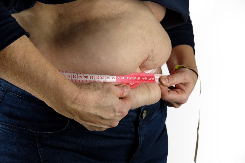
In a Journal of the American Geriatrics Society analysis of nationally representative data from 6905 US adults aged 65 or older followed between 2011–2024, higher body mass index (BMI) was associated with lower mortality risk, whereas higher waist circumference, a marker of abdominal obesity, was independently associated with higher mortality risk after accounting for BMI.
Compared with normal weight, overweight males and males with class I and II obesity (BMI of 30 to less than 40 kg/m2) had 46% and 51% lower risks of mortality, respectively. Additionally, a high waist circumference was also associated with a 24% higher mortality risk in males. Similar patterns were observed in females.
When BMI and waist circumference were evaluated together, being overweight was associated with lower mortality risk regardless of waist circumference among males. Class I and II obesity also remined associated with lower mortality risk exclusively among males with elevated waist circumference. In contrast, being underweight, regardless of waist circumference, and having normal BMI with elevated waist circumference were both associated with higher mortality risk in both males and females.
“BMI and waist circumference provide complementary information about health risk in older adults,” said study co-author, Bryan Blissmer, PhD, of the University of Rhode Island. “Our findings suggest that considering both measures together, rather than relying on BMI alone, may improve mortality risk assessment and support more informed clinical decision-making.”
Technique developed for treating Parkinson’s disease also disrupts growth of glioblastoma
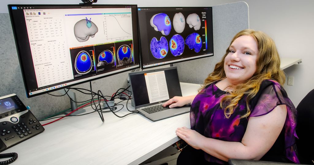
Postdoctoral researcher Erin Iredale, who has worked on intratumoral modulation therapy since her undergraduate degree, hopes the proposed electric fields treatment will one day be used for patients with brain cancer. (Christopher Kindratsky/Western Communications)
More than a decade ago, Dr Matthew Hebb was treating patients with Parkinson’s disease using deep brain stimulation by implanting tiny electrodes into the brain and delivering electrical signals to control tremors. He wondered if the same basic technology could be used against brain cancer.
Hebb, neurosurgery professor at Western University’s Schulich School of Medicine & Dentistry, took tumour samples removed during surgery back to his laboratory, implanted electrodes and stimulated the cancer cells. The tumours responded.
That unexpected observation set in motion years of research into what is now called Intratumoral Modulation Therapy, or IMT – an original approach that uses low-amplitude electric fields to disrupt the growth of glioblastoma, one of the most aggressive and difficult-to-treat brain cancers.
Now, a Western-led research team has taken another step toward potentially bringing the technology from the laboratory to patients.
The latest study, published in Neuro-Oncology Advances, shows that IMT can safely deliver stronger, dynamic electric fields directly to a glioblastoma tumour while significantly slowing brain tumour growth, in an animal model.
The research team, which included Hebb, physics and astronomy professor Eugene Wong, medical biophysics professor Terry Peters, anatomy and cell biology professor Susanne Schmid and postdoctoral researcher Erin Iredale, observed an eight-fold reduction in tumour growth measured through bioluminescence and a five-fold reduction in tumour volume measured by magnetic resonance imaging (MRI) after seven days of treatment.
For Iredale, the first author on the study and has worked on the IMT project since her undergraduate degree, the results represent another important step toward a treatment she hopes could one day help patients.
“It’s so interdisciplinary,” said Iredale. “We need everyone from different fields, with their own expertise, to come together to solve this huge problem in health care.”
Different kind of electrical treatment
Glioblastoma is a devastating cancer that begins in the cells or tissues of the brain. Even with current treatments, including surgery, radiation and chemotherapy, patients diagnosed with the disease have a median survival of just more than a year. One of the challenges is that glioblastoma cells divide rapidly despite aggressive conventional treatments, leading to recurrence mainly near the site of the surgery. IMT takes direct aim at that process.
Rather than using electricity to acutely burn or destroy the tumour, the treatment delivers chronic low-amplitude electric fields that interfere with the way cancer cells divide.
“When we put this electric field on those cells, it prevents them from dividing properly,” said Iredale. “So, they’re kind of stalled in their cell division process.”
The exact biological mechanisms are still being investigated, but the team has repeatedly observed reduced tumour growth when the electric fields are applied.
The idea grew from Hebb’s early experiments and then expanded as physicists and biomedical researchers joined the project. Deep brain stimulation normally operates at frequencies designed to produce a neurological response. For cancer treatment, however, the goal is different.
The researchers increased the frequency so the stimulation could target the tumour without producing unwanted effects in normal brain tissue. The result is a treatment designed to destroy the cancer while leaving the surrounding brain largely undisturbed.
Finding the sweet spot
Iredale joined the IMT project in 2016 as an undergraduate student when she was studying medical physics and applied mathematics. She was drawn to the possibility of combining physics and mathematics with the practical goal of helping patients – an interest that eventually led to a PhD in medical biophysics, where her research focused on developing a treatment-planning system for IMT.
Iredale’s work in the Hebb lab has helped address one of the central challenges of treating a tumour inside the brain: precisely controlling where the electric field goes and how strong it is.
The latest study, done in rats, marks the first time the team used multiple electrodes in a living brain to create a dynamic electric field. Three electrodes were implanted around the tumour. By shifting the phase of the electrical signals delivered by each electrode, the researchers created an electric field that rotates over time.
The approach helps cover the tumour more completely, reducing the possibility of ‘cold spots’ where cancer cells might escape treatment.
“We’re basically triangulating the tumour,” said Iredale.
“We’re using the electrodes to target very specific areas, making sure the electrical stimulation reaches the tumour while delivering the right amount of energy to each spot.”
– Western postdoctoral researcher Erin Iredale
The researchers used computational modelling to determine how the fields would be distributed through the brain and then confirmed those predictions with direct electrical measurements. Importantly, the treatment produced no neurological adverse effects or imaging evidence of brain injury.
From computer models to the clinic
The new treatment-planning system, devised by Iredale, is designed to eventually help physicians personalise IMT for individual patients. A physician could provide a patient’s MRI, and the system would calculate where electrodes should be implanted and what stimulation parameters should be used to provide the necessary tumour coverage.
The system currently relies on traditional computational methods, including an optimisation algorithm developed by Iredale. Artificial intelligence and machine learning could potentially be incorporated in the future.
The research has progressed from studies in cancer cells to animal models, and while further work will be required before the treatment can be tested in people, the team is already developing a prototype with the goal of moving toward a first-in-human clinical trial.
“In five to 10 years, I would hope to see IMT go through an initial clinical trial to test its efficacy against glioblastoma,” said Iredale. “From there, we could start to see it become part of the treatment options available to patients with brain cancer.”
Phase 1/2 trial of a targeted treatment regimen showed promising activity and manageable side effects in patients with HR-positive, HER2-positive cancer
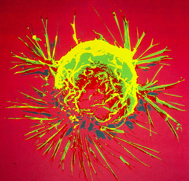
Colourised scanning electron micrograph of a breast cancer cell. Credit: NIH
Researchers at the Icahn School of Medicine at Mount Sinai found that a chemotherapy-free combination of four targeted therapies showed encouraging results in patients with HR-positive, HER2-positive metastatic breast cancer, offering a potential first-line treatment option that may be more convenient for some patients and avoids chemotherapy.
The findings, published in the Journal of the National Cancer Institute, come from the multicentre phase 1/2 ASPIRE clinical trial, which evaluated anastrozole, palbociclib, trastuzumab, and pertuzumab as first-line treatment for patients with hormone receptor (HR)-positive, HER2-positive metastatic breast cancer. The regimen combines endocrine therapy with medicines that target two key pathways that drive tumour growth, potentially allowing patients to avoid chemotherapy in the initial treatment setting.
“This study demonstrates that a chemotherapy-free treatment approach can produce durable responses while maintaining a manageable safety profile for many patients with this subtype of metastatic breast cancer,” said senior author Amy Tiersten, MD, Professor of Medicine (Hematology and Medical Oncology) and Clinical Director of Breast Medical Oncology at the Icahn School of Medicine at Mount Sinai. “Although these findings need to be confirmed in larger randomised clinical trials, they suggest that some patients may eventually have an effective alternative to chemotherapy as their first treatment.”
Breast cancer is the most frequently diagnosed cancer in women, and approximately 10 percent of breast cancers are HR-positive and HER2-positive. While current first-line treatments often combine HER2-targeted therapy with chemotherapy, chemotherapy can cause significant short- and long-term side effects that affect patients’ quality of life.
The ASPIRE trial enrolled patients with previously untreated HR-positive, HER2-positive metastatic breast cancer across five clinical sites affiliated with Mount Sinai, NYU Langone Health, and Columbia University. Researchers first established the optimal dose of palbociclib before evaluating the four-drug combination in 29 patients.
In the trial, 97 percent of patients experienced clinical benefit during the first six months of treatment. Patients lived a median of nearly 25 months before their disease progressed; median overall survival had not yet been reached at the time of the analysis. After a median follow-up of approximately 39 months, nearly 93 percent of participants were still alive. Several patients also experienced durable responses to treatment, including one who has remained on the regimen for more than six years.
The treatment’s side effects were consistent with those expected from the medications used. The most common were neutropenia (having low levels of a type of white blood cell called neutrophils in your blood), low white blood cell counts, diarrhea, and anemia. Only one patient discontinued treatment because of side effects, and no treatment-related deaths occurred.
“The challenge in treating this type of breast cancer is balancing effectiveness with quality of life,” said Rima Patel, MD, Assistant Professor of Medicine (Hematology and Medical Oncology) at the Icahn School of Medicine and first author of the study. “Many patients, particularly older adults or those with other medical conditions, may not be ideal candidates for chemotherapy. A targeted regimen that can be given primarily through oral medication and subcutaneous injection could offer a more convenient option while reducing some of chemotherapy’s burden.”
Researchers caution that the study was relatively small and did not compare the regimen directly with current standard treatments. Because of those limitations, the findings should be viewed as hypothesis-generating until confirmed in larger randomized trials.
The investigators are planning additional studies to determine whether the regimen can improve outcomes compared with current standard-of-care treatment.
Of 1357 devices authorised by the US FDA, only 3 were evaluated on clinical effectiveness
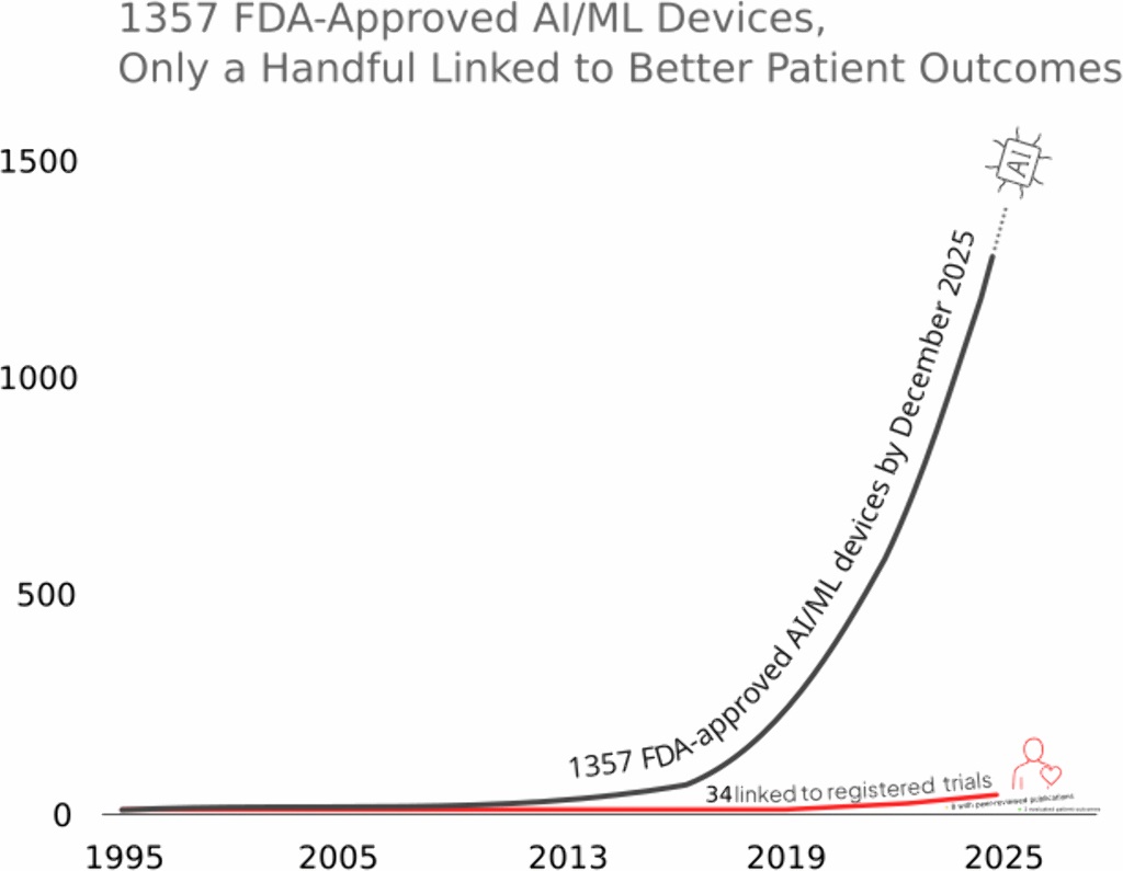
Growth of FDA-cleared AI/ML-enabled medical devices from 1995 to December 2025. Of 1,357 cleared devices, only 34 were linked to registered clinical trials and only 3 were evaluated for patient-centred outcomes. (Fig 1 of the article.), Credit: Abulibdeh R, Cajas Ordóñez SA, Celi LA, Gorijavolu R, Izath N, Markussen Lunde T, 2026, PLOS Digital Health, CC-BY 4.0 (https://creativecommons.org/licenses/by/4.0/)
A new analysis shows that, of 1357 artificial intelligence (AI)-based medical devices authorised by the US Food and Drug Administration (FDA) for use in patient care, only three had been tested on whether they actually improve patients’ health. Rawan Abulibdeh of the University of Toronto, Canada, and colleagues present these findings in the open access journal PLOS Digital Health on August 19, 2026.
New AI devices increasingly inform clinical care, such as systems that aid surgical planning, calculate cardiovascular risks, and guide interpretation of mammograms and other imaging. In order to be authorised for use in the US, AI devices typically only need to show “substantial equivalence” to an existing authorised device, and developers are not required to demonstrate whether new AI devices help people live healthier lives – with benefits shared equitably across diverse subgroups.
To deepen understanding of this topic, Abulibdeh and colleagues investigated how all 1357 AI devices authorised by the FDA as of December 5, 2025, had been evaluated in patients prior to authorization.
They found that only 34 of the devices had been included in registered clinical trials, with results posted for 12 and peer-reviewed manuscripts published for 12. Only 3 devices had been tested on patient-centred outcomes, such as death rates, strokes, hospitalizations, and quality of life. Most studies were conducted in highly resourced healthcare systems, and most excluded key patient subgroups, such as pregnant women, adults over 75, and non-English speakers.
The researchers suggest that structural barriers such as financial incentives and logistical challenges discourage developers from testing AI devices on patient outcomes, resulting in greater emphasis on speedy development than on rigor. They discuss how this framework could allow new tools to amplify existing disparities in healthcare and how it could lead to patients in low- and middle-income countries becoming inadvertent test populations for under-studied AI devices, as many countries rely on higher-income countries’ authorisation decisions.
On the basis of their findings, the researchers conclude that existing policies for AI medical device authorization should be redesigned. They propose a novel, three-phase framework that includes demonstration of effectiveness across diverse patient subgroups and healthcare settings.
The authors add: “We expected the evidence base to be thin, but not this thin. Out of 1357 AI devices the FDA has cleared for use in patient care, only three have been tested on whether patients actually live longer or better. Clearance tells you a device resembles something already on the market. It does not tell you it helps anyone.”
New Emory University research suggests that higher vitamin D supplement intake may be associated with better cognitive function in adults with an elevated risk of dementia.
In a study of 54 adults with sleep disturbances and mild cognitive impairments (MCI), two hallmarks of early dementia, participants taking at least 5000 IU of vitamin D daily showed better cognitive function compared to those who didn’t.
The study, recently published in Sleep Medicine, found that participants who reported taking 5000 IU or more of vitamin D daily scored more than 13% higher on the Montreal Cognitive Assessment (MoCA) than those who didn’t, even after accounting for other factors.A screening tool to detect risk of developing dementia, MoCA is widely used to assess memory and thinking skills. In this study, lower daily doses of vitamin D were not associated with higher cognitive scores.
The study also emphasises the significance of timing. MCI is an intermediate stage between normal cognitive aging and dementia.
“In older adults experiencing both sleep disturbance and mild cognitive impairment, this may represent a critical window for intervention, when cognitive changes are emerging, but opportunities to support brain health may remain,” says Victoria Pak, senior author of the study.
“Identifying accessible and modifiable factors, such as vitamin D supplement intake, during the earlier stages of cognitive decline may become increasingly important, particularly as rates of Alzheimer’s disease continue to rise,” adds Pak, associate professor at Emory University’s Nell Hodgson Woodruff School of Nursing.
In addition to the timing of the intervention, the study also found that cognitive performance did not differ based on the form of vitamin D – D2, typically derived from plants or fungi, or D3, produced after sun exposure or consuming animal-based foods.
Vitamin D, an essential nutrient, is not only necessary for muscle and nerve function, but also influences sleep quality and sleep-wake cycles. Additionally, 50% of those with moderate to severe Alzheimer’s disease report having sleep disturbances, indicating a bidirectional relationship between sleep deprivation and cognitive decline.
While vitamin D deficiencies have been implicated in sleep disorders, such as insomnia and more nighttime awakenings, this preliminary study is the first to assess the relationship between vitamin D supplement intake and cognitive function in a high-risk population with both MCI and sleep disturbances.
In the study, investigators analysed 2003–2018 data from 1044 US women participating in the National Health and Examination Survey: 174 with and 870 without cervical cancer. With these data, the team looked for an association between cervical cancer and the Composite Dietary Antioxidant Index (CDAI) – a validated composite score that reflects an individual’s intake level of dietary antioxidants, based on dietary recall interviews.
After adjusting for other factors that might affect cancer risk, the researchers found that each 1-unit increase in the CDAI was associated with a 11.4% lower prevalence of cervical cancer. Participants in the highest CDAI tertile had a 55.8% lower prevalence of cervical cancer than those in the lowest tertile. Additional analyses indicated a linear inverse association between the CDAI and the prevalence of cervical cancer.
“These findings suggest that dietary antioxidant intake may play a potential role in the prevention of cervical cancer. However, further prospective studies are needed to confirm this association and clarify the underlying mechanisms,” the authors wrote.
An overlooked factor can limit sweat’s cooling effect, ASU researchers discover
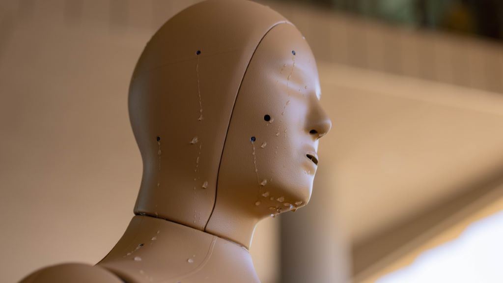
ANDI the sweating manikin helps ASU researchers discover the secrets of sweating. Photo by Samantha Chow/Arizona State University
The purpose of perspiration was a mystery in 1775, when English physician Charles Brian Blagden and a few inquisitive friends experimented on themselves by spending time in rooms heated to over 230 degrees Fahrenheit (110°C).
Although the temperatures were hot enough to cook raw meat placed in those rooms, Blagden noted how human bodies resisted the heat. Their core body temperatures remained nearly constant as the sweat poured out and evaporated.
The science of sweating has come a long way in 250 years. Yet a surprising amount remains unexplored, according to Konrad Rykaczewski, an associate professor of engineering at Arizona State University.
In a new study, Rykaczewski and colleagues have uncovered an unrecognised physical process that can dramatically change how effectively sweat cools the body in hot, dry and windless weather.
“It turns out that the impact is huge,” Rykaczewski said. “It can change how much sweat evaporates from your skin by over 50%.”
Why this research matters
Research is the invisible hand that powers America’s progress. It unlocks discoveries and creates opportunity. It develops new technologies and new ways of doing things.
Learn more about ASU discoveries that are contributing to changing the world and making America the world’s leading economic power at researchmatters.asu.edu.
Understanding the mechanics of sweating is vital to accurately predict heat strain and cooling efficiency in different situations. Scientists use this knowledge to guide recommendations for heat-stressed workers and to design cooling clothing and heat-management systems for first responders, soldiers and athletes.
Unlike Blagden’s crew in 1775, the ASU researchers didn’t subject anyone to oven-like conditions in their experiments. Instead, they used ANDI, a customised manikin rigged throughout its body with sensors to measure heat loss and heat gain and covered with pores that drip simulated sweat in response to rising heat.
The evaporation of sweat into water vapor is what cools the body. Air movement speeds evaporation and enhances cooling. The ASU researchers focused on an overlooked aspect of the cooling process: how temperature and humidity change the buoyancy of air near the skin.
When the outside temperature is hotter than a person’s skin, the air very close to the skin cools, becomes denser and drifts downward. But when the weather is hot and dry, humidity from evaporating sweat creates an opposing upward flow because humid air is lighter than dry air.
Experiments showed that at temperatures around 105 F with low humidity and no wind, the opposing currents near the skin can completely cancel each other, stopping airflow that could help sweat evaporate. In the absence of wind, this phenomenon leads to increased body heat storage and significantly elevated skin and core temperatures. Commonly used human heat-balance models don’t take this into account and will underestimate how hot the body becomes in hot, arid environments with little airflow.
The researchers measured the impact using models of sweat evaporation combined with simulations of the human body working to control its temperature. Neglecting humidity-driven buoyancy effects led to an underprediction of core body temperature rise by nearly 2 F for a person at rest in still air after two hours of heat exposure.
“This is really important for indoor settings or places with very little air movement,” Rykaczewski said. “Think about a tent, or a partially enclosed worksite or an unfinished building.”
Rykaczewski published the findings in Science Advances on Aug 19 with first author Shri Viswanathan, who completed his PhD at ASU in May, and eight others at the university.
The competing buoyancy effect is familiar to engineers working on heat exchange problems in electronics, “but it’s been overlooked in terms of the human body,” Rykaczewski said. “I think the reason is that most people working in this area don’t come from an engineering background. It really shows the benefit of having engineers and physiologists working together in interdisciplinary research.”
Breaking a sweat for research
Proving how the effect applies to sweating human bodies was no easy task. The ASU researchers performed dozens of experiments using ANDI. They developed a computer model and ran about 100 sweating simulations representing a wide range of conditions.
“There are multiple heat-transfer pathways involved, so isolating each one, making sure we could replicate it computationally and then combining them all into one model took a tremendous amount of effort,” Rykaczewski said.
Last year, the ASU researchers uncovered hidden details about how sweat rises and spreads over the skin. Those experiments required volunteers to don a bodysuit lined with tubes that circulate hot or cold water to warm or cool the wearer. The researchers observed how sweat first saturated the skin’s outermost layer, then collected in shallow pools around pores and spread into a thin, connected film. After a first round of sweat evaporated, it left a light salt residue. When the volunteers were heated again, that residue helped new sweat wick across the surface more rapidly, allowing a film to form without the earlier pooling stage.
A thin film can expose more sweat to the air, potentially making evaporation more efficient. The findings also suggest that sweating may behave differently across the body, where hair, sweat-gland density and skin structure vary.
Plenty of perspiration questions remain unanswered. The ASU team is exploring how sweat can either cling to the body and evaporate or run and drip off before it can evaporate and how that effects body cooling. The researchers are planning studies of the interactions between skin, sweat and clothing textiles. They see opportunities to improve clothing design so that it cools people more effectively by improving sweat evaporation.
“The bigger question is how you manage that sweat and what kinds of materials you can put next to the skin to optimise cooling,” Rykaczewski said.
In field studies across Arizona, the researchers are measuring how different populations experience and respond to extreme heat using advanced environmental sensing platforms combined with their improved models of sweat evaporation and thermoregulation. Their insights are supporting the design of heat adaptation measures and safer buildings and outdoor spaces.
The science of sweating was not a specialty Rykaczewski envisioned when he began his research career.
“It’s funny because it’s kind of an icky topic, but it’s also fascinating. You can spend your entire career doing highly specialised research that only 20 experts care about. But not sweating, because everybody sweats.”
An international study led by King’s College London has found that proposed new obesity definitions give greater insight into the ill health of metabolic bariatric surgery candidates compared with Body Mass Index (BMI).
The study, published in Jama Open Network, found that people considered for metabolic bariatric surgery had marked variation in disease burden – the number and severity of diseases – and operation-related risks, despite having similar BMI.
However, by reviewing clinical data from thousands of bariatric surgery candidates and applying new obesity definitions, the researchers were able to better understand their underlying health – which could have important implications for patient treatment and surgery prioritisation in the future.
Led by Professor Francesco Rubino, Chair of Metabolic and Bariatric Surgery at King’s College London, the researchers reviewed retrospective clinical data from 2,316 surgical candidates across four specialist centres – King’s College Hospital in the UK, and centres in France, Spain and Brazil.
They applied the new obesity definitions: clinical obesity, where there is clear evidence that excess fat, or adiposity, is causing organ damage, and preclinical obesity, where organ function is preserved despite excess adiposity.
From the surgical candidates, 73.8% had clinical obesity and 26.2% had preclinical obesity. Despite both groups of patients having similar BMI, patients with clinical obesity had far greater surgery-related and cardiovascular risks. They also had an overall high chance of death.
These findings suggest that distinguishing between clinical and preclinical obesity reveals key information about the health status and risks to surgery candidates that BMI alone cannot capture.
This study shows that the distinction between clinical and pre-clinical obesity is clinically meaningful even among surgical candidates with very high BMI levels, because BMI alone cannot tell us who has active disease.”Professor Francesco Rubino, senior author and Chair of Metabolic and Bariatric Surgery at King’s College London
He continued: “It is now essential that future surgical studies and registries systematically report patients’ clinical or pre-clinical obesity status, so that surgical safety, effectiveness and cost-effectiveness can be interpreted in the appropriate clinical context.”
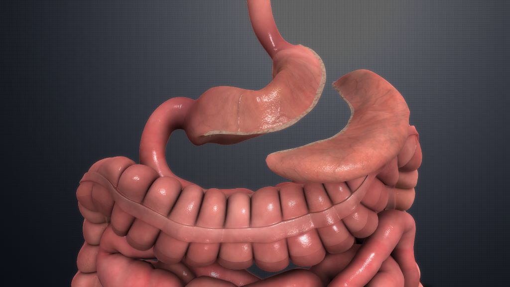
Traditionally, obesity has been classified primarily using BMI, a measure of weight relative to height that provides limited information about whether excess fat is actually affecting a person’s health. BMI levels have historically played a central role in determining eligibility and priority for metabolic bariatric surgery – operations of the stomach that help people lose weight and fix health issues, such as type 2 diabetes.
In 2025, the Lancet Diabetes & Endocrinology Commission on Clinical Obesity proposed a new diagnostic framework. This distinguished between clinical obesity, where there is clear evidence that excess fat, or adiposity, is leading to organ dysfunction, from preclinical obesity, where organ function is preserved but future health risk is increased.
In the latest study, the researchers found that these differences in disease status were not reflected in BMI. In the UK centre, for example, BMI was roughly 47.5 among patients with clinical obesity and 48.5 among those with preclinical obesity. Yet those with clinical obesity were approximately 10 years older and had substantially higher risk of death, cardiovascular risk and operation-related risk.
The researchers argue that for patients with clinical obesity, surgery primarily represents treatment of established disease. On the other hand, for those with preclinical obesity, its key goal may instead be to reduce future health risk. Recognising this distinction could help clinicians plan surgery, as well as select and prioritise surgical candidates.
It also saves around 44 units of blood products for every 100 patients treated
Photo by Charliehelen Robinson on Pexels
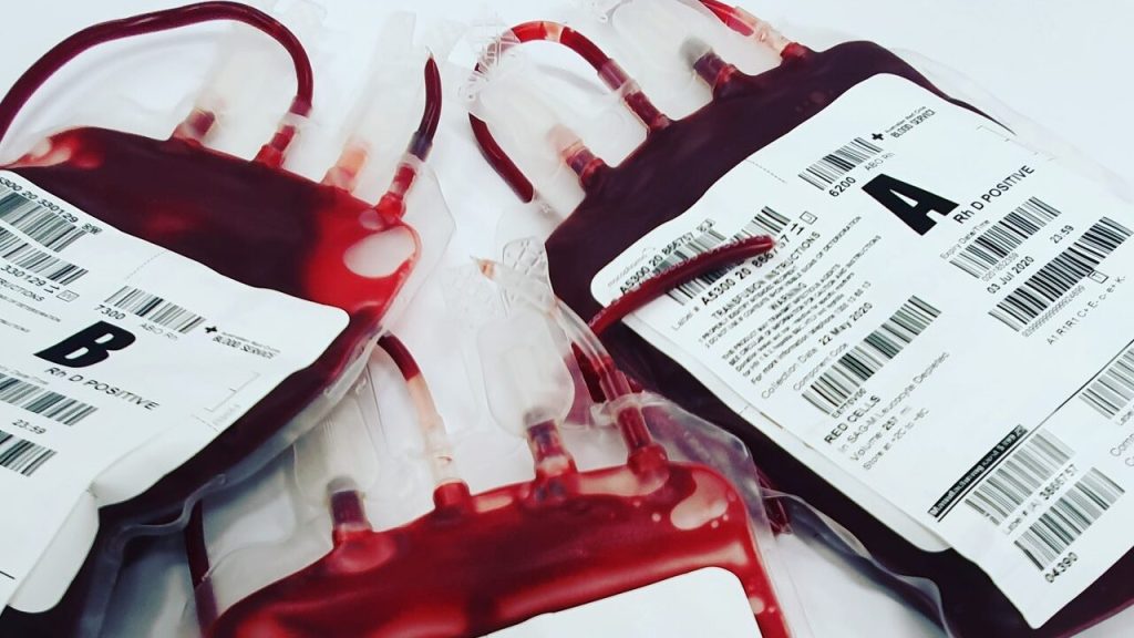
Giving intravenous iron to patients with anaemia before heart surgery reduces the need for a red blood transfusion and results in an extra day at home in the first 90 days after surgery, finds a clinical trial published by The BMJ today.
Intravenous iron also saved approximately 44 units of blood products for every 100 patients treated.
A third of patients undergoing cardiac surgery are anaemic and 20-50% receive a blood transfusion after surgery, which is linked to increased risks of complications, longer hospital stays, and death after surgery.
Previous studies suggest that intravenous iron before surgery boosts haemoglobin levels (the protein in red blood cells that carries oxygen around the body) and may reduce the need for transfusion, but there is currently no evidence to show that this improves outcomes that matter to patients.
To address this gap, researchers enrolled 955 adults with anaemia (average age 66 years; 60% male) undergoing elective cardiac surgery across 33 hospitals in 10 countries between 15 July 2016 and 15 December 2023.
Patients with inherited blood disorders, those having kidney dialysis or who had intravenous iron given in the previous four weeks were excluded. Other factors such as age, sex, ethnicity, and pre-existing conditions were also taken into account.
Participants were randomly assigned to either intravenous iron or placebo 1-26 weeks before surgery and the number of days alive and at home up to 90 days after surgery was recorded. Other outcomes included red cell transfusion and postoperative complications.
Of 921 patients assessed, the average number of days alive and at home up to 90 days after surgery in patients assigned to intravenous iron was 81 and in patients receiving placebo was 80.
Red blood cell transfusions were given to 262 patients (61%) in the iron group and 302 patients (68%) in the placebo group during their hospital stay. There were no differences in major complications or length of hospital stay.
The researchers acknowledge several limitations. For example, they enrolled patients with anaemia but did not require confirmation of absolute iron deficiency and say a one day difference in a 90 day recovery period is in itself a very small treatment effect.
However, study strengths included a low drop-out rate and measurement of patient-centred outcomes such as quality of recovery, days at home, and quality of life. Findings were also consistent after further analyses, suggesting they are robust.
As such, they conclude: “This study shows that intravenous iron repletion in patients with anaemia before cardiac surgery increased preoperative haemoglobin concentration, reduced the need for red cell transfusion, and resulted in an extra day at home in the first 90 days after surgery.”
Intravenous iron also saved approximately 44 units of blood products for every 100 patients treated, they add.
UCT’s Professor Liesl Zühlke has won a global women in science award for her groundbreaking work in improving care for children with cardiovascular disease. (Photo: Supplied)
By Sue Segar for Spotlight
Long before she became a pioneering scientist, Professor Liesl Zühlke insisted on seeing her tonsils after they were removed when she was just five years old. Today, paediatric cardiology is close to her heart, and her research has helped answer critical questions in the entire pipeline of heart disease.
In the mid-2000s, while working as a trainee in paediatric cardiology at the Red Cross War Memorial Children’s Hospital, Liesl Zühlke helped treat a 12-year-old patient who had an unusual congenital heart condition.
“His heart was so weak that he was dying,” she says. “He was thin, short of breath and very tired. He’d had multiple admissions to hospital and numerous health folders, tied together with elastic. On the front of his folder, the letters DNR (meaning do not resuscitate) were written in black khoki-pen.”
Zühlke recalls requesting an ultrasound of the heart, among other tests that eventually led to a diagnosis of a coronary artery anomaly. “This is a structural heart defect, present at birth, where the major coronary artery supplying the heart itself has the wrong origin, resulting in oxygen-poor blood supplying the heart muscle, causing it to die,” she explains. (See this Cleveland Clinic page for more on the condition.)
Fortunately, the defect is treatable with cardiac surgery to move the coronary artery to the correct place. Within a year of having this operation, the boy’s heart function had returned to normal.
A journey to research
This encounter was something of a turning point in Zühlke’s life. Till then, she’d only done clinical work, but the case piqued her interest in this particular lesion.
This experience, she says, was a lesson in looking beyond the obvious, re-examining a patient with a fresh approach, and asking new questions. “By thinking differently, we changed the trajectory of his life and many others.”
Zühlke did a small study looking at 30 cases of this lesion, and found it was more common than thought. “Our work led us to being able to diagnose this condition much earlier, before heart damage could happen, and children with the lesion could get surgery earlier. It made me realise why research and evidence-based medicine are so important … if one wants to impact not just the patient in front of you but multiple patients.”
A lifetime steeped in medicine
Zühlke, who was born and raised in Athlone, Cape Town, was from childhood fascinated about the inner workings of the human body.
“My siblings remember me cutting up my dolls and doing transplants with pieces of chicken,” she recalls with amusement.
When she had her tonsils removed at a tender five years old, she insisted on seeing the fleshy lymph node masses.
“My grandmother worked as a domestic worker in a residence at the University of Cape Town (UCT) and told us stories about students and what was happening there. Apparently, from when I was three or four, I said I wanted to study medicine,” says Zühlke.
It thus came as no surprise that she ended up studying medicine and surgery at the UCT. In 2015, she completed her PhD on the outcomes of asymptomatic and symptomatic rheumatic heart disease, which was supervised by the late Professor of Cardiology Bongani Mayosi. She calls him her “academic father” and “the man who saw something in me that I didn’t”.
In 2019, Zühlke was the first woman in South Africa to be appointed as a full professor in paediatric cardiology.
“It was always going to be paediatrics,” she says, adding “there was always a kid on my hip during ward rounds. With children, it’s not about status but about caring, and meeting them at their level.”
Losing patients, Zühlke says, is part of any doctor’s experience, “but it’s terrible telling a mother their child hasn’t got a normal heart and that there’s a possibility of death. To do that with compassion … is what we’re put in this world for.”
She says while her work started with research into rheumatic heart disease, her focus later evolved. “I started looking more at the reasons why we have the outcomes we do; the overarching determinants of why people are diagnosed late, why they don’t have enough treatment, and enough surgery; and why they have poorer outcomes.”
Today, Zühlke is vice-president of the South African Medical Research Council, and at Red Cross she is director of the Children’s Heart Disease Research Unit and a paediatric cardiologist in the Division of Paediatric Cardiology.
What types of things go wrong in children’s hearts?
Heart disease in children refers to any problem with the structure or work of the heart, explains Zühlke. There are two main types: congenital heart disease which relates to structural problems with the heart that are present at birth; and acquired heart disease that develops later.
“The heart is complete between six and eight weeks in utero. With congenital heart disease, the structural abnormalities are there already,” she says.
“The heart can be viewed as a house with four rooms or chambers, with walls that separate them, and veins or arteries going in or out to allow blood to move from one chamber to the next, through four valves or doors. There can be problems with any of those, for example, a hole in the heart, which is one of the most common things we see. If there’s a hole in one of the walls separating these chambers, then oxygen-rich and oxygen-poor blood can mix, making the heart and lungs work harder,” Zühlke says.
She says that a much more serious problem is when, instead of being born with four parts of the heart, a child is born with three or there’s obstruction to blood flow to the body. “This critical cardiac lesion requires surgery to reorganise the circulation. No surgery almost certainly means death. The range of structural abnormalities ranges from mild to critical. Congenital heart disease is the most common birth defect, affecting one in 50 children,” Zühlke says.
Explaining rheumatic heart disease, she says: “This is acquired later in life, particularly in children between the ages of five and fifteen, as a consequence of an infection called Streptococcus pyogenes (Strep A) which causes a sore throat or a skin infection and sets off an abnormal immune response. The body attacks and damages the tissue in the heart, an acute illness follows called Rheumatic fever and the permanent heart valve damage which results is known as Rheumatic Heart Disease.”
Zühlke believes not enough studies have been done to prove categorically what the prevalence of heart disease is in children in South Africa. “It’s hard to do a true birth prevalence study. But we’ve done a lot of work (through extrapolating numbers from other studies) to fill in the data gap showing that the prevalence in South Africa is similar to other parts of the world.” She says congenital heart disease is within the top five killers of children and more common than any other birth defects combined.
Unclear causes
The causes of congenital heart disease could include genetic changes, like chromosomal disorders like Down syndrome; maternal health issues like diabetes, obesity or infections; or exposure to smoke, alcohol or restricted medications during pregnancy.
The causes of acquired heart disease are similarly varied, ranging from environmental causes to and infections. It can take many forms. Kawasaki disease is a childhood illness that causes inflammation in the walls of blood vessels, resulting in fever, rashes and swelling; cardiomyopathy is a disease of the heart muscle that makes it harder for the heart to pump blood to the rest of the body. HIV-associated heart disease comes about because people living with HIV face a higher risk of developing cardiovascular disease.
The symptoms of heart disease, Zühlke says, include rapid breathing, an increased heart rate, and excessive sleepiness. Babies can be pale or blue around the fingers, toes or lips, and can tire when drinking or have problems gaining weight.
There are several possible diagnostics, but they are not always available in the public healthcare system. These include heart ultrasound, electrocardiograms, X-rays, and checking the amount of oxygen in the blood soon after birth, with a pulse oximetry test.
In addition, Zühlke says that some provinces such as the Northern Cape and Mpumalanga lack a paediatric cardiologist to make the diagnosis. “There are also issues with referrals to get diagnosed patients to centres for timeous attention. Sadly, people are dying without even knowing they had heart disease,” she says.
What we can do differently
Asked what could be done differently in the public sector to improve early diagnosis of heart issues, she says pulse oximetry screening is a simple, cost-effective, efficient test to rule out critical congenital heart disease. Zühlke says there are plans to incorporate it into the Road to Health Booklet used by health professionals and parents to monitor a child’s health and development.
“It wouldn’t be difficult to add pulse oximetry screening as a check before a baby is discharged from a hospital. It’s been well proven that it works. All that’s needed is a probe and somebody to do it; and it can tick a box on the child’s Road-to-Health chart.
“But that requires money because you need a particular newborn probe to do it … and people are busy in the clinics,” she says.
On rheumatic heart disease, Zühlke says it’s “unethical” that an entirely preventable disease is still such a problem in SA and Africa. “We need research, funding, and advocacy to change that, improving diagnosis, treatment and long-term care. Also, we need to make sure there’s a vaccine to treat strep A.”
First prize, she says, would be to enable immediate diagnosis of rheumatic fever. “Currently, there’s no one test to say a person has acute rheumatic fever.”
“Next, we need a different way of treating it (the current penicillin injection is extremely painful for children); and getting a vaccine in countries where it actually matters. In the meantime, the goal is to ensure people living with the disease get access to care.”
Zühlke says she’s following the work on a vaccine for rheumatic heart disease closely and is involved in a vaccine collaborative which is working to ensure that when the vaccine becomes available, it is accessible and affordable.
‘Some of them were my heart patients as children’
Meanwhile, her “varied, mad, interesting” life sees her overseeing numerous intra- and extramural research projects for the SAMRC and directing her childrens heart research unit at UCT. She still works in a multi-disciplinary clinic for women with cardio-vascular disease in Groote Schuur’s maternity centre which she loves.
“Some of them were my heart patients as children,” Zühlke says, adding that transitional care is a priority “because patients with congenital and rheumatic heart disease become adults, we need to ensure that they can get the best possible life-long care.”
Her big vision has always been “to integrate research into the clinical space, so that as you see patients, it goes into the research, and as you research, it goes back to the patient.” Zühlke adds: “Being part of the research landscape at the SAMRC is deeply gratifying. What I’d like to see, most importantly, is childhood onset heart disease recognised as an entity, part of policy … and in the national action plans.”
She is the 2026 laureate for Africa and the Arab States in the L’Oréal-UNESCO For Women in Science International Awards for her pioneering work in improving care for children with cardiovascular disease, particularly rheumatic heart disease. Given to the top five female scientists globally, the distinguished award recognised her for “combining cutting-edge science with social justice” and “turning the fight against childhood heart disease into a public policy priority”.
Zühlke is married to Alexander Zühlke, a plastic surgeon at Tygerberg Hospital in Cape Town, and together they have two children.