South Africa is facing a major health transition. While the average life expectancy has nearly doubled over the past century, the quality of those additional years, commonly referred to as one’s ‘healthspan,’ remains under threat from non-communicable diseases (NCDs). This threat calls for a renewed national focus on prevention and early intervention to address the rapidly growing challenge.
The Healthspan Imperative
According to health data presented at the 2025 Momentum Healthcare Insights Summit, NCDs such as heart disease, cancer, diabetes and neurodegenerative disorders now account for 51% of all deaths in South Africa. In contrast, communicable diseases make up around 40% and non-natural causes (like accidents and violence) account for a mere 9%. This shift reflects a global trend, although some challenges remain; infectious diseases become more manageable, while chronic conditions associated with lifestyle and ageing take centre stage.
Damian McHugh, Chief Marketing Officer at Momentum Health
“Medical advances have added years to our lives, but not necessarily life to our years. More and more, there is growing evidence to support the fact that prevention offers the greatest potential to reduce the burden as well as cost of chronic disease and improve quality of life,” says Damian McHugh, Chief Marketing Officer at Momentum Health.
Prevention Outperforms Treatment
The summit highlighted compelling evidence that prevention is more effective than treatment for advanced disease. For example, research from the American Cancer Society shows that tobacco control measures, such as smoking bans and taxes, have prevented 3.8 million lung cancer deaths in the United States since 1970. The most effective way to save lives from late-stage lung cancer has not been through treatment, but through reducing or eliminating smoking altogether.
The same principle applies to other chronic diseases. Managing risk factors such as high blood pressure, obesity, high blood glucose, and abnormal cholesterol can actively prevent or delay the onset of disease. These factors are strongly influenced by behaviours such as a lack of physical activity, poor nutrition, unmanaged stress levels, and even excessive alcohol use or smoking.
Investing in regular health check-ups and preventative care can mitigate the risk of serious health problems, ultimately reducing the incidence of costly, advanced illnesses. Simple lifestyle changes, such as prioritising rest and recovery, making time to connect with loved ones, maintaining balanced nutrition, practicing mindfulness, and engaging in regular exercise not only promotes physical and mental health, but can also translate into significant long-term savings in healthcare costs.
“Making quality healthcare more accessible, while enabling and rewarding healthy, preventative habits can lead to complete physical and mental health and wellbeing. Investing in access and wellbeing is a powerful pathway to realising more wealth and more health for more South Africans,” says McHugh.
Measuring and Managing Healthspan within the South African Context
Momentum Health’s data reveals that many South Africans are living longer, but the average age of medical scheme beneficiaries has increased by nearly three years over the past decade, and the proportion of pensioners in medical schemes is rising. Without proactive measures, our nation’s ailing healthcare system will face increasing claims and costs as the population ages and chronic diseases become more prevalent.
Momentum Health believes that the solution lies in taking measures to improve access to both quality medical care and reliable health information and empowering individuals to take responsibility for their own health.
South Africa’s rising NCD burden is not inevitable. With early detection, healthy lifestyle choices, and consistent engagement with preventative healthcare, individuals can not only extend their lifespan but also improve the years lived in good health.
“Prevention isn’t just a personal choice; it’s a public health imperative. By investing in wellness now, we can reduce the future burden on our healthcare system and help more South Africans enjoy longer, healthier lives,” concludes McHugh.
He speaks in measured tones – calm, reflective, deliberate. But when Dr Dumisani Bomela describes the future he envisions, the words carry power. For the CEO of the Hospital Association of South Africa (HASA), healthcare is not just a profession. It is a promise rooted in dignity, equity and access to every South African.
Q: Dr Bomela, what drew you to medicine and what keeps you committed to healthcare in South Africa? A: I have always seen healthcare as an act of service. As a doctor, you learn to see beyond symptoms, to understand the person behind the diagnosis. As a leader at HASA, I take that same approach. Our work is about people. About making sure that every South African can get quality care when they need it.
Q: HASA represents South Africa’s private hospital sector. Why is this sector important to the country’s overall health system? A: Private hospitals are a cornerstone of healthcare in South Africa. We treat millions of patients each year. More than that, HASA members invest heavily in medical training, advanced technology and infrastructure. We are strategic partners in the national system, that makes our sector a vital national asset.
Q: How does HASA contribute to economic development beyond just healthcare? A: Healthcare is a growth engine. HASA members are major employers, from doctors and nurses to technicians and support staff. We support local communities and stimulate investment. When healthcare systems are strong, economies thrive – and so do people.
Q: What is HASA’s stance on universal health coverage? A: We believe every person has the right to choose their provider and to receive high-quality care. That is why we support reforms that strengthen the system and build equity. HASA members are ready to work side by side with the government to make that vision a reality. Our hospital groups bring deep experience, including in some cases from geographies where universal healthcare systems operate, and strong infrastructure to the table.
Q: What kind of leadership do you believe is needed in South African healthcare today? A: We need leaders who listen. Who understand not just policy, but people. Leadership in healthcare must be grounded in compassion and collaboration. At HASA, we strive to lead by example, building trust, fostering partnerships, and always remembering that every system ultimately affects human lives.
Q: How do HASA hospitals stay at the forefront of medical technology and innovation? A: By investing intentionally. Our members understand that modern medicine is not static, it is constantly evolving. We equip our hospitals with advanced diagnostic and treatment tools. But technology alone is not enough. We also invest in people – training nurses, specialists and support teams to lead with excellence.
Q: In a country facing complex health challenges, how do you stay hopeful? A: Hope grows where there is action. Across our hospitals, I see incredible work being done every day – surgeons saving lives, nurses comforting families, teams innovating to improve care. We are proving, together, that with collaboration and commitment, South Africa’s health system can be strong, inclusive and world-class.
Q: What gives you the greatest sense of pride in your work with HASA? A: Honestly, it is seeing the impact private hospitals have. When families walk out of our hospitals healed. When professionals grow into health leaders. When communities feel their well-being is supported. These outcomes remind us why the work matters. My pride does not come from titles; it comes from knowing we are making a real, human difference every single day.
Many diseases affect men and women differently. Asthma tends to strike men earlier in life, yet more women develop asthma as they get older. Parkinson’s is more common in men, but Alzheimer’s is more common in women.
The differences are even more stark when it comes to autoimmune disease. Women are around two and a half times more likely than men to develop multiple sclerosis and nine times more likely to develop lupus.
Why would some diseases strike one sex more than another? And why do some tissues, such as the lungs and brain, seem especially vulnerable to these sex-based differences?
To answer these questions, scientists at La Jolla Institute for Immunology (LJI) are leading new research into how our immune cells defend specific parts of the body.
“In just the last two years, LJI scientists have uncovered a whole new body of information about how the immune systems of men and women are very different,” says Saphire. “We’re looking at what is genetically encoded in our XX or XY chromosomes, and how hormones like oestrogen and testosterone affect what is genetically programmed into our immune cells.”
In the paper, the researchers define biological sex (in an immunology context) as the presence of XX chromosomes in females and XY chromosomes in males. “Every cell in your body is either XX or XY,” says Saphire. “That X chromosome has many, many immune-related genes. Women have two copies of each. That gives them, in a sense, twice the palette of colours to paint from in formulating an immune response. It can also give them a stronger immune response for those genes that are doubly active – active in both copies simultaneously.
Sex hormones are important for much more than reproductive function. Immune cells can also sense hormones such as oestrogen and testosterone and use them to determine which genes to turn on or off and which ones to turn on more brightly or dim. This means similar immune cells can do different things, depending on whether that cell is from a male or a female.
Further, female cells vary in which of their two copies of X is “turned on.” As a result, women have organs with a collage, or mosaic, of immune cells that work differently in different tissues. This innate “variety” of immune cells appears to be an effective way to ward off infectious disease (women are better than men at fighting off pathogens such as SARS-CoV-2).
But scientists have also found that having more genes from X chromosomes may predispose women to autoimmune disease. This increased X chromosome “dosage” is closely linked to a higher risk for autoimmune diseases such as Sjögren’s syndrome and scleroderma.
New research into sex-based immune system differences is also critical for developing new cancer immunotherapies, Sharma explains.
“We’re increasingly understanding how sex-based differences affect disease outcomes. When it comes to medicine, one size doesn’t fit everybody,” says Sharma, who directs LJI’s Center for Sex-Based Differences in the Immune System. “This is leading to new research, particularly in the cancer field, toward precision medicine. We’re asking how a person’s individual immune system is contributing to controlling that cancer through immunotherapy.”
Saphire and Sharma also highlight environmental factors, such as nutrition and chemical exposures, that may add to the complex interplay of chromosomes and sex hormones. Men and women also appear to have some signature differences in their skin and gut microbiomes.
The researchers hope these foundational discoveries can lead to medical advances for all, and they’re working with collaborators across the country to move this research forward. “It takes a team to translate these findings,” says Sharma.
In response to US funding cuts for South African health services and research projects, National Treasury has provided the National Department of Health with hundreds of millions of rands in emergency funds. Spotlight and GroundUp look at how precisely the government intends to spend this money.
Health Minister Dr Aaron Motsoaledi recently announced that National Treasury had released roughly R753 million to help plug the gap left by US funding cuts to South Africa’s health system. Another R268 million is also being released in the following two years for researchers that lost their US grants.
But this may only constitute the first round of emergency funds from government, according to sources we spoke to. The health department is planning on submitting a bid for an additional allocation later on, which will be considered by Treasury. But this will likely only be approved if the first tranche of funding is properly used.
So how is the money supposed to be used? To find out, we spoke with officials from the National Treasury, the National Department of Health and the South African Medical Research Council (SAMRC).
Money for provinces is for saving jobs at government clinics
The current tranche of money comes from Treasury’s contingency reserve, which exists partially to deal with unforeseen funding shortfalls. It was released in terms of Section 16 of the Public Finance Management Act.
Of the R753 million that’s been announced for this year, Motsoaledi stated that R590 million would be going to provincial health departments via the District Health Programme Grant – a conditional grant for funding the country’s public health efforts, particularly HIV, TB, and other communicable diseases. Such conditional grants typically give the health department more say over how provincial departments spend money than is the case with most other health funding in provinces.
To explain how government officials arrived at this figure, it’s worth recapping what services the US previously supported within provinces.
Prior to Donald Trump becoming US president on 20 January, the US Agency for International Development (USAID) had financed health programmes in specific districts with high rates of HIV. These districts were scattered across all South Africa’s provinces, save for the Northern Cape.
The funds were typically channelled by USAID to non-governmental organisations (NGOs), which used the money to assist the districts in two ways.
The first is that NGOs would hire and deploy health workers at government clinics. The second is that the NGOs would run independent mobile clinics and drop-in centres, which assisted so-called key populations, such as men who have sex with men, sex workers, transgender people, and people who inject drugs.
In response, the health department began negotiations with Treasury to get emergency funding to restore some of these services. As part of its application, the health department submitted proposals for each province, which specified how much money was needed and how it would be used. (Though this only took place after significant delay and confusion).
Since Treasury couldn’t afford to plug the entire gap left by the US funding cuts, the provincial-level proposals only requested money for some of the services that had been terminated. For instance, funding was not requested for the key populations health centres. Instead, the priority was to secure the jobs that had been lost at government health facilities.
As such, the total amount that was requested from Treasury for each province was largely calculated by taking the total number of health workers that NGOs had hired at clinics and working out how much it would cost to rehire them for 12 months.
Rather than paying the NGOs a grant to deploy these workers as was done by USAID, the health department proposed hiring them directly. This meant that they calculated their wages according to standard government pay scales, which is less than what these workers would have earned from the NGOs.
The total came to just under R1.2 billion for all the provinces combined.
Treasury awarded roughly half of this on the basis that the money would be used to finance these wages for six months, rather than 12. This amounts to the R590 million for provinces that was announced by Motsoaledi.
If all goes smoothly and this money is used effectively to hire these staff over the next six months, then a new tranche of Section 16 funding could be released in order to continue hiring them. Funds might also be released to fund the key populations health sites.
A concern, however, is that the money may just be used by provinces to augment their ordinary budgets. If the funds aren’t actually used to respond to the US cuts, then it is much less likely that more emergency funding will be released.
At this stage, it is too early to tell how provinces will use the money, particularly given that it appears that at least some of them haven’t gotten it yet.
Spotlight and GroundUp sent questions to several provincial health departments. Only the Western Cape responded. The province’s MEC for Health and Wellness, Mireille Wenger, said that the funds have not yet been received by her department, but that once they were, they would be directed to several key priority areas, including digitisation of health records, and the strengthening of the primary healthcare system.
It’s thus not clear whether the province will be using any of the funds to employ health staff axed by US-funded NGOs. In response to a question about this, Wenger stated that “further clarity is still required from the National Department of Health and National Treasury regarding the precise provincial allocations and conditions tied to the additional funding”.
What about research?
Of the R753 million that’s been released for this year, R132 million has been allocated to mitigate the funding cuts for research by US federal institutions, primarily the National Institutes for Health (NIH). Unlike USAID, the NIH is not an aid body. It provides grants to researchers who are testing new treatments and medical interventions that ultimately benefit everyone. These grants can be awarded to researchers in the US or abroad as part of a highly competitive application process.
Researchers in South Africa are awarded a few billion rands worth of grants from the NIH each year, largely due to their expertise in HIV and TB. But over the last few months, much of this funding has been terminated or left in limbo. (See a detailed explanation of the situation here).
The R132 million issued by Treasury is supposed to assist some of these researchers. It will be followed by another R268 million over the following two years. The Gates Foundation and Wellcome Trust are chipping in an additional R100 million each – though in their case, the funds are being provided upfront.
All of this money – R600 million in total – is being channelled to the SAMRC, which will release it to researchers via a competitive grant allocation system.
According to SAMRC spokesperson Tendani Tsedu, they have already received the R132 million from Treasury, though they are still “finalizing the processes with the Gates Foundation and Wellcome Trust for receipt of [their donations]”.
The SAMRC is also in negotiation with a French research body about securing more funds, though these talks are ongoing.
In the meantime, the SAMRC has sent out a request for grant applications from researchers who have lost their US money. The memo states: “Applicants may apply for funding support for up to 12 months to continue, wind down or complete critical research activities and sustain the projects until U.S. funding is resumed or alternative funds are sourced.”
“The plan,” Tsedu said, “is to award these grants as soon as possible this year.”
Professor Linda-Gail Bekker, CEO of the Desmond Tutu Health Foundation, told us that the hope is that the grants could fill some of the gaps. “This is a bridge and it is certainly going to save some people’s jobs, and some research,” she said, but “it isn’t going to completely fill the gap”.
Indeed, the SAMRC has made clear that its grants aren’t intended to replace the US funding awards entirely. This is unsurprising given that the money that’s being made available is a tiny fraction of the total grant funding awarded by the NIH.
It’s unlikely that research projects will continue to operate as before, and will instead be pared down, said Bekker.
“It’s going to be about getting the absolute minimum done so you either save the outcome, or get an outcome rather than no outcome,” she said.
In other cases, the funds may simply “allow you to more ethically close [the research project] down,” Bekker added.
For some, this funding may also have come too late. Many researchers have already had to lay off staff. Additionally, patients who had been on experimental treatments may have already been transitioned back into routine care. It’s unclear how such projects could be resumed months later.
In response, Tsedu stated: “For projects that have already closed as a result of the funding cuts, the principal investigator will need to motivate whether the study can be appropriately resurrected if new funds are secured.”
The SAMRC has established a steering committee which will adjudicate bids. They will be considering a range of criteria, Tsedu said, including how beneficial the research might be for the South African health system, and how heavily the project was impacted by the US funding cuts. They will also consider how an SAMRC grant could “be leveraged for future sustainability of the project, personnel or unit”, added Tsedu.
An endless back and forth
The job of the SAMRC steering committee will likely be made a lot more complicated by the erratic policy changes within the NIH. On 25 March, the body sent a memo to staff – leaked to Nature and Bhekisisa – instructing them to hold all funding awards to researchers in South Africa. After this, numerous researchers in the country said they couldn’t renew their grants.
However, last month, Science reported that a new memo had been sent to NIH staff which said that while South African researchers still couldn’t get new grants, active awards could be resumed.
Since then, some funds appear to be trickling back into the country, but certainly not all. For instance, Spotlight and GroundUp spoke to one researcher who had two active NIH awards before the cuts. He stated that one of these was resumed last month, while the other is still paused.
Bekker also told us that she had heard of one or two research grants being resumed in the last week, though she said the bulk of active awards to South Africa are still pending.
“Where people are the prime recipients [of an NIH grant] without a sub awardee, there seems to be a queue and backlog but some [of those awards] are coming through,” said Bekker. “But how long this is going to take and when it might come through, we’re waiting to hear.” She said a strategy might be to apply for the SAMRC bridging funding and “if by some miracle the [NIH funding is resumed]” then researchers could then presumably retract their SAMRC application.
In the meantime, health researchers will have to continue spending their time working out how to respond to the abrupt and increasingly confusing changes to funding guidelines that have dogged them since Trump assumed office.
“It’s such a dreadful waste of energy,” said Bekker. “If we were just getting on with the research, it would be so much better.”
Gait analysis and pain measures show that subtly adjusting the angle of the foot during walking may reduce knee pain caused by osteoarthritis, an approach which may also slow progression of the incurable condition.
Led by a team of researchers at NYU Langone Health, the University of Utah, and Stanford University, a new study explored whether changing the way patients position their feet when walking could lessen extra loading, helping to treat the disease.
For the investigation, the scientists tested this intervention in 68 men and women with mild to moderate knee osteoarthritis and then used advanced MRI scans to track how well it worked.
The results suggest that those trained to angle their feet slightly inward or outward from their natural alignment experienced slower cartilage degeneration in the inner part of their knee compared with those who were encouraged to walk more frequently without changing their foot position. The study is reported online in The Lancet Rheumatology.
“Although our results will have to be confirmed in future studies, they raise the possibility that the new, noninvasive treatment could help delay surgery,” said study co-lead author Valentina Mazzoli, PhD.
Dr. Mazzoli, an assistant professor in the Department of Radiology at NYU Grossman School of Medicine, notes that the earlier patients receive a knee replacement, the more likely they are to require additional procedures in the future.
The findings also revealed that those who adjusted their foot angle reduced their pain score by 2.5 points on a 10-point scale, an effect equivalent to that of over-the-counter pain medications. By contrast, those who did not change their gait reduced their pain scores by little more than a point.
“Altogether, our findings suggest that helping patients find their best foot angle to reduce stress on their knees may offer an easy and fairly inexpensive way to address early-stage osteoarthritis,” added Dr. Mazzoli.
About one in seven Americans have some form of osteoarthritis, commonly in the inner side of the knee, according to the U.S. Centers for Disease Control and Prevention. A leading cause of disability, the disease is often managed with pharmaceutical pain relievers, physical therapy, and in the most severe cases, knee-replacement surgery. Experts believe that excess loading can over time contribute to the condition.
Past research has offered little evidence that changes in gait can effectively reduce knee pain caused by osteoarthritis, says Dr. Mazzoli. Some previous trials trained all participants to adopt the same foot angle and found no relief, while others did not compare the intervention to a control group or only followed the participants for a month.
The new study is the first to show that tailoring each patient’s foot angle to their unique walking pattern can alleviate the disease’s symptoms in the long term and may slow cartilage breakdown, the authors say.
Dr Mazzoli adds that this technique may have a significant advantage over pharmaceutical painkillers. These drugs, she says, do not address the underlying disease and can cause liver and kidney damage, stomach ulcers, and other unwanted side effects when taken for long periods.
For the study, the research team recorded the participants walking on a treadmill at a specialized gait-assessment laboratory. A computer program simulated their walking patterns and calculated the maximum loading that occurred in the inner side of their knees. Next, the team generated computer models of four new foot positions—angled inward or outward by either 5 or 10 degrees—and estimated which option reduced loading the most.
The patients were then randomly divided into two groups. Half were trained in six sessions to walk with their ideal angle, while the other half were instructed to continue walking naturally. Pain scores and MRI scans were taken at the beginning of the study period and one year after the intervention.
Study findings showed those who adjusted their gait reduced the maximum loading in the knees by 4 percent, while those who kept their normal walking pattern increased their loading by more than 3 percent.
“These results highlight the importance of personalizing treatment instead of taking a one-size-fits-all approach to osteoarthritis,” said Dr. Mazzoli. “While this strategy may sound challenging, recent advances in detecting the motion of different body parts using artificial intelligence may make it easier and faster than ever before.”
While the authors relied on a specialized laboratory for the new study, AI software that estimates joint loading using smartphone videos is now available and can allow clinicians to perform a gait analysis in the clinic.
The researchers next plan to test whether these tools can indeed identify the most effective walking method for osteoarthritis patients, says Dr. Mazzoli. They also plan to expand their study to people with obesity.
Discovery could help pave the way for a prosthetic device to restore blink function lost to injury or disease
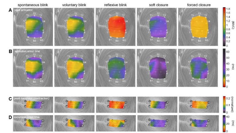
Muscle activation and movement patterns over time across the upper and lower eyelids, shown under different actions. Credit: Anatomical Engineering Group/UCLA
A blink of an eye is vital to protecting the eye by keeping it from drying out. This simple function seems natural and instantaneous, but is it?
Now, a team of UCLA biomechanical engineers and ophthalmologists has uncovered new details about the muscle that controls blinking, offering a pathway toward developing blink-assisting prostheses. Published in PNAS, the study found that the orbicularis oculi – the muscle that controls eyelid movement – contracts in complex patterns that vary by action and move the eyelid in more than just a simple up-and-down motion.
The researchers studied how this muscle behaves differently across various actions including spontaneous blinks, protective rapid closures and squeezed shut-eye motions.
“The eyelid’s motion is both more complex and more precisely controlled by the nervous system than previously understood,” said study corresponding author Tyler Clites, an assistant professor of mechanical and aerospace engineering at the UCLA Samueli School of Engineering. “Different parts of the muscle activate in carefully timed sequences depending on what the eye is doing. This level of muscle control has never been recorded in the human eyelid. Now that we have this information in rich detail, we can move forward in designing neuroprostheses that help restore natural eyelid function.”
In experiments with volunteers, the researchers looked at five different ways the eyes close:
Spontaneous blink: An automatic, unconscious blink that occurs regularly to keep the eye lubricated
Voluntary blink: An intentional blink, as when someone is asked to blink on command
Reflexive blink: A rapid, involuntary blink triggered to protect the eye from a collision
Soft closure: A gentle, slow eyelid descent, similar to the beginning of sleep
A forced closure: A deliberate squeezing of the eyelids tightly shut
To record activity in the orbicularis oculi with high precision, an ophthalmic surgeon inserted tiny wire electrodes into the eyelid. The researchers then used a motion-capture system to track eyelid movement in ultraslow motion. These tools allowed the team to measure subtle differences in eyelid movement, including speed, direction, and which part of the muscle initiated the action.
Video of spontaneous blink – dynamic muscle activation patterns and eyelid kinematics. Credit: Anatomical Engineering Group/UCLA
“People can lose the ability to blink due to a stroke, tumour, infection or injury. The condition is painful in the short term and can damage the eyes enough to cause vision loss,” said study co-author Dr Daniel Rootman, an associate professor of ophthalmology at the David Geffen School of Medicine at UCLA and director of the UCLA Orbital Disease Center. “We know that a small electric pulse can stimulate the orbicularis oculi muscle to move, but designing one that works well has been elusive. What we now have is a good roadmap to such a device, including where exactly to place electrodes, how to time them, and how strong the pulse should be. These guidelines could help pave the way for the development and clinical testing of such a device, with the ultimate goal of providing real relief for patients.”
With this fundamental knowledge of eyelid biomechanics in hand, the researchers can now work on refining a prototype neuroprosthesis to assist people with blinking.
“Understanding how the eyelid works is crucial to designing an accurate stimulation pattern for a prosthesis, as well as for diagnostic purposes,” said study first author Jinyoung Kim, a UCLA mechanical engineering doctoral student and member of Clites’ research group, the Anatomical Engineering Group at UCLA. “We are more than excited to bridge this gap and move forward to work with patients who have facial paralysis and help improve their lives.”
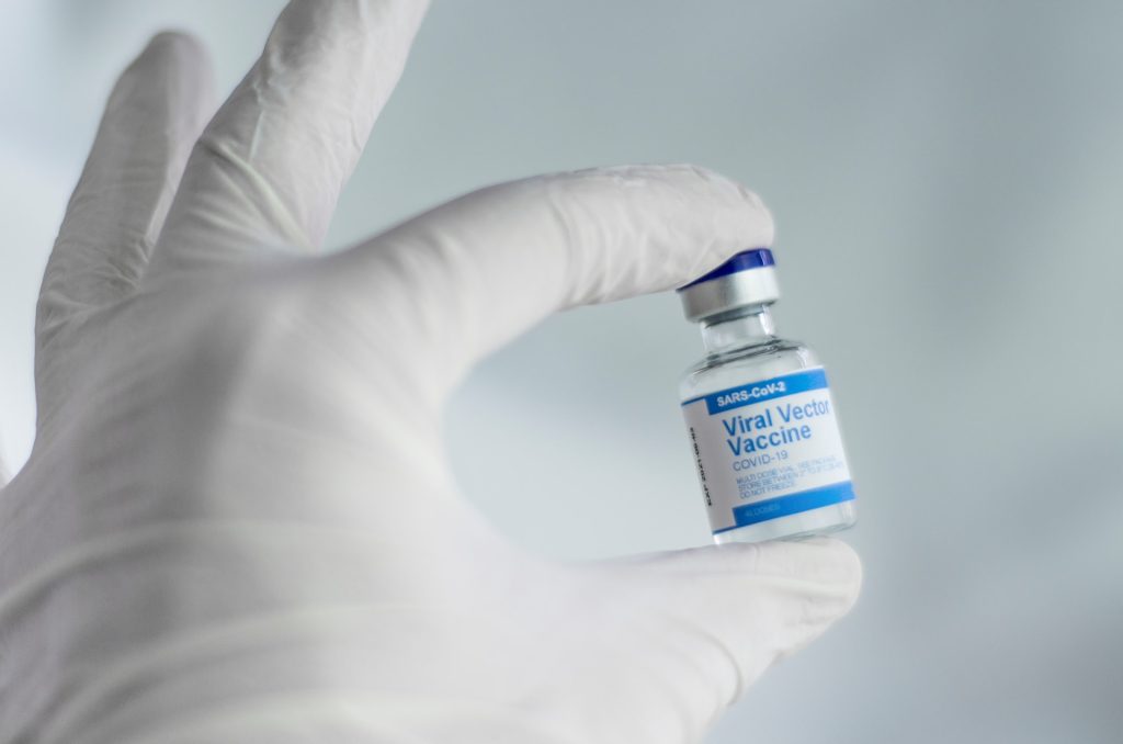
Thanks to vaccinations against SARS-CoV-2 in the period 2020-2024, 2.533 million deaths were prevented at the global level, one death was avoided for every 5400 doses of vaccine administered. The 82% of the lives saved by vaccines involved people vaccinated before encountering the virus, 57% during the Omicron period, and 90% involved people aged 60 years and older. In all, vaccines have saved 14.8 million years of life (one year of life saved for 900 doses of vaccine administered).
These are some of the data released in an unprecedented study published in the journal Jama Health Forum and coordinated by Prof Stefania Boccia, Professor of General and Applied Hygiene at Università Cattolica, with contributions from Dr Angelo Maria Pezzullo, Researcher in General and Applied Hygiene, and D. Antonio Cristiano, a medical resident in Hygiene and Preventive Medicine. The two researchers spent a period at Stanford University, collaborating directly with the group of Professor John P.A. Ioannidis, director of the Meta-Research Innovation Center (METRICS), in the context of the project “European network staff eXchange for integrAting precision health in the health Care sysTems- ExACT” funded by the European Research Excellence Programme RISE project-Marie Slodowska Curie and coordinated by Professor Stefania Boccia.
Professor Boccia and Dr Pezzullo explain: “Before ours, several studies tried to estimate lives saved by vaccines with different models and in different periods or parts of the world, but this one is the most comprehensive because it is based on worldwide data, it also covers the Omicron period, it also calculates the number of years of life that was saved, and it is based on fewer assumptions about the pandemic trend.”
The experts studied worldwide population data, applying a series of statistical methods to figure out who among the people who became ill with COVID did either before or after getting vaccinated, before or after Omicron period, and how many of them died (and at what age). ‘We compared this data with the estimated data modeled in the absence of COVID vaccination and were then able to calculate the numbers of people who were saved by COVID vaccines and the years of life gained as a result of them,’ Dr Pezzullo explains.
It also turned out that most of the saved years of life (76%) involved people over 60 years of age, but residents in long-term care facilities contributed only with 2% of the total number. Children and adolescents (0.01% of lives saved and 0.1% of life years saved) and young adults aged 20-29 (0.07% of lives saved and 0.3% of life years saved) contributed very little to the total benefit.
Professor Boccia concludes: ‘These estimates are substantially more conservative than previous calculations that focused mainly on the first year of vaccination, but clearly demonstrate an important overall benefit from COVID-19 vaccination over the period 2020-2024. Most of the benefits, in terms of lives and life-years saved, have been secured for a portion of the global population who is typically more fragile, the elderly’.
Researchers at Children’s Hospital of Philadelphia (CHOP), along with several academic partners, announced the primary results of the Pediatric KIDney Stone (PKIDS) trial, the largest comparative effectiveness study of surgical interventions for children and adolescents with kidney stones. The CHOP-led PKIDS trial, with two published studies, marks a significant breakthrough by offering stronger evidence for treating stones of varying sizes, including new information on patient experiences after surgery, thus reducing uncertainty and empowering informed decision-making for patients, caregivers, and physicians.
Kidney stones were once largely a disease that affected adults. However, kidney stones in children have been on the rise in recent decades, doubling the likelihood that a child will develop a kidney stone. As a result, CHOP founded the PKIDS Care Improvement Network in 2019, which now includes 31 sites in the United States and Canada.
Ureteroscopy (an endoscopic outpatient procedure), shockwave lithotripsy (a noninvasive outpatient procedure) and percutaneous nephrolithotomy (a minimally invasive surgery with a short hospital stays) are the procedures used to treat children and adults with kidney stones. Most children and adolescents with kidney and ureteral stones are treated with ureteroscopy despite uncertainty of which procedure is more effective and their impact on patients’ lives. As pioneers in urology, CHOP leaders aimed to enhance pediatric patient and caregiver decision-making for kidney stone surgeries while enabling urologists to adopt techniques for optimal outcomes, including patient-selected experiences.
In the first study in JAMA Network Open, Tasian and his team enrolled 1142 patients aged 8 to 21 with kidney and/or ureteral stones between 2020 and 2023 at all 31 sites in the United States and Canada. Researchers evaluated ureteroscopy against shockwave lithotripsy and found that shockwave lithotripsy was associated with less pain and fewer urinary symptoms compared with those who had ureteroscopy. No meaningful differences were detected in stone-free rates for the procedures.
In the other CHOP-led study in JAMA Network Open, Jonathan S. Ellison, MD, an Associate Professor of Urology at the Medical College of Wisconsin and Pediatric Urologist at Children’s Wisconsin, and the PKIDS team compared percutaneous nephrolithotomy (PCNL) and ureteroscopy. That study found that for children with larger stones, PCNL not only cleared more stones effectively but also led to a better overall recovery experience than ureteroscopy.
Overall, the authors emphasised the post-surgery experiences of children, noting that quality of life factors, such as the loss of school time for children and work time for caregivers, are crucial in determining effective treatment options. While the authors are planning further research, they also hope these findings will lead to immediate improvements for families.
“The PKIDS trial demonstrated that ureteroscopy and shockwave lithotripsy remove stones equally well and that patients having shockwave lithotripsy recover more quickly after surgery with less pain and fewer urinary symptoms. Our findings provide new information that allow for tailored approaches to kidney stone treatment for children and their families,” said Gregory E. Tasian, MD, MSc, MSCE, Director of the PKIDS network and an attending pediatric urologist in the Division of Urology at Children’s Hospital of Philadelphia. “Although future clinical trials are important, we hope that clinical practice guidelines will consider outcomes that matter to patients.”
The novel exercise involves applying a pneumatic cuff to restrict the flow of blood. Credit: University of South Australia
New research from the University of South Australia is offering fresh hope to people living with rheumatoid arthritis (RA).
Evaluating the effectiveness of a novel form of exercise – blood flow restricted resistance training – among people with RA, researchers found that this alternative workout method not only improved their strength and physical performance, but also reduced their pain.
Blood flow restricted resistance training involves placing a pneumatic cuff – much like a blood pressure cuff – around the top of the working limb. The cuff is then inflated so that it restricts blood flow out of the limb, creating a highly metabolic environment which forces the muscles to work harder, even when using lighter weights or less effort.
The Arthritis Australia-funded study is the first to trial blood flow restricted resistance training on both the upper and lower limbs in people with RA, using five exercises – leg press, machine hamstring curl, machine knee extension, cable tricep extension, and cable bicep curl – with gradually increasing weights.
All participants in the study reported that they “liked” the programme, and the group showed clear improvements in strength, movement and pain levels.
Lead researcher UniSA’s Dr Hunter Bennett says the training offers a practical and achievable option for people with RA.
“RA can cause a loss of muscle mass and strength, which affects day-to-day activities, independence, and increases the risk of falls and fractures,” he says.
“Resistance training is one of the best ways to rebuild that strength, but for people with RA, using heavy weights can be difficult or harmful due to pain, fatigue or injury risk. This is where blood flow restricted resistance training can help.”
Dr Bennett says this approach is ideal for people who need to do resistance exercises but find it hard to lift weights.
“Many people with health conditions are understandably deterred by exercise, yet it is often one of the best things they can do to improve their condition,” he says.
And while this exercise might look unusual, the research shows that it works.
“This kind of training could be a game-changer for people with rheumatoid arthritis.
“It offers a way to build strength and reduce pain without pushing through discomfort – and that’s incredibly empowering for people who’ve often been limited by their condition.”
While this was a small-scale trial, researchers say the results are promising and lay the foundations for a larger trial comparing blood flow restricted resistance exercise to more traditional exercise approaches.
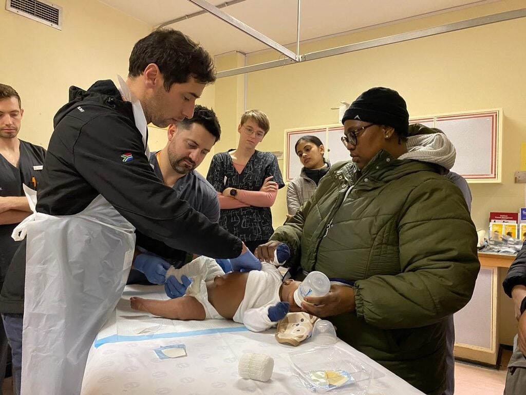
Dr Saul Kaplan (left) stands next to Dr Kenny Beck, with mother Zukiswa Panyaza as her baby receives a full leg cast at the clubfoot clinic in Tygerberg Hospital’s Division of Orthopedic Surgery, while medical students observe. (Photo: Sue Segar/Spotlight)
By Sue Segar
When Karen Mara Moss’s son was diagnosed with clubfoot, she travelled to the US in search of a life-changing treatment. She made a promise to bring it home and two decades on, her non-profit is at the heart of a remarkable success story.
“I looked at those tiny feet. They were turned over and rigidly pointing inwards,” recalls Karen Mara Moss about the day her son Alex was born in 2003.
For her, the memory is as vivid today as it was then. Within moments of his birth, a concerned obstetrician commented on Alex’s feet. Then the paediatrician diagnosed Alex with bilateral clubfoot, a condition in which a baby is born with one or both feet twisted inward and downward.
“I remember thinking: Will he walk with a limp? Will people mock him?” Moss tells Spotlight. “It was a traumatic time.”
She says the paediatrician told her not to worry. “He said they’d have to cut his feet and straighten them, and it would all be perfect,” says Moss.
Despite having several prenatal scans and tests, the condition had not been picked up before birth.
The most common form of clubfoot present at birth is idiopathic clubfoot, medically known as talipes equinovarus. It is when a baby’s foot is pointed in and down because the tissues connecting the muscles to the bone are shorter than usual, leading to pain and reduced mobility if left untreated, according to a review study published in The Lancet medical journal. In most cases, the cause of this congenital anomaly which ranges from mild to severe, is unknown, baby boys are twice as likely to be born with clubfoot as baby girls, and about half of children with clubfoot have it in both feet. Globally, an estimated 176 000 babies are born with the condition every year.
Eight days after Alex was born, Moss says she met with a paediatric orthopaedic surgeon. She says he told her he’d fixed many clubfeet using the Kite method and even had one patient playing first-team rugby. The Kite method was developed in the 1930s and uses manipulations and castings to achieve a sequential and gradual full correction of the forefoot, then the hindfoot, and finally, the ankle. After the casting is done, the baby wears a special splint to keep the feet pointing slightly outward and upward, but, critically, many would also require further surgery.
Back then, the standard of care for clubfoot was surgical management, says Dr Pieter Maré, an orthopaedic surgeon who heads up the clubfoot clinic at Greys Hospital in Pietermaritzburg, Kwazulu-Natal. “The reality was that a large number of children required extensive surgery before the Ponseti method,” he says.
Moss followed the doctor’s advice, and during that first appointment, she says he began applying casts up to Alex’s knees. “He started wrenching Alex’s foot, holding the back, whilst pushing the front of the foot, and plastering the foot. Alex was blue in the face from screaming. I was crying while holding him down,” she says.
Another way
But after two months and seven casts, she says there was little improvement in Alex’s feet. That is when Moss began searching for answers herself. Doing research on the internet, she discovered the University of Iowa Children’s Hospital website, where she read about a technique developed by Dr Ignacio Ponseti, which he claimed could help children have pain-free, functional feet without surgery.
The Ponseti method was developed in the 1950s but only became more widely used in the United States in the 1990s, and later in much of the rest of the world. The technique uses gentle manipulations and plaster casts to correct the midfoot, hindfoot, and forefoot simultaneously, while the ankle is treated afterwards. In some cases, before the last cast is applied, it may require a percutaneous tenotomy which is a minimally invasive procedure to cut the heel cord that is resistant to stretching. A brace is then fitted the same day as the last cast is taken off.
“The Kite method was developed to correct clubfoot but over time it was realised that this method was using the wrong anatomical methods,” explains Professor Anria Horn, a consultant orthopaedic surgeon at the Red Cross Children’s Hospital in Cape Town.
“There are multiple joints in the foot and the Kite method was, effectively, manipulating the wrong joint in an attempt to bring about the correction in the foot. Ponseti discovered that the manipulation should occur at a different joint,” she says.
Back in 2003, Moss emailed Ponseti, and a few days later called his office. “I was put through to a man with a Spanish accent. He said he’d read my email, and that he’d seen the photos I sent of my son’s feet; that what we’d done was not the way his method worked. He suggested I go to Iowa because nobody in South Africa was practising his method,” she says.
Not long after this, Moss and her husband travelled with ten-week-old Alex for 10 000 miles from sunny South Africa to an unseasonable snowstorm in Iowa.
The idea of travelling to a foreign country to see a “special” doctor that one read about on the internet, with a treatment carrying his name, may raise red flags for some. There are after all no shortage of quacks out there exploiting vulnerable people with just this type of story of an underutilised treatment. Ponseti, however, was a serious scientist and, even by 2003, his method had performed well in several studies and had been quite widely adopted by doctors in the United States.
Moss says in that first visit, Ponseti eventually did a cast all the way up Alex’s leg. “He looked like a little turtle with his legs sticking out. By the time he’d done the second cast, Alex was asleep,” Moss recalls.
“Dr Ponseti’s normal protocol was to remove the cast every week, then re-manipulate the foot into a different position, and reapply the cast. For out-of-town patients, he accelerated the treatment and changed the cast every five days,” she adds.
After just one cast, Alex’s foot looked different, says Moss. “They did another cast, and five days later, it was time for the third cast. Dr Ponseti took the second cast off and then did the percutaneous tenotomy, as well as the third and final cast.”
After this procedure and with Alex now in his final casts, they were told they could return to South Africa and take the casts off three weeks later. Moss said an orthotist measured Alex’s feet before the tenotomy and gave her instructions on how to fit the clubfoot brace he would wear for four years while sleeping.
Three weeks later, back home, Moss soaked the casts off and started to put the brace on at night. She says Alex’s feet were straight.
‘A parting gift’
On her final day in Iowa, Moss recalls Ponseti telling her: “You’re the first South African that’s ever been here. Please go back home and tell the doctors not to operate on clubfoot”. He gave her his book, copies of his research papers, and CDs demonstrating his casting method – a parting gift that would shape the course of her life.
Determined to share her what she had learnt, Moss created a website to provide information on clubfoot. The website gained traction and soon she started getting requests from parents across southern Africa for help to access the Ponseti method.
At the time, Moss says she knew of only one doctor using the method, whom she recommended parents consult. “I’d met him soon after my return to South Africa in 2003 and had lent him Dr Ponseti’s book and papers. He’d then gone to the US to attend a Ponseti training workshop and started using the method. I was sending everyone to him.”
The founding of STEPS
Moss realised the best solution was to bring the training directly to South Africa. In 2005, despite having no experience in running a non-profit organisation, she founded STEPS, driven by her commitment to introduce the Ponseti method across the country.
Moss says STEPS held its first two-day Ponseti training course in 2006, with about 60 paediatric orthopaedic surgeons attending. “Three Ponseti experts came from Canada, Brazil and the UK to give the training. They taught a lot of theory and used bone models to demonstrate the method,” she says.
The second STEPS Ponseti workshop in 2007 focused on public health facilities. Moss says the training took place at the Charlotte Maxeke Johannesburg Academic Hospital.
Partly due to the workshops, partly due to the strength of the accumulating scientific evidence, the method caught on in the country. In 2012, the South African Paediatric Orthopaedic Society officially endorsed the method. A Cochrane Review published in the same year found that, while the available evidence was far from complete, it did indicate that the method works well. Cochrane Reviews are a highly regarded type of study that attempts to assess evidence from all randomised clinical trials relating to a specific medical question.
“The Ponseti method has become the gold standard for the treatment of idiopathic clubfoot,” stated an article published in the World Journal of Orthopedics in 2014. And according to the Lancet study cited earlier, “the Ponseti method is widely recognised as an effective conservative treatment approach for clubfoot that avoids corrective surgery in over 90% of cases”.
Today, Horn says the Kite method isn’t used in South Africa any more, having been replaced by the Ponseti method. “STEPS has played a big part in promoting the Ponseti method in South Africa, as well as providing training, workshops and conferences and supporting clubfoot clinics across the country. Our job would have been much harder without the support that STEPS provides,” she adds.
Ponseti in the public sector
Given the equipment and know-how involved, making the Ponseti method available in South Africa’s public sector was a challenge. In 2013, Moss launched a support programme to help government clinics offer the treatment. STEPS began by partnering with just six clinics. With support from donors, they recruited staff to visit each clinic weekly to guide families or trained someone on-site to do so. They also provided educational materials to help raise awareness. Over time, this led to STEPS helping develop standard systems and processes for running the clinics, making care more consistent and accessible. When some clinics couldn’t provide braces, STEPS arranged for it to be donated.
Today, STEPS has 48 partner clinics across South Africa, ranging from a tiny rural clinic in Lusikisiki in the Eastern Cape to bigger clinics in Gauteng and the Western Cape. “Lusikisiki might see three patients a week, and Chris Hani Baragwanath Academic Hospital could see 80. They all open once a week, except for some small, rural clinics,” Moss says.
Based on stats that STEPS collected, Moss estimates that at least 2 000 children are born every year with clubfoot in South Africa. Through the help of her organisation, she says: “More than 20 500 children have accessed effective treatment. We’ve … distributed 22 628 clubfoot braces. In 2024, we supported 4 592 children at partner clinics in different stages of the four-year treatment protocol.”
Moss adds that STEPS has conducted over 20 training sessions across South Africa, Namibia, Botswana, and the Seychelles, with more than 2 000 healthcare professionals. “Parents were bringing their children over the border as they couldn’t access treatment back home. We worked with the ministries of health in those countries to teach the Ponseti method there,” she says.
Though separated by an ocean, Moss says she stayed in close contact with Ponseti. She says the last time they saw each other was at a clubfoot symposium in Iowa in 2007. Two years later, he passed away at the age of 95.
“I felt as if I’d lost a member of my family,” Moss says. “He was the master, and he inspired me in my work to improve the lives of children born with clubfoot.” She said she would always carry the ache of missing him, but bringing his method to South Africa, just as she had promised, was something that gave her a deep sense of purpose and peace.
That promise, purpose and peace started with Alex who is today in his final year of a Bachelor of Commerce degree and who, in his own words, “enjoys being active outdoors with my friends”, likes playing padel, and going on hikes.
*This article is part of Spotlight’s 2025 Women in Health series, featuring the remarkable contributions of women to healthcare and science.