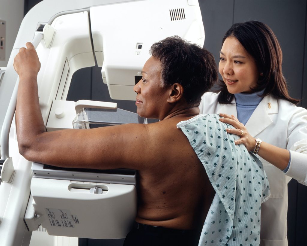
In a recent study published in Pathogens and Immunity, researchers issue a call to action over how rising antifungal resistance is worsening the problem of invasive fungal infections.
Fungal infections have become more than just Epidemiological data published inMicrobial Cell indicates that a rise in severe fungal infections has resulted in over 150 million cases annually and almost 1.7 million fatalities globally.
Skin contact with microorganisms found in soil or on hard surfaces, such as common shower facilities, or exposure to infected pets, can result in fungal infections known as dermatomycoses. Rashes, itching, burning and skin irritation are among the symptoms of fungal infection.
Thomas McCormick and Mahmoud Ghannoum, professors of dermatology at the Case Western Reserve University School of Medicine and affiliated with University Hospitals Cleveland Medical Center, explained extent of the problem. “This is not just an issue that affects individual patients,” McCormick said.
“The World Health Organization has recognised it as a widespread threat that has the potential to impact entire healthcare systems if left unchecked.”
Based on their findings, the researchers issued precautions and a “call to action” for the medical community to help protect people from multidrug-resistant fungi, starting with awareness and education.
“Healthcare providers must prioritise the use of diagnostic tests when faced with an unknown fungal infection,” Ghannoum said.
“Early detection can make all the difference in improving patient outcomes.”
Patients treated with medications to protect the immune system after cancer and transplant procedures are more vulnerable to fungal infections – making them especially more vulnerable to infections from drug-resistant fungi, the researchers said.
The emergence of multidrug-resistant fungal species, such as Candida auris and Trichophyton indotineae, is especially troubling and requires urgent attention, they reported.
In a study recently published in Emerging Infectious Diseases, Ghannoum’s research team and the Centers for Disease Control and Prevention (CDC), detailed a case that demonstrated Trichophyton indotineae, in addition to becoming drug-resistant, was also sexually transmissible.
To address the growing health concern, McCormick and Ghannoum suggest several measures:
Increased awareness and education: Raising awareness in the general healthcare setting to obtain a more accurate understanding of the rise of antifungal-resistant infections.
Diagnostic Testing: Routine use of diagnostic tests can guide appropriate treatment strategies.
Antifungal Susceptibility Testing (AST): Improving insurance reimbursement rates for AST and increasing the number of qualified laboratories with the capacity to perform these tests.
Call to Action: Addressing the emerging challenge of antifungal resistance involves concerted efforts from healthcare professionals, researchers, policymakers and the pharmaceutical industry to develop and implement strategies for managing and preventing antifungal resistance.
“The ultimate goal of these measures,” Ghannoum said, “is to improve the quality of patient care by ensuring effective treatment and preventing further escalation of the problem.”
Forming social bonds facilitates effective communication and neural synchronisation across individuals of different social status within a group
When small hierarchical groups bond, neural activity between leaders and followers aligns, promoting quicker and more frequent communication, according to a study published on March 19th in the open-access journal PLOS Biology by Jun Ni from Beijing Normal University, China, and colleagues.
Social groups are often organised hierarchically, where status differences and bonds between members shape the group’s dynamic. To better understand how bonding influences communication within hierarchical groups and which brain regions are involved in these processes, the researchers recorded 176 three-person groups of human participants (who had never met before) while they communicated with each other, sitting face-to-face in a triangle. Participants wore caps with fNIRS (functional near-infrared spectroscopy) electrodes to non-invasively measure brain activity while they communicated with their group members. Each group democratically selected a leader, so each group of three ultimately included one leader and two followers. After strategising together, groups played two economic games designed to test their willingness to make sacrifices to benefit their group (or harm other groups).
Experimenters assigned some triads to go through a bonding session, where they were grouped according to colour preferences, given uniforms, and led through an introductory chat session to build familiarity. Bonded groups spoke more freely and bounced between speakers more frequently and rapidly, relative to groups that didn’t experience this bonding session. This bonding effect was stronger between leaders and followers than between two followers. Neural activity in two brain regions linked to social interaction, the right dorsolateral prefrontal cortex (rDLPFC) and the right temporoparietal junction (rTPJ), aligned between leaders and followers if they had bonded. The authors state that this neural synchronisation suggests that leaders may be anticipating followers’ mental states during group decision-making, though they acknowledge that their findings are restricted to East Asian Chinese individuals communicating via text (without non-verbal cues), whose culture emphasises group cohesion and commitment towards group leaders.
The authors add, “Social bonding increases information exchange and prefrontal neural synchronisation selectively among individuals with different social statuses, providing a potential neurocognitive explanation for how social bonding facilitates the hierarchical structure of human groups.”
A new study by researchers at UC Davis Health found human brains are getting larger. Study participants born in the 1970s had 6.6% larger brain volumes and almost 15% larger brain surface area than those born in the 1930s. The researchers hypothesise that the increased brain size may lead to an increased brain reserve, potentially reducing the overall risk of age-related dementias.
“The decade someone is born appears to impact brain size and potentially long-term brain health,” said first author Charles DeCarli, a distinguished professor of neurology and director of the UC Davis Alzheimer’s Disease Research Center.
“Genetics plays a major role in determining brain size, but our findings indicate external influences – such as health, social, cultural and educational factors – may also play a role.”
75-year study reveals brain changes between generations
The researchers used brain magnetic resonance imaging (MRIs) from participants in the Framingham Heart Study (FHS). The community-based study was launched in 1948 in Framingham, Massachusetts, to analyse patterns of cardiovascular and other diseases.
The original cohort consisted of 5209 men and women between the ages of 30 and 62. The research has continued for 75 years and now includes second and third generations of participants.
The MRIs were conducted between 1999 and 2019 with FHS participants born during the 1930s through the 1970s.
The brain study consisted of 3226 participants (53% female, 47% male) with an average age of about 57 at the time of the MRI.
The research led by UC Davis compared the MRIs of people born in the 1930s to those born in the 1970s.
It found gradual but consistent increases in several brain structures.
For example, a measure that looked at brain volume (intracranial volume) showed steady increases decade by decade.
For participants born in the 1930s, the average volume was 1234mL, but for those born in the 1970s, the volume was 1321 mL, or about 6.6% greater volume.
Cortical surface area showed an even greater increase over the decades.
Participants born in the 1970s had an average surface area of 2104cm2 compared to 2056cm2 for participants born in the 1930s — almost a 15% increase in volume.
The researchers found brain structures such as white matter, gray matter and hippocampus (a brain region involved in learning and memory) also increased in size when comparing participants born in the 1930s to those born in the 1970s.
Larger brains may mean lower incidence of dementia
Although the numbers are rising with America’s aging population, the incidence of Alzheimer’s – the percentage of the population affected by the disease – is decreasing.
A previous study found a 20% reduction in the incidence of dementia per decade since the 1970s.
Improved brain health and size may be one reason why.
“Larger brain structures like those observed in our study may reflect improved brain development and improved brain health,” DeCarli said.
“A larger brain structure represents a larger brain reserve and may buffer the late-life effects of age-related brain diseases like Alzheimer’s and related dementias.”
One of the study’s strengths is the design of the FHS study, which allows the researchers to examine brain imaging of three generations of participants with birthdates spanning almost 80 years.
A limitation is that non-Hispanic white participants make up the majority of the FHS cohort, which is not representative of the U.S. population.
An analysis of Swedish data, where the definition of triple negative breast cancer (TNBC) differs from that used internationally, brings additional insights to on ongoing discussion in the scientific community. The study was presented at the 2023 European Society for Medical Oncology (ESMO) meeting and is now published in Lancet Regional Health – Europe.
The Swedish definition of TNBC differs from the international version in that it also includes tumours with low expression of the Oestrogen Receptor (ER) biomarker, ie in 1–9% of tumour cells. Internationally, ER-low breast cancer is classified as hormone-sensitive and treated differently from TNBC patients. This is despite previous studies demonstrating that the majority of ER-low tumours are molecularly similar to ER-zero, the latter completely without expression of ER, and meta-analyses that show no survival benefit from endocrine therapy in ER-low tumours.
The Swedish population-based study included all women diagnosed with TNBC in Sweden during 2008–2020 using the National Quality Register for Breast Cancer. Patient and tumour characteristics, treatment and survival in patients with low ER expression was compared to patients with no ER tumour expression.
The study identified and included 5655, and 560 patients (10%) were defined as ER-low and 5095 (90%) as ER-zero. The data demonstrated there are only small differences in tumour characteristics, no differences in response to neoadjuvant chemotherapy and no significant differences in prognosis.
“The international cut-off for ER-positivity and thus the definition of TNBC as only completely ER-negative is now increasingly questioned. ER-low tumours behave like ER-zero tumours and should be treated as such. On the basis of real-world data, the Swedish cutoff for hormone receptor positivity appears to be more clinically relevant. A changed international definition would give patients with ER-low expressing breast cancer the same treatment options as in TNBC, within studies and in clinical routine,” says study leader Dr Irma Fredriksson.
The study was carried out in collaboration with the pharmaceutical company MSD.
Anorexia nervosa, a mental health disorder in which people dangerously restrict their eating or purge their stomachs soon after a meal, is one of the deadliest psychological diseases. Yet, the neural mechanisms behind this have remained unclear, and therapies are limited.
Scientists have been tailing a lead for years, though. They’ve known that the disorder is often associated with anxiety and depression, hinting that the biological basis for anorexia could be regulated by neurons somewhere in the brain region that controls emotion – the amygdala.
That’s exactly where Haijiang Cai, a University of Arizona associate professor in the Department of Neuroscience and BIO5 Institute member, and his team found it: Anorexia is caused by a combination of two subregions in the amygdala, according to new research published in Cell Reports.
One knot of neurons in the central nucleus of the amygdala curbs appetite when a person gets full, feels nauseous or tastes something bitter. The other is in the oval region of the bed nucleus of the stria terminalis, which also halts eating due to inflammation and sickness.
Cai and his research team found that when they destroyed a certain type of brain cell, called PKC-delta neurons, in both of these regions, they could prevent anorexia development.
They also found that PKC-delta neurons become more active in response to eating during the anorexia development. What’s more, when they artificially activated these neurons, they caused a suppression in eating habits and increased exercise.
“This study suggests two important insights to treat anorexia,” Cai said. “One is that we need to target multiple brain regions to develop therapies. We also need to treat multiple conditions. For example, maybe one drug will target nausea and another drug target will target inflammation, and you have to combine them, like a cocktail therapy, to have better therapeutic effects.”
The team relied on mice models for their research.
“There’s no animal model that can mimic human disease completely, but this is as close as we can get,” Cai said. “For example, there are multiple common features, including a warped body image, a very low body weight, limited food intake and excessive exercise. We can’t know if an animal has a warped body image, but we can measure the other three features.”
One future step – since researchers cannot destroy neurons for human treatment – is to develop a method to silence the neurons temporarily, using drugs or some other method to test if that can prevent anorexia development or speed up recovery for people who have already developed the disorder.
With the rise in gestational diabetes and metabolic disorders during pregnancy, metformin is also being prescribed more frequently. Although it is known that the oral antidiabetic agent can cross the placental barrier, the impacts on the brain development of the child are largely unknown. Now, researchers have been able to demonstrate in a mouse model that although metformin has positive effects in pregnant animals, it does not in the offspring. The researchers, from German Institute of Human Nutrition Potsdam-Rehbrücke (DIfE), published their findings in Molecular Metabolism.
Around one in six pregnant women worldwide are affected by gestational diabetes. According to the Robert Koch Institute, 63 000 women in Germany were affected by the disease in 2021, and the trend is increasing. Excessively high blood sugar levels during pregnancy are associated with negative consequences for mother and child. It increases the risk of affected women developing type 2 diabetes later on and their children have a higher risk of developing metabolic disorders and being overweight.
Long-term effect of metformin on offspring is unclear
The placenta-crossing oral antidiabetic agent metformin has been gaining importance as an alternative to insulin administration when lifestyle changes fail to treat gestational diabetes. But there are currently only a few studies on the long-term effects of metformin on the health of offspring. It is known that metformin has an impact on the AMPK signaling pathway, which regulates the networking of nerve cells during brain development.
The interdisciplinary team of DIfE researchers led by Junior Research Group Leader Dr Rachel Lippert therefore grappled with two central questions:
Firstly, is metformin treatment only beneficial for the mother or also the child?
Secondly, does metformin treatment lead to long-term negative physiological changes in the offspring, especially in connection with the development of neuronal circuits in the hypothalamus, a critical region in the regulation of energy homeostasis?
Mouse models shed some light
To answer the key questions, the researchers used two mouse models with high-fat or control diets to represent the main causes of gestational diabetes, ie, severe obesity of the mother before pregnancy and excessive weight gain during pregnancy. The antidiabetic treatment of female mice and their offspring took place during the lactation period as this corresponds to the third trimester of a human pregnancy in terms of brain development.
The mice were treated with insulin, metformin, or a placebo, with dosage based on standard human treatments. The research team collected data on the body weight of the mice, analysed various metabolic parameters and hormones, and examined molecular signaling pathways in the hypothalamus.
Maternal metabolic state is crucial
“As a result of antidiabetic treatment in the early postnatal period, we were able to identify alterations in the weight gain and hormonal status of the offspring, which were critically dependent on the metabolic state of the mother,” explains Lippert. Furthermore, sex-specific changes in hypothalamic AMPK signalling in response to metformin exposure were also observed. Together with the metformin-induced shift in the examined hormone levels, the results indicate that the maternal metabolic state must be taken into account before starting the treatment of gestational diabetes.
Focusing on prevention
According to Rachel Lippert, treatment of gestational diabetes in future could entail developing a medication that is available for all and does not cross the placenta. “Given the increasing prevalence, education about gestational diabetes and preventive measures are of vital importance. If we can find a way to manage lifestyle and diet more proactively, we are in a better position to exploit the potential of gestational diabetes treatment,” says Lippert.
Researchers have discovered that meningiomas – the most common type of brain tumour in humans and dogs – are extremely similar genetically. These newly discovered similarities will allow doctors to use a classification system that identifies aggressive tumours in both humans and dogs, while also opening the door for new and exciting collaborations between human and animal medicine. The researchers, from Texas A&M School of Veterinary Medicine & Biomedical Sciences (VMBS), Baylor College of Medicine and Texas Children’s Hospital, published their findings in the scientific journal Acta Neuropathologica.
Until now, the lack of reliable and viable experimental models has been a barrier to understanding the biology of and developing effective treatments for these brain tumours.
“The discovery that naturally occurring canine tumours closely resemble their human counterparts opens numerous avenues for exploring the biology of these challenging tumors,” said Dr. Akash Patel, an associate professor of neurosurgery at Baylor College of Medicine and principal investigator at the Jan and Dan Duncan Neurological Research Institute (Duncan NRI) at Texas Children’s Hospital.
“It also provides opportunities for developing and studying novel treatments applicable to both humans and dogs.”
The study was led by Patel; Dr Jonathan Levine, a VMBS professor and head of the Department of Small Animal Clinical Sciences (VSCS); and Dr Tiemo Klisch, assistant professor at Baylor College of Medicine and principal investigator at Duncan NRI. VSCS assistant professor Dr Beth Boudreau was a key collaborator.
For the project, the team analysed 62 canine meningiomas from 27 dog breeds and discovered that the tumours shared remarkable similarities to the same kinds of tumours when they occur in humans.
This is the largest study to date of the gene expression profiles of canine meningiomas.
Watching the signs
The new discovery was made possible by building on recent work conducted by Patel’s team, as well as previous work by Levine and Boudreau that explored gliomas, another type of brain tumour.
In 2019, Patel and others at Baylor College of Medicine and Texas Children’s Hospital found that they could classify meningiomas in humans into three biologically distinct subtypes – MenG A, B, and C – by analysing their RNA.
The new classification system can predict patient outcomes with greater accuracy than the standard tissue sample analysis.
“Because RNA shows how a tumour’s genes activate, it allows researchers to accurately predict how a tumour will behave – whether it will be aggressive or if it’s going to respond to certain therapies,” Levine said.
“We ended up agreeing to provide Patel with canine tumor samples we had worked years and years to archive, to see if he could isolate the RNA, which is not always easy to do,” Levine said.
“He was able to produce this very robust dataset that showed a similar pattern structure to human tumours. Our team also provided Dr Patel with key clinical outcome data, including responses to certain treatments.”
Onward to clinical trials
Now that the researchers have established a connection between tumors across the two species, they can begin preparations for clinical trials, which can take several years to plan and fund.
“We’re really interested in creating wins for both human and animal medicine,” Levine said.
“For example, we hope to give dog owners access to therapy that’s not available anywhere else in the world through clinical trials. At the same time, that information will also inform the next step of human trials.”
Incidentally, a separate group of researchers from the University of California, Davis, conducted a similar study with matching conclusions about meningiomas in dogs and people and published its work in the same journal.
The two research groups look forward to collaborating in the future to develop tumour treatments for both species.
A research consortium led by Nestlé Research in Switzerland and the Yong Loo Lin School of Medicine, National University of Singapore (NUS Medicine) made a recent discovery that the natural molecule trigonelline – present in coffee, fenugreek, and also in the human body – can help to improve muscle health and function. The researchers published their findings in Nature Metabolism.
In an international collaboration among the University of Southampton, University of Melbourne, University of Tehran, University of South Alabama, University of Toyama and University of Copenhagen, the work builds on a previous collaborative study that described novel mechanisms of human sarcopenia.
Sarcopenia is a condition where cellular changes that happen during ageing gradually weaken the muscles in the body and lead to accelerated loss of muscle mass, strength and reduced physical independence.
One important problem during sarcopenia is that the cellular cofactor NAD+ declines during ageing, while mitochondria, the energy powerhouses in our cells, produce less energy.
The study team discovered that levels of trigonelline were lower in older people with sarcopenia.
Providing this molecule in pre-clinical models resulted in increased levels of NAD+, increased mitochondrial activity and contributed to the maintenance of muscle function during ageing.
NAD+ levels can be enhanced with different dietary precursors like the essential amino acid L-tryptophan (L-Trp), and vitamin B3 forms such as nicotinic acid (NA), nicotinamide (NAM), nicotinamide riboside (NR) and nicotinamide mononucleotide (NMN).
Assistant Professor Vincenzo Sorrentino from the Healthy Longevity Translational Research Programme at NUS Medicine added, “Our findings expand the current understanding of NAD+ metabolism with the discovery of trigonelline as a novel NAD+ precursor and increase the potential of establishing interventions with NAD+-producing vitamins for both healthy longevity and age-associated diseases applications.”
Nutrition and physical activity are important lifestyle recommendations to maintain healthy muscles during ageing. “We were excited to discover through collaborative research that a natural molecule from food cross-talks with cellular hallmarks of ageing. The benefits of trigonelline on cellular metabolism and muscle health during ageing opens promising translational applications,” said Jerome Feige, Head of the Physical Health department at Nestlé Research.
The brain is an incredibly complex and active organ that uses electricity and chemicals to transmit and receive signals between its sub-regions. Researchers have explored various technologies to directly or indirectly measure these signals to learn more about the brain. Functional magnetic resonance imaging (fMRI), for example, allows them to detect brain activity via changes related to blood flow.
Yen-Yu Ian Shih, PhD, professor of neurology and associate director of UNC’s Biomedical Research Imaging Center, and his fellow lab members have long been curious about how neurochemicals in the brain regulate and influence neural activity, blood flow, and subsequently, fMRI measurement in the brain.
A new study by the lab has confirmed their suspicions that fMRI interpretation is not as straightforward as it seems.
“Neurochemical signalling to blood vessels is less frequently considered when interpreting fMRI data,” said Shih, who also leads the Center for Animal MRI. “In our study on rodent models, we showed that neurochemicals, aside from their well-known signalling actions to typical brain cells, also signal to blood vessels, and this could have significant contributions to fMRI measurements.”
Their findings, published in Nature Communications, stem from the installation and upgrade of two 9.4-Tesla animal MRI systems and a 7-Tesla human MRI system at the Biomedical Research Imaging Center.
When activity in neurons increases in a specific brain region, blood flow and oxygen levels increase in the area, usually proportionate to the strength of neural activity. Researchers decided to use this phenomenon to their advantage and eventually developed fMRI techniques to detect these changes in the brain.
For years, this method has helped researchers better understand brain function and influenced their knowledge about human cognition and behaviour. The new study from Shih’s lab, however, demonstrates that this well-established neuro-vascular relationship does not apply across the entire brain because cell types and neurochemicals vary across brain areas.
Shih’s team focused on the striatum, a region deep in the brain involved in cognition, motivation, reward, and sensorimotor function, to identify the ways in which certain neurochemicals and cell types in the brain region may be influencing fMRI signals.
For their study, Shih’s lab controlled neural activity in rodent brains using a light-based technique, while measuring electrical, optical, chemical, and vascular signals to help interpret fMRI data. The researchers then manipulated the brain’s chemical signalling by injecting different drugs into the brain and evaluated how the drugs influenced the fMRI responses.
They found that in some cases, neural activity in the striatum went up, but the blood vessels constricted, causing negative fMRI signals. This is related to internal opioid signaling in the striatum. Conversely, when another neurochemical, dopamine, predominated signaling in striatum, the fMRI signals were positive.
“We identified several instances where fMRI signals in the striatum can look quite different from expected,” said Shih. “It’s important to be mindful of underlying neurochemical signaling that can influence blood vessels or perivascular cells in parallel, potentially overshadowing the fMRI signal changes triggered by neural activity.”
Members of Shih’s lab, including first- and co-authors Dominic Cerri, PhD, and Lindsey Walton, PhD, travelled to the University of Sussex in the United Kingdom, where they were able to perform experiments and further demonstrate the opioid’s vascular effects.
They also collected human fMRI data at UNC’s 7-Tesla MRI system and collaborated with researchers at Stanford University to explore possible findings using transcranial magnetic stimulation, a procedure that uses magnetic fields to stimulate the human brain.
By better understanding fMRI signaling, basic science researchers and physician scientists will be able to provide more precise insights into neural activity changes in healthy brains, as well as in cases of neurological and neuropsychiatric disorders.
A massive and long-awaited study of an experimental tuberculosis vaccine has kicked off in South Africa. Marcus Low reports.
Photo by National Cancer Institute
By Marcus Low for Spotlight
The first jabs in a much-anticipated clinical trial of an experimental tuberculosis (TB) vaccine have been administered at a clinical trial site at the University of the Witwatersrand in Johannesburg. Up to 20 000 people are anticipated to take part in the study, according to study sponsor, the Bill and Melinda Gates Medical Research Institute (Gates MRI).
The study will be conducted at 60 different sites in South Africa, Zambia, Malawi, Mozambique, Kenya, Indonesia, and Vietnam. The researchers estimate that between 50% and 60% of the study participants will be in South Africa.
The experimental vaccine called M72/AS01E (M72 for short) made waves in 2018 and 2019 when it was found to be around 50% effective at preventing people with latent TB infection from falling ill with TB over a three-year period in a phase 2b clinical trial. In June 2023, it was announced that, after some delays, $550 million in funding had been secured for a phase 3 study of the vaccine. Medicines or vaccines are typically only registered and brought to market after being shown to be safe and effective in large, phase 3 clinical trials.
While most cases of TB can be cured using a combination of four antibiotics for four or six months, TB rates are declining relatively slowly and it is widely thought that an effective vaccine would help bring TB rates down much more quickly. The World Health Organization estimates that at the level of protection seen in the phase 2b trial, the vaccine could potentially save 8.5 million lives and prevent 76 million people from falling ill with TB over a 25-year period. The one TB vaccine we already have, called bacille Calmette-Guerin (BCG), is over a century old and only provides limited protection against severe illness for children and no protection for adolescents or adults.
“Reaching Phase 3 with an urgently needed TB vaccine candidate is an important moment for South Africans because it demonstrates that there is a strong local and global commitment to fight a disease that remains distressingly common in our communities,” said Dr Lee Fairlie, national principal investigator for the trial in South Africa, in a media statement released by Gates MRI.
“South Africa also has considerable experience with TB- and vaccine-related clinical trials and a strong track record for protecting patient safety and generating high quality data essential for regulatory approvals.”
Fairlie is also the Director of Maternal and Child Health at the Wits Reproductive Health and HIV Institute at Wits University.
The initial response from TB activists was positive.
“TB Proof (a South African TB advocacy group) is delighted that the M72 phase 3 trial has been launched,” the organisation’s Ruvandhi Nathavitharana and Ingrid Schoeman told Spotlight. “Having an effective TB vaccine is critical for TB elimination efforts.”
While he said it is good to finally see the phase 3 trial of M72 get underway, Mike Frick, TB co-director at Treatment Action Group, a New York-based TB advocacy organisation, went on to say:
“The fact that we had to wait so long between phase II and phase III says everything one needs to know about the headwinds – financial, political, commercial – that TB research is up against.”
How the study will work
Half of the up to 20 000 study participants will receive the M72 jab and the other half a placebo. The vaccine is administered as two intramuscular injections given a month apart. After being jabbed, study participants, all aged 15 to 44, will be followed for four years from the date of the first study participant being enrolled to see if they fall ill with TB.
“The plan is to complete enrolment in 2 years,” Fairlie and Alemnew Dagnew, clinical lead for the trial, told Spotlight in response to written questions. They explained that the actual duration of the trial will depend on how long it takes for 110 people in the study to fall ill with TB. According to the Gates MRI statement, the study is expected to take around five years to complete.
According to Fairlie and Dagnew, the majority of study participants (around 18 000 people) will be people who are HIV negative and who have latent TB infection – that is to say people who have TB bacteria in their lungs, but who are not ill with TB. Latent TB infection is thought to be very common in South Africa and only around 10% of people with latent infection ever fall ill with TB. In the study, latent infection will be tested for using a type of test called an IGRA (Interferon-Gamma Release Assay).
Around 1000 HIV negative people with no TB infection will also be recruited to the study. This is being done to make sure the vaccine is safe and effective in this group of people – while latent infection will be tested for in the study, in the real world such testing may not always be feasible prior to vaccination.
It is anticipated that 1000 of the 20 000 study participants will be people living with HIV. Establishing how well the vaccine works in people living with HIV is important since around 13% of people in South Africa are living with HIV and HIV substantially increases the risk of falling ill with TB. The main phase 2b study of M72 did not include people living with HIV although another phase 2 study looked specifically at the safety and immunogenicity of M72 in people living with HIV – according to Fairlie and Dagnew, “that trial “was completed and supported the inclusion of such participants in a phase 3 trial”.
Smaller than previously thought
When funding for the phase 3 trial was announced last year, it was estimated that 26 000 people would participate in the study. That number has now been revised down to 20 000.
“As a result of ongoing discussions between the institute and our funders, the decision was taken to review the study protocol with the intent of simplifying the study given its size and complexity. This will not affect the safety of the trial. It is common to continue to refine a protocol. We found a way to expedite the study that would potentially allow us to offer the public health impact of this vaccine to those in need sooner. All partners, including the trial funders, are fully aligned to the protocol refinements,” Fairlie and Dagnew explained to Spotlight.
“Some assumptions used to inform the design of the first protocol were deemed overly conservative, so the clinical team used slightly less conservative assumptions on vaccine efficacy and TB incidence rate, thus allowing for a reduction in the number of participants in the trial, while still retaining the primary goal of confirming the safety and efficacy of the M72/AS01-E-4 vaccine for prevention of TB, guided by the final results of the phase 2b study completed several years ago.”
Planning for access
The development of M72 has taken a somewhat unusual path – with the pharmaceutical company GSK leading development up to the end of phase 2b and then largely passing the baton to Gates MRI with the conclusion of a licensing deal in 2020. GSK has come in for some criticism for not moving more quickly after the initial publication of the phase 2b results in 2018. A ProPublica article published last year suggested that the development of M72 slowed because GSK were focussing on more profitable vaccines.
According to the Gates MRI statement, GSK continues to provide technical assistance to the Gates MRI, supplies the adjuvant component of the vaccine for the phase 3 trial, and will provide the adjuvant post licensure should the trial be successful. An adjuvant is an agent included in the vaccine that improves the immune response elicited by the vaccine – in the case of M72/AS01E the AS01E refers to the adjuvant made by GSK.
This ongoing dependence on a single company for the adjuvant has some activists worried. “We are concerned about reports that scaling this vaccine may be difficult due to limited availability of the vaccine adjuvant. Access for everyone who needs it should be part of the early phases of the research process – not an afterthought,” said Nathavitharana and Schoeman.
“The press release announcing the study’s start in several places refers to the ‘complexity’ of ‘developing and ensuring access’ to a new vaccine. Part of the unspoken complexity here is the opaque licensing deal GSK and Gates MRI signed in 2020 in which GSK gave rights to develop and commercialise M72 to Gates MRI while retaining control over the AS01E adjuvant,” Frick told Spotlight. “There are legitimate concerns that the fine print of this arrangement could work against equitable access, but terms of the licence remain unknown to the public.”
When asked about supply concerns, Gates MRI told Spotlight: “Gates MRI collaboration with GSK includes provisions to ensure there is sufficient supply of adjuvant for the clinical development and first adoption in low-income countries with high TB burden, at an affordable price, should the vaccine candidate be successful in phase 3 trials and approved for use. For broader implementation, GSK has committed to working with its partners to ensure there is sufficient supply.”
Disclosure: The Gates MRI is a non-profit subsidiary of the Bill and Melinda Gates Foundation. Spotlight receives funding from the Bill and Melinda Gates Foundation. Spotlight is editorially independent and a member of the South African Press Council.