According to the results of a new study published inDevelopmental Medicine & Child Neurology, early identification and treatment of patients with spinal muscular atrophy (SMA) can greatly reduce the total financial costs associated with the condition.
A genetic disorder, SMA is characterised by progressive muscle weakness, reduced tone with associated destruction of alpha motor units. There are four main subtypes of spinal muscular atrophy defined by the age of onset and severity with type 0 presenting in utero and causing death within the first months of life and type 4 in adulthood, causing mild weakness and no effect on lifespan. Understanding the underlying pathophysiology, subtypes, and emerging treatments is key to treating patients with spinal muscular atrophy effectively.
Analysing the data of 149 SMA patients, (93 untreated, 42 treated after symptoms arose, and 14 treated after early diagnosis), the total societal cost was lower in untreated patients (due to high drug costs in treated patients), but costs were lower for treated patients who were identified by newborn screening than for treated patients identified due to the development of symptoms.
“These data are important as they are issued from a real-life prospective collection. They demonstrate clearly that as long as the decision to reimburse treatments for SMA has been made, newborn screening becomes a no-brainer—not only because it gives patients a much better future, but also because it saves a significant amount of money for the taxpayer,” said senior author Laurent Servais, PhD, of the University of Liege, in Belgium and the University of Oxford, in the UK. “Using these data issued from the real world, we are working currently on a model that estimates the lifetime cost of the different strategies.”
Christelike Maatskappy Raad Noord (CMRN), an NGO in Gauteng which focuses on children’s welfare through the use of social workers, has been defunded by the government. This amounts to just over half of its funding, according to marketing manager Anya le Cornu. Other NGOs have also had their funding cut, she said, as heard via the Auditor General’s office.
This comes in the wake of the COVID pandemic as CMNR had to cope with continuing to deliver services amidst lockdowns. If other NGOs are similarly impacted, . Founded in 1936, CMRN aims to eradicate child abuse and neglect, providing a wide range of child protection service from its 16 centres.
The NGO assists a large number of families of children: 6000 beneficiaries received material or skill support in 2020–2021, its Child Protection Awareness campaign reached 14 500 people, 622 children were protected through the legal system, and 900 children received speech or play therapy.
However, these services are obviously under threat from the significant loss of provincial government funding, which at R7 million, accounted for 53% of its income.
In order to cope, CMNR has been forced to restructure, reducing costs wherever possible. Unfortunately, it has having to slash its social workers from 28 to 17 as of 1 July.
Due to the lack of subsidy and other challenges, areas such as statutory work may be impacted.
According to le Cornu, CMRN will try and secure funding through every means possible. “We will maintain and strengthen our relationship with the NG church, our other funding partner,” she says. “We will also continue with our marketing and fundraising initiatives. Professional fees will also be applied where possible. We will also reach out to schools and other institutions where part time social work services are needed and contract these services out to generate an income stream.”
The organisation remains hopeful despite these challenges. “We do wish to have a good relationship with the Department of Social Development and would apply for government funding in specific programs where the objectives of these programs are aligned to our own and the communities we serve,” says le Cornu.
“The CMR North believes that we will survive this crisis and hope to be a beacon of light for other NGOs who might suffer the same fate. It is our passion to continue bringing hope to the vulnerable and we see these events as an opportunity to re-invent our services so that they can have a broader and positive impact in the communities we serve.”
Hopefully, additional funding can be found so that CMRN can continue to provide its services, but if this is part of a wider pattern, people in South Africa who are most in need and depend on these services will suffer the most.
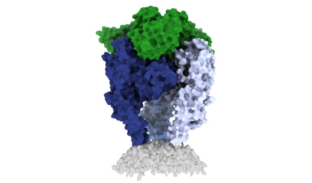
Scientists from La Jolla Institute for Immunology and the Institut Pasteur have shed light on the structure of the rabies virus glycoprotein, seen here. Credit: Heather Callaway, Ph.D., LJI
In a new study, researchers from La Jolla Institute have unveiled one of the first high-resolution looks at the rabies virus glycoprotein in its vulnerable ‘trimeric’ form. These new images, published in Science Advances, may open up a new vaccine for the deadly virus.
The CDC estimates that 59 000 people die from rabies virus every year, with 40% of those bitten by rabid animals being under 15. Some victims, especially kids, don’t realise they’ve been exposed until it is too late. The intense rabies treatment regimen is not widely available and the average $3800 is out of the reach of less well-off families.
Rabies vaccines, rather than treatments, are much more affordable and easier to administer. But according to Professor Erica Ollmann Saphire, PhD, of the La Jolla Institute, lead researcher of the new study, those vaccines also come with a massive downside.
“Rabies vaccines don’t provide lifelong protection. You have to get your pets boosted every year to three years,” she said. “Right now, rabies vaccines for humans and domestic animals are made from killed virus. But this inactivation process can cause the molecules to become misshapen – so these vaccines aren’t showing the right form to the immune system. If we made a better shaped, better structured vaccine, would immunity last longer?”
“The rabies glycoprotein is the only protein that rabies expresses on its surface, which means it is going to be the major target of neutralising antibodies during an infection,” said LJI Postdoctoral Fellow Heather Callaway, PhD, the study’s first author.
“Rabies is the most lethal virus we know. It is so much a part of our history – we’ve lived with its spectre for hundreds of years,” added Prof Saphire. “Yet scientists have never observed the organisation of its surface molecule. It is important to understand that structure to make more effective vaccines and treatments – and to understand how rabies and other viruses like it enter cells.”
Shapeshifting Rabies virus evades antibodies
Why rabies vaccines don’t provide long-term protection is still unclear, but they do know that its shape-shifting proteins are a problem.
The rabies glycoprotein has sequences that unfold and flip upward when needed, like a Swiss Army knife. The glycoprotein can shift back and forth between pre-fusion (before fusing with a host cell) and post-fusion forms. It can also come apart, changing from a trimer structure (where three copies come together in a bundle) to a monomer (one copy by itself).
This shapeshifting can make rabies invisible to human antibodies, which are built to recognise a single site on a protein. They cannot follow along when a protein transforms to hide or move those sites.
The new study gives scientists a critical picture of the correct glycoprotein form to target for antibody protection.
Capturing the glycoprotein at last
Over the course of three years, Callaway worked to stabilise and freeze the rabies glycoprotein in its pre-fusion form.
Callaway paired the glycoprotein with a human antibody, which helped her pinpoint one site where the viral structure is vulnerable to antibody attacks. The researchers then captured a 3D image of the glycoprotein using cutting-edge cryo-electron microscope equipment at LJI.
The new 3D structure highlights several key features researchers hadn’t seen before. Importantly, the structure shows the fusion peptides, the way they appear in real life. These two important sequences link the bottom of the glycoprotein to the viral membrane, but project into the target cell during infection. Getting stable image of these sequences is challenging: other rabies researchers have had to cut them off to try to get images of the glycoprotein.
Dr Callaway solved this problem by capturing the rabies glycoprotein in detergent molecules. “That let us see how the fusion sequences are attached before they snap upward during infection,” said Prof Saphire.
Now that scientists have a clear view of this viral structure, they can better design vaccines to create antibodies with a better picture of the targt.
“Instead of being exposed to four-plus different protein shapes, your immune system should really just see one – the right one,” said Dr Callaway. “This could lead to a better vaccine.”
Preventing a family of viruses
More images are needed of rabies virus and its relatives together with neutralising antibodies, and could reveal common antibody targets for lyssaviruses, which can also infect humans and animals. According to Dr Callaway, scientists are working on solving several of these structures, which could reveal antibody targets that lyssaviruses have in common.
“Because we didn’t have these structures of the rabies virus in this conformational state before, it’s been hard to design a broad-spectrum vaccine,” Dr Callaway said.
Scientists have harnessed a computational approach usually used in oil exploration to search for cures for rare genetic diseases such cystic fibrosis. By using the method to analyse the spatial relationships between different variants of a protein, instead of the relationships between test wells across an oil field, the researchers can obtain valuable information on how disease affects a protein’s underlying shape and how drugs can restore that shape to normal.
The new method, detailed in the journal Structure, runs with just a few gene sequences collected from people with disease. Then, it determines how the structure of each corresponding variant protein is associated with its function, and how this functional structure can affect pathology and be repaired by therapeutics. To test the techniques, the researchers showed why existing drugs for cystic fibrosis fall short of curing the disease.
“This is an important step forward for treating rare diseases,” said senior author William Balch, PhD, professor of Molecular Medicine at Scripps Research. “The fact that we can get so much information from a few gene sequences is really unprecedented.”
Studies on inherited diseases often rely on the precise three-dimensional shape of a protein affected by disease. But genetic diseases can be caused by thousands of gene variants, some of which destabilise or change the protein shape in ways that make isolating the protein for further investigation much more difficult than usual.
Prof Balch, with Scripps Research senior staff scientist Chao Wang and staff scientist Frédéric Anglés, instead wanted to use natural variation to their advantage. So the group developed a method called variation-capture (VarC) mapping to analyse the natural array of gene sequences which exist in the human population and determine the mechanism by which they each changed a protein’s structure to cause disease.
Among other statistical tools, Prof Balch’s group integrated the methods that oil companies use to draw inferences about the location of an oil reservoir using only a small number of test wells. With only a few gene sequences, this let the researchers determine the most likely structural mechanisms driving function for each variant leading to disease, as well as model how drugs impacted those structural functions.
In the case of cystic fibrosis, disease is caused by genetic variants in the cystic fibrosis transmembrane conductance regulator (CFTR), leading to a buildup of mucus in the lungs. More than 2000 variants of the CFTR gene have been identified, and many of these variants were known to have very different effects on the CFTR protein, but it has been difficult to compare and contrast these variants to guide how patients with different variants should be treated differently in the clinic.
“When you want to treat patients, you really have to appreciate that different therapeutics might target different variants in completely different ways, and that’s why our approach that looks at many different variants all at once is so powerful,” explained Wang. “Our approach not only reveals how these variants contribute to each patient’s biology, but also connects them in a way that each variant can inform how to manage the others.”
The researchers input about 60 genetic variants found in the cystic fibrosis population into their VarC program. The analysis captured how each amino acid residue talks to every other residue to generate function, and revealed that most of the cystic fibrosis patients had the same net effect on the protein: an unstable inner core.
When the program modelled how existing cystic fibrosis drugs impacted the structures, the researchers discovered that, despite the drugs’ effect on CFTR structure, none of them effectively stabilised the protein’s hidden inner core. This was like how the location of an oil reservoir in a complex landscape can be revealed by test wells.
Now that the researchers better understand the structural deficiencies in CFTR in cystic fibrosis patients, they say that the job of developing an effective drug to fix it is much easier. Potential compounds can be modelled in advance of lab experiments for their effect on the inner core of the CFTR protein.
“In most drug discovery, you throw thousands of compounds at a protein and see which ones change it, often without fully understanding the mechanism,” said Prof Balch. “To fix a thing, you must first understand the problem.”
Already, his team is applying the method to other rare genetic diseases, as well as pursuing new drugs to treat cystic fibrosis.
The world’s first complete study of living donor uterine transplantation, published in the journal Fertility and Sterility, has found that it is an effective, safe method to remedy infertility when a functioning uterus is lacking.
After seven of the study’s nine transplants, in vitro fertilisation (IVF) treatment ensued. In this group of seven women, six (86%) became pregnant and gave birth. Three had two children each, making the total number of babies nine.
In terms of what is known as the ‘clinical pregnancy rate’, the study also showed good IVF results. The probability of pregnancy per individual embryo returned to a transplanted uterus was 33%, about the same as for typical IVF.
Participants followed up
Few cases were studied, the researchers observed, but the material is the world best and included extensive, long-term follow-ups of participants’ physical and mental health.
None of the donors had pelvic symptoms but, in a few, the study describes mild, partially transient symptoms in the form of discomfort or minor swelling in the legs.
After four years, health-related quality of life in the recipient group as a whole was higher than in the general population. Neither members of the recipient group nor the donors had levels of anxiety or depression that required treatment.
Growth and development of the children were monitored as well, up to age two and is, accordingly, the longest child follow-up study conducted to date in this context. Further monitoring is planned to adulthood.
Good health in the long term
“This is the first complete study that’s been done, and the results exceed expectations in terms both of clinical pregnancy rate and of the cumulative live birth rate,” said study leader Mats Brännström, professor of obstetrics and gynaecology at Sahlgrenska Academy, University of Gothenburg.
“The study also shows positive health outcomes: The children born to date remain healthy and the long-term health of donors and recipients is generally good too.”
The first birth after uterine transplantation took place in Gothenburg in 2014. Another seven births followed, within the framework of the same research project, before anyone outside Sweden gave birth following uterine transplantation.
The research group has since passed on its methods and techniques through direct knowledge transfer to several research centres outside Sweden. By the end of 2021, there were an estimated 90 uterine transplants worldwide, of which 20 had been done in Sweden. Worldwide, some 50 children have been born after uterine transplantation.
Oestrogen may protect diabetes patients from cardiomyopathy, according to research published in Circulation: Heart Failure. The study showed that severe insulin resistance in the heart causes cardiomyopathy and death in male mice, but also showed that oestrogen protected female mice.
“Cardiovascular disease is the major cause of morbidity and mortality for diabetic patients,” explained Shaodong Guo, PhD, primary investigator for the study at Texas A&M.
“Previous studies have shown that while there’s a lower instance of both cardiovascular disease and Type 2 diabetes in premenopausal women than in their age-matched male counterparts, these incidences rise sharply after female menopause,” Prof Guo said.
This indicates that the ovaries and ovarian hormones, such as oestrogen, may protect from Type 2 diabetes and cardiovascular diseases, he said.
The study investigated the role of ovaries and oestrogen in cardiac function and energy metabolism with mice who had the the cardiac insulin receptor substrate, IRS, modified or suppressed to mimic cardiac insulin resistance.
“Our previous studies reported that impaired cardiac insulin signalling with loss of insulin receptor substrate IRS1 and IRS2 genes leads to death of male mice,” he said. “In this study, we wanted to know how the removal of the ovaries might affect cardiomyopathy in female mice and also what other impacts the loss of insulin receptors might have on energy metabolism and mitochondrial function.”
Insulin resistance and signaling
About 90–95% of patients with Type 2 diabetes suffer from insulin resistance, a risk factor for heart failure. In healthy tissue, insulin binds to insulin receptors, activating a network of intracellular signalling pathways. Disruptions in these signalling pathways have been linked to mitochondrial dysfunction, cardiomyopathy, and impaired glucose and fatty acid metabolism, among other health issues.
In this study, mice lacking IRS developed dilated cardiomyopathy, and analysis showed lowered activity of genes important for mitochondrial function and energy metabolism.
The study fills in some blanks in understanding the role of insulin and estrogen signaling in mitochondrial function.
Study findings
Guo said there were four important findings from the study:
All female mice that lacked insulin receptor substrates survived for more than a year.
Female mice without insulin receptor substrates were less likely to experience severe cardiac dysfunction and death if they had ovaries. If the mice also lacked ovaries but received oestrogen, it prolonged their lifespans. Doses of oestrogen also protected IRS-altered male mice from heart dysfunction.
Guo said oestrogen also prevents cardiomyopathy induced by loss of cardiac insulin receptor substrates.
“And removal of the ovaries leads to the death of female cardiac IRS1 and IRS2 double genes knockout mice if there is no reintroduction of oestrogen,” he said.
Loss of IRS1 and IRS2 genes in heart tissue disrupts cardiac energy metabolism, gene activity involved in mitochondrial function, and whole-body energy metabolism. However, oestrogen partially reverses these effects.
Oestrogen is important for healthy cellular signalling pathways and promotes mitochondrial function.
Prof Guo said the study shows that oestrogen enhances cardiac function, promotes energy metabolism, prevents cardiomyopathy and prolongs survival in both male and ovariectomy female mice lacking the insulin receptor substrates.
“This study provides evidence for the gender difference for the incidence of cardiovascular disease and implies that oestrogen replacement therapy is feasible for the treatment of diabetic cardiomyopathy through enhancement of mitochondrial function and energy metabolism,” he said. “It also reveals some of the signalling pathways that may be potential therapeutic targets for the prevention or treatment of cardiovascular diseases in patients with Type 2 diabetes.”
Guo also noted that diet could also play a role with oestrogens in foods.
“The study implies that food-derived oestrogens or phytoestrogens may play similar roles to oestrogen, as observed in mice,” he said. “This may help us reshape our knowledge of nutrient and food sciences related to plant hormones that can modulate chronic metabolic diseases such as Type 2 diabetes and associated cardiovascular complications.”
A Swedish court has convicted Paolo Macchiarini, a formerly lauded trachea surgeon, of causing bodily harm to a patient through negligence during a highly experimental stem-cell trachea transplant. For this, the court handed down a two-year probational sentence. He was acquitted of assault charges on two other patients; all three died in the months and years after the surgeries.
In 2010, Macchiarini was hired by the Karolinska Institute (KI) and the Karolinska University Hospital to support Sweden’s regenerative medicine innovation. His specialty was replacing damaged tracheae with artificial ones that combined stem cells with polymer scaffolds or decellularised donor tracheae. Starting in 2011, he began operating on patients as an experimental life-saving measure but his work at at KI was suspended in 2013 after the second of his three patients died. However, he continued performing surgeries in Russia.
Yet there were already hints that something was amiss even before the first surgeries. In 2011, another academic, Pierre Delaere of UZ Leuven in Belgium accused Macchiarini of misrepresenting research findings in published articles. In 2012, Macchiarini was arrested in Italy and charged with fraud and attempted extortion.
By 2014, after the death of his first patient, three separate allegations were raised of scientific misconduct in reporting the cases. He would later be cleared of these, but in 2016 a TV documentary called ‘The Experiment’ described the suffering and deaths patients of failed artificial tracheas transplants, and raised many issues concerning care and research ethics. The severe public backlash caused KI to launch another investigation into Macchiarini, amid an upheaval which saw a string of resignations and an overhaul of hiring and ethics. He was found to have falsified his CV, and published papers with false or misleading data that were subsequently retracted. By March, he had been fired and criminal charges filed against him.
BBC News reported that at least seven people had died following the surgeries. In 2018, KI found seven researchers guilty of academic misconduct. Swedish authorities decided to reopen investigations into the three deaths.
Matthias Corbascio, a cardiac surgeon at KI who testified in the trial, told SVT Nyheter that he doesn’t believe justice has been done. “My reaction is that it is very meager. It is a terrible scandal and terrible for the patients’ families that he could get away so easily,” he said.
Chief judge Bjoern Skaensberg said the court had agreed with prosecutors that the surgery had not been consistent with “science and proven experience”. However, he told public broadcaster SVT that it had concluded that “two of the interventions were justifiable, but not the third”.
The court had found that all three patients had suffered serious bodily injury, Judge Skaensberg said. But Macchiarini was cleared of assault as no intent to harm had been proven.
Macchiarini had always denied any wrongdoing, arguing that the transplants were aimed at saving the patients’ lives.
However, whistleblower Dr Matthias Corbascio told SVT that the verdict was a scandal and there had never been any chance of the operations succeeding.
The suspended sentence means he will be on probation for the next two years.
Researchers have tested a new potato processing technique designed to slow down the digestion of potato starch. Their experiments show that the approach prevents certain digestive enzymes from reaching the potato starch as quickly, leading to a more controlled release of dietary glucose.
Foods with lower glycaemic index have a variety of health benefits, such as reducing the risk of cardiovascular disease.
“There is a perception that potato foods are unhealthy because eating a large amount of some potato foods can cause a rapid increase in blood sugar, which is a risk for people with diabetes or those who want to control body weight,” said Amy Lin, PhD, the study’s principal investigator at A*STAR. “Our team revealed that toggling the accessibility of two digestion enzymes – α-amylase and mucosal α-glucosidase – in the small intestine is a successful strategy to make dietary glucose slowly and continuously release from potatoes.”
The findings are being presented online at NUTRITION 2022 LIVE ONLINE, the flagship annual meeting of the American Society for Nutrition.
The researchers cut potatoes into cubes and blanched them in hot water with a food grade ingredient for 30 minutes., though they did not disclose what the ingredient was.
An enzyme barrier of pectin
This process causes a reaction with pectin, a water-soluble fibre in potatoes, creating a gelling structure which makes a porous barrier between starch granules and digestive enzymes. The size of the barrier’s pores can can be determined by the processing method to moderate how quickly α-amylase is able to penetrate the potato parenchyma cells and degrade starch to small molecules. Converting starch molecules to glucose relies on mucosal α-glucosidase, which is too big to penetrate those pores. Therefore, the elevation of dietary glucose of processed potatoes depends on the how quickly small starch molecules leach out of parenchyma cells and are digested by mucosal α-glucosidase.
“Without our treatment, enzymes move freely in and out of cells, and starch is quickly degraded by both enzymes and rapidly converted to glucose,” said Dr Lin. “The treatment allows the starch to be slowly degraded to prevent a spike in glycemia and then fully converted to glucose to meet our energy and nutritional needs.”
The technique is not designed to prevent the potato from being digested, but rather to slow digestion to avoid a rapid increase in blood sugar. Researchers say the modification could also help consumers feel full for a longer period after eating the treated potatoes, helping to avoid overeating.
Researchers report that the method performed well in tests with a simulated digestion process in the laboratory. Treatment increased the fraction of the starch that is considered slowly digestible from 10% to 35% and significantly reduced the ability for the enzyme a-amylase to access starch within the cell walls.
But how do they taste?
Since the process essentially pre-cooks the potatoes, treated potatoes are not shelf-stable but could be frozen and then cooked or further processed for dishes such as roasted potatoes, hash browns, soups or stir-fry, researchers say. Initial taste tests had good results in terms of digestibility and texture.
As a next step, the researchers are preparing to further test impacts on digestibility in a clinical trial. They also plan to study whether a similar approach could be used to improve other staple foods.
Infected cell covered with SARS-CoV-2 viruses. Source: NIAID
Findings from a new study reported in The Lancet has found the risks of developing long COVID are greatly reduced (by ~50% to 75%) as a result of Omicron infection compared to Delta infection.
The study, the first of its kind to report on long COVID risk associated with Omicron, highlights the speed with which app-based health surveillance can provide insights. These have further been shown to be consistent and replicable.
A major strength of the study was the ability to log a wide range of symptoms with the app. Limitations of the self-reported data include no direct testing of infectious variants (here assumed from national data) and no objective measures of illness duration. There was insufficient data to estimate the odds of long COVID in unvaccinated individuals. Finally, to enable swift reporting, the period of assessment of omicron cases was slightly shorter than for the delta variant, and assessment of longer durations of long COVID (eg, >12 weeks) was not possible.
In this case-control observational study, the researchers took self-reported data from the COVID Symptom Study app.
Participants were selected on a positive COVID test after SARS-CoV-2 after vaccination, at least one log per week in the app for at least 28 days after testing positive, and no previous SARS-CoV-2 infections before vaccination. The researchers identified 56 003 UK adults first testing positive between Dec 20, 2021, and March 9, 2022, when the Omicron variant became dominant. Similarly, researchers identified 41 361 UK adult cases first testing positive between June 1, 2021, and Nov 27, 2021, during the period of Delta dominance.
Both symptomatic and asymptomatic infections were considered, and, for the Omicron period, only participants testing positive before Feb 10, 2022, were included, to ensure all participants had at least 28 days for symptom reporting after testing positive. The researchers adjusted for socioeconomic deprivation and other factors.
In both periods, female participation was higher than male participation (55% for Omicron and 59% for Delta cases). Delta and Omicron cases had similar age (mean age 53 years) and prevalence of comorbidities (around 19%). Among Omicron cases, 2501 (4.5%) of 56 003 people experienced long COVID and, among Delta cases, 4469 (10.8%) of 41 361 people experienced long COVID. Cases were stratified by time from vaccination to first infection. They found that, for all vaccine timings, Omicron cases were less likely to experience long COVID, with an odds ratio ranging from 0.24 to 0.50.
However, the researchers noted that the the absolute number of people with long COVID at a certain time depends on the pandemic curve. Considering the UK Omicron peak of more than 350 000 new symptomatic COVID cases per day estimated on March 26, 2022, by the ZOE app model, with 4% of cases being long COVID, future numbers with long COVID will inevitably rise.
Fentanyl is one of the most commonly used analgesics in the hospital and, in rodents, may have lasting sensorimotor and behavioural impacts. A study published in the British Journal of Anaesthesia has shown that fentanyl can induce changes similar to autism-like behaviours in young mice.
Fentanyl, a mu-opioid receptor agonist, is a potent synthetic opioid, which, similar to morphine, produces analgesia but is 50 to 100 times more potent. A dose of only 100 micrograms can produce equivalent analgesia to approximately 10mg of morphine. However, fentanyl exhibits vastly different properties and pharmacokinetics. Clinically, its most common use is as a sedative in intubated patients and severe cases of pain in patients with renal failure due to its primarily hepatic elimination. Fentanyl’s side effects are similar to those of heroin.
However, whether the use of fentanyl is associated with the development of autism is not known. An animal study led by investigators at Massachusetts General Hospital (MGH), Shanghai 10th People’s Hospital, and the University of Pennsylvania e. The findings are
Research by other groups has shown that N-methyl-D-aspartate receptor dysfunction contributes to autism. Variations in Grin2a and Grin2b, the genes encoding GluN2A and GluN2B subunits of N-methyl-D-aspartate receptor, are associated with autism. In addition, the anterior cingulate cortex of the brain is affected in autism.
In this current study, the research team reported that fentanyl induces autism-like behaviors in young male and female mice via activating the mu-opioid receptor in the anterior cingulate cortex. Further, these fentanyl-induced autism-like behaviors appear partially due to the hypermethylation-mediated reduction of Grin2b expression in the anterior cingulate cortex of mice.
“Because the anterior cingulate cortex is a hub for mediating social information, we focused on the expression of Grin2b in that area,” says Yuan Shen, MD, PhD, the paper’s senior author and a professor of Psychiatry at Shanghai 10th People’s Hospital. “We found fentanyl decreased expression of Grin2b in the anterior cingulate cortex. The overexpression of Grin2b prevents fentanyl-induced autism-like behavior in the mice. These findings suggest a potential mechanism to prevent or treat the autism-like behavior,” says Shen.
The group used an open field test (in which a mouse can walk inside a box) and an elevated plus-maze (in which a mouse can walk on an elevated platform) to observe the anxiety and stereotyped behaviours of mice. Using a three-chamber social preference test (where a mouse can interact with another mouse), they also assessed potential social deficits. “We used these tests because impaired social interaction, stereotyped behaviours, and anxiety are the key feature of autism-like behaviours in mice,” said Zhihao Sheng, co-first author of the paper. Sheng is a graduate student at Shanghai 10th People’s Hospital.
“However, the changes of mice in these behavioral tests do not equal autism in humans. These behavioral tests are only used to study the autism-like behaviors in mice because they can demonstrate certain features of behavior changes similar to the manifestation of autism,” said co-first author Qidong Liu, PhD, assistant professor at Shanghai 10th People’s Hospital.
Co-senior author Zhongcong Xie, MD, PhD, added: “There is no current evidence that fentanyl is associated with a similar effect in humans and the outcome of the animal study is not an indication to avoid fentanyl in clinical anesthesia. However, the outcome will promote further research, including clinical investigations, to determine the potential neurobehavioral influence of opioids on brain development.”