Day: June 20, 2022

Paediatric Kidney Transplants without Immunosuppressive Drugs
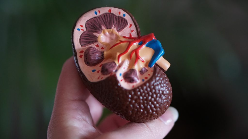
Anatomic model of a kidney
Photo by Robina Weermeijer on Unsplash

Stanford Medicine physicians have developed a way to provide paediatric kidney transplants without immunosuppressive drugs. Their key innovation is a safe method to transplant the donor’s immune system to the patient before surgeons implant the kidney.

The medical team has dubbed the two-transplant combination a “dual immune/solid organ transplant,” or DISOT. The first three DISOT cases, all performed at Lucile Packard Children’s Hospital Stanford were described in the New England Journal of Medicine, accompanied by an an editorial about the research.

The Stanford innovation removes the possibility that the recipient will experience immune rejection of their transplanted organ, the most common reason for a second organ transplant The new procedure also rids recipients of the substantial side effects of a lifetime of immune-suppressing medications, including increased risks for cancer, diabetes, infections and hypertension.

“Safely freeing patients from lifelong immunosuppression after a kidney transplant is possible.”

Alice Bertaina, MD, PhD, report’s lead author, associate professor of paediatrics, Stanford University

The first three DISOT patients were children with a rare immune disease, but the team is expanding the types of patients who could benefit. The protocol received FDA approval on May 27, 2022, for treating patients with a variety of conditions that affect the kidneys. Dr Bertaina anticipates that the protocol will eventually be available to many people needing kidney transplants, starting with children and young adults, and later expanding to older adults. The researchers also plan to investigate DISOT’s utility for other types of solid-organ transplants.

The scientific innovation from Dr Bertaina’s team has another important benefit: It enables safe transplantation between a donor and recipient whose immune systems are genetically half-matched, meaning children can receive stem cell and kidney donations from a parent.

The advance is especially meaningful for Jessica and Kyle Davenport of Muscle Shoals, Alabama. Their two children, both born with a rare and potentially deadly immune disease, are among the first recipients of DISOT: 8-year-old Kruz received transplants from Jessica, while his 7-year-old sister, Paizlee, received transplants from Kyle.

“They’ve healed and recovered, and are doing things we never thought would be possible,” said Jessica Davenport. After years of helping Kruz and Paizlee cope with severe immune deficiency and its attendant infection riskk as well as kidney dialysis, she and her husband are thrilled that their children have more normal lives.

The idea of transplanting a patient with their organ donor’s immune system has been around for decades, but it has been difficult to implement. Transplants of stem cells from bone marrow provide the patient with a genetically new immune system, as some of the bone marrow stem cells mature into immune cells in the blood. First developed for people with blood cancers, stem cell transplants carry the risk of the new immune cells attacking the recipient’s body, a potentially lethal complication called graft-versus-host disease.

Source: Stanford Medicine

Early Intervention in Spinal Muscular Atrophy is Key

Photo by National Cancer Institute on Unsplash

According to the results of a new study published in Developmental Medicine & Child Neurology, early identification and treatment of patients with spinal muscular atrophy (SMA) can greatly reduce the total financial costs associated with the condition. 

A genetic disorder, SMA is characterised by progressive muscle weakness, reduced tone with associated destruction of alpha motor units. There are four main subtypes of spinal muscular atrophy defined by the age of onset and severity with type 0 presenting in utero and causing death within the first months of life and type 4 in adulthood, causing mild weakness and no effect on lifespan. Understanding the underlying pathophysiology, subtypes, and emerging treatments is key to treating patients with spinal muscular atrophy effectively.

Analysing the data of 149 SMA patients, (93 untreated, 42 treated after symptoms arose, and 14 treated after early diagnosis), the total societal cost was lower in untreated patients (due to high drug costs in treated patients), but costs were lower for treated patients who were identified by newborn screening than for treated patients identified due to the development of symptoms. 

“These data are important as they are issued from a real-life prospective collection. They demonstrate clearly that as long as the decision to reimburse treatments for SMA has been made, newborn screening becomes a no-brainer—not only because it gives patients a much better future, but also because it saves a significant amount of money for the taxpayer,” said senior author Laurent Servais, PhD, of the University of Liege, in Belgium and the University of Oxford, in the UK. “Using these data issued from the real world, we are working currently on a model that estimates the lifetime cost of the different strategies.” 

Source: Wiley

Child Welfare NGO One of Many Defunded by Government

Photo by Chayene Rafaela on Unsplash

Christelike Maatskappy Raad Noord (CMRN), an NGO in Gauteng which focuses on children’s welfare through the use of social workers, has been defunded by the government. This amounts to just over half of its funding, according to marketing manager Anya le Cornu. Other NGOs have also had their funding cut, she said, as heard via the Auditor General’s office.

This comes in the wake of the COVID pandemic as CMNR had to cope with continuing to deliver services amidst lockdowns. If other NGOs are similarly impacted, . Founded in 1936, CMRN aims to eradicate child abuse and neglect, providing a wide range of child protection service from its 16 centres.

The NGO assists a large number of families of children: 6000 beneficiaries received material or skill support in 2020–2021, its Child Protection Awareness campaign reached 14 500 people, 622 children were protected through the legal system, and 900 children received speech or play therapy.

However, these services are obviously under threat from the significant loss of provincial government funding, which at R7 million, accounted for 53% of its income.

In order to cope, CMNR has been forced to restructure, reducing costs wherever possible. Unfortunately, it has having to slash its social workers from 28 to 17 as of 1 July.

Due to the lack of subsidy and other challenges, areas such as statutory work may be impacted.

According to le Cornu, CMRN will try and secure funding through every means possible. “We will maintain and strengthen our relationship with the NG church, our other funding partner,” she says. “We will also continue with our marketing and fundraising initiatives. Professional fees will also be applied where possible. We will also reach out to schools and other institutions where part time social work services are needed and contract these services out to generate an income stream.”

The organisation remains hopeful despite these challenges. “We do wish to have a good relationship with the Department of Social Development and would apply for government funding in specific programs where the objectives of these programs are aligned to our own and the communities we serve,” says le Cornu.

“The CMR North believes that we will survive this crisis and hope to be a beacon of light for other NGOs who might suffer the same fate. It is our passion to continue bringing hope to the vulnerable and we see these events as an opportunity to re-invent our services so that they can have a broader and positive impact in the communities we serve.”

Hopefully, additional funding can be found so that CMRN can continue to provide its services, but if this is part of a wider pattern, people in South Africa who are most in need and depend on these services will suffer the most.

A Crystal Clear Look at Rabies Opens up New Vaccines

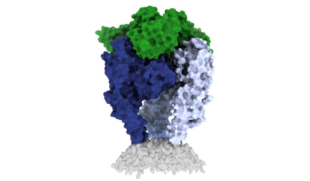
Scientists from La Jolla Institute for Immunology and the Institut Pasteur have shed light on the structure of the rabies virus glycoprotein, seen here. Credit: Heather Callaway, Ph.D., LJI

In a new study, researchers from La Jolla Institute have unveiled one of the first high-resolution looks at the rabies virus glycoprotein in its vulnerable ‘trimeric’ form. These new images, published in Science Advances, may open up a new vaccine for the deadly virus.

The CDC estimates that 59 000 people die from rabies virus every year, with 40% of those bitten by rabid animals being under 15. Some victims, especially kids, don’t realise they’ve been exposed until it is too late. The intense rabies treatment regimen is not widely available and the average $3800 is out of the reach of less well-off families.

Rabies vaccines, rather than treatments, are much more affordable and easier to administer. But according to Professor Erica Ollmann Saphire, PhD, of the La Jolla Institute, lead researcher of the new study, those vaccines also come with a massive downside.

“Rabies vaccines don’t provide lifelong protection. You have to get your pets boosted every year to three years,” she said. “Right now, rabies vaccines for humans and domestic animals are made from killed virus. But this inactivation process can cause the molecules to become misshapen – so these vaccines aren’t showing the right form to the immune system. If we made a better shaped, better structured vaccine, would immunity last longer?”

“The rabies glycoprotein is the only protein that rabies expresses on its surface, which means it is going to be the major target of neutralising antibodies during an infection,” said LJI Postdoctoral Fellow Heather Callaway, PhD, the study’s first author.

“Rabies is the most lethal virus we know. It is so much a part of our history – we’ve lived with its spectre for hundreds of years,” added Prof Saphire. “Yet scientists have never observed the organisation of its surface molecule. It is important to understand that structure to make more effective vaccines and treatments – and to understand how rabies and other viruses like it enter cells.”

Shapeshifting Rabies virus evades antibodies

Why rabies vaccines don’t provide long-term protection is still unclear, but they do know that its shape-shifting proteins are a problem.

The rabies glycoprotein has sequences that unfold and flip upward when needed, like a Swiss Army knife. The glycoprotein can shift back and forth between pre-fusion (before fusing with a host cell) and post-fusion forms. It can also come apart, changing from a trimer structure (where three copies come together in a bundle) to a monomer (one copy by itself).

This shapeshifting can make rabies invisible to human antibodies, which are built to recognise a single site on a protein. They cannot follow along when a protein transforms to hide or move those sites.

The new study gives scientists a critical picture of the correct glycoprotein form to target for antibody protection.

Capturing the glycoprotein at last

Over the course of three years, Callaway worked to stabilise and freeze the rabies glycoprotein in its pre-fusion form.

Callaway paired the glycoprotein with a human antibody, which helped her pinpoint one site where the viral structure is vulnerable to antibody attacks. The researchers then captured a 3D image of the glycoprotein using cutting-edge cryo-electron microscope equipment at LJI. 

The new 3D structure highlights several key features researchers hadn’t seen before. Importantly, the structure shows the fusion peptides, the way they appear in real life. These two important sequences link the bottom of the glycoprotein to the viral membrane, but project into the target cell during infection. Getting stable image of these sequences is challenging: other rabies researchers have had to cut them off to try to get images of the glycoprotein.

Dr Callaway solved this problem by capturing the rabies glycoprotein in detergent molecules. “That let us see how the fusion sequences are attached before they snap upward during infection,” said Prof Saphire.

Now that scientists have a clear view of this viral structure, they can better design vaccines to create antibodies with a better picture of the targt.

“Instead of being exposed to four-plus different protein shapes, your immune system should really just see one – the right one,” said Dr Callaway. “This could lead to a better vaccine.”

Preventing a family of viruses

More images are needed of rabies virus and its relatives together with neutralising antibodies, and could reveal common antibody targets for lyssaviruses, which can also infect humans and animals. According to Dr Callaway, scientists are working on solving several of these structures, which could reveal antibody targets that lyssaviruses have in common.

“Because we didn’t have these structures of the rabies virus in this conformational state before, it’s been hard to design a broad-spectrum vaccine,” Dr Callaway said.

Source: La Jolla Institute for Immunology

Oil Exploration Software Reveals why Cystic Fibrosis Drugs Fail

Photo by WORKSITE Ltd. on Unsplash

Scientists have harnessed a computational approach usually used in oil exploration to search for cures for rare genetic diseases such cystic fibrosis. By using the method to analyse the spatial relationships between different variants of a protein, instead of the relationships between test wells across an oil field, the researchers can obtain valuable information on how disease affects a protein’s underlying shape and how drugs can restore that shape to normal.

The new method, detailed in the journal Structure, runs with just a few gene sequences collected from people with disease. Then, it determines how the structure of each corresponding variant protein is associated with its function, and how this functional structure can affect pathology and be repaired by therapeutics. To test the techniques, the researchers showed why existing drugs for cystic fibrosis fall short of curing the disease.

“This is an important step forward for treating rare diseases,” said senior author William Balch, PhD, professor of Molecular Medicine at Scripps Research. “The fact that we can get so much information from a few gene sequences is really unprecedented.”

Studies on inherited diseases often rely on the precise three-dimensional shape of a protein affected by disease. But genetic diseases can be caused by thousands of gene variants, some of which destabilise or change the protein shape in ways that make isolating the protein for further investigation much more difficult than usual.

Prof Balch, with Scripps Research senior staff scientist Chao Wang and staff scientist Frédéric Anglés, instead wanted to use natural variation to their advantage. So the group developed a method called variation-capture (VarC) mapping to analyse the natural array of gene sequences which exist in the human population and determine the mechanism by which they each changed a protein’s structure to cause disease.

Among other statistical tools, Prof Balch’s group integrated the methods that oil companies use to draw inferences about the location of an oil reservoir using only a small number of test wells. With only a few gene sequences, this let the researchers determine the most likely structural mechanisms driving function for each variant leading to disease, as well as model how drugs impacted those structural functions.

In the case of cystic fibrosis, disease is caused by genetic variants in the cystic fibrosis transmembrane conductance regulator (CFTR), leading to a buildup of mucus in the lungs. More than 2000 variants of the CFTR gene have been identified, and many of these variants were known to have very different effects on the CFTR protein, but it has been difficult to compare and contrast these variants to guide how patients with different variants should be treated differently in the clinic.

“When you want to treat patients, you really have to appreciate that different therapeutics might target different variants in completely different ways, and that’s why our approach that looks at many different variants all at once is so powerful,” explained Wang. “Our approach not only reveals how these variants contribute to each patient’s biology, but also connects them in a way that each variant can inform how to manage the others.”

The researchers input about 60 genetic variants found in the cystic fibrosis population into their VarC program. The analysis captured how each amino acid residue talks to every other residue to generate function, and revealed that most of the cystic fibrosis patients had the same net effect on the protein: an unstable inner core.

When the program modelled how existing cystic fibrosis drugs impacted the structures, the researchers discovered that, despite the drugs’ effect on CFTR structure, none of them effectively stabilised the protein’s hidden inner core. This was like how the location of an oil reservoir in a complex landscape can be revealed by test wells.

Now that the researchers better understand the structural deficiencies in CFTR in cystic fibrosis patients, they say that the job of developing an effective drug to fix it is much easier. Potential compounds can be modelled in advance of lab experiments for their effect on the inner core of the CFTR protein.

“In most drug discovery, you throw thousands of compounds at a protein and see which ones change it, often without fully understanding the mechanism,” said Prof Balch. “To fix a thing, you must first understand the problem.”

Already, his team is applying the method to other rare genetic diseases, as well as pursuing new drugs to treat cystic fibrosis.

Source: Scripps Research Institute