Why People With Autism May Be More Likely To Get Parkinson’s Disease
Dopamine transporters in the brain could be early biomarkers for the potential development of Parkinson’s disease

Researchers at the University of Missouri may have uncovered a clue explaining why young adults with autism are roughly six times more likely to develop Parkinson’s disease later in life.
In a recent study, the researchers found that some young adults with autism show abnormalities in dopamine transporters, tiny molecules in the brain that recycle unused dopamine, on brain scans that are typically used to diagnose older adults with Parkinson’s disease.
Future research could help determine whether the health of dopamine transporters could be an early warning sign of Parkinson’s disease developing later in life.
“While the loss of these dopamine transporters can be biomarkers for Parkinson’s disease, no one had ever thought to look at them in the context of young adults with autism, so hopefully this work can help us explore if there is a potential link going forward,” David Beversdorf, a professor in the School of Medicine and College of Arts and Science, said. “There has been previous work looking into the total amount of dopamine in the brains of people with autism, but we took a new approach by looking at abnormalities in terms of how dopamine is processed in a specific part of the brain called the basal ganglia via these dopamine transporters.”
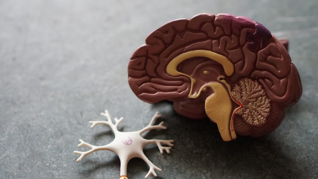
Dopamine under the spotlight
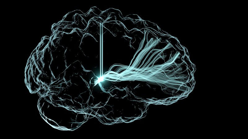
Dopamine is a neurotransmitter involved in numerous body functions, such as memory, pleasure, motivation, behaviour and attention. Of particular interest to Beversdorf, a clinician at the Thompson Center for Autism and Neurodevelopment, is that dopamine also helps control muscle movement as well as cognition.
Beversdorf, who collaborated with lead author Nanan Nuraini on the study, originally wanted to know whether certain repetitive behaviors common in some young adults with autism, such as hand-flapping or rocking back and forth, were linked with abnormalities in dopamine transporters.
While he did not notice patterns in that regard, what he found surprised him.
Beversdorf looked at Dopamine Transporter (DaT) brain scans of 12 young adults with autism.
Four different nuclear medicine specialists examined the scans. All of them agreed that two of the 12 young adults had abnormal dopamine transporters and that eight appeared normal. They disagreed on the remaining two.
“Since these DaT scans are typically used to diagnose or evaluate older adults with Parkinson’s disease, the appearance of abnormalities in some young adults with autism was very surprising, so we should look into this topic more going forward,” Beversdorf said. “While it’s too early to jump to conclusions, hopefully our work raises awareness about the importance of monitoring the brain health of young adults with autism as they age.”
Next, Beversdorf hopes to study a broader range of people with autism by conducting more DaT scans across different age groups.
“The earlier we can identify those who might be at greater risk for getting Parkinson’s disease down the road, the sooner we can discuss preventative measures, including whether certain medications could potentially slow down the progression of disease,” Beversdorf said.
Source: University of Missouri