
Day: November 5, 2026

How Canada Lost its Measles Elimination Status Highlights Threat of Immunity Gaps

90% of cases occurred in historically under-vaccinated communities
Findings offer insights for countries aiming to retain or regain their elimination status 
Photo by Gustavo Fring on Pexels

The outbreak that contributed to Canada losing its measles elimination status in November 2025 was concentrated in historically under-vaccinated communities with close social networks across Canada, the US, Mexico, and beyond, creating opportunities for cross-border transmission, concludes a study published by The BMJ.

The researchers say the findings highlight the ongoing risk of measles resurgence in countries that have previously successfully controlled the disease, and underscore the importance of culturally tailored community engagement to build trust and confidence in vaccination and prevent future outbreaks.

Canada achieved measles-free status in 1998 after introducing a two-dose vaccine schedule and a national catch-up programme for school-aged children, but lost it in November 2025 after a measles outbreak that involved several provinces. In Ontario, the outbreak disproportionately affected historically under-vaccinated communities – groups with historically low vaccine coverage and close social networks across Canada, the US, and Mexico.

To better understand the course of this outbreak and its impact, researchers analysed measles cases reported to Ontario’s public health surveillance system during the outbreak period (28 October 2024 to 6 October 2025).

The outbreak consisted of 2377 cases (2064 confirmed and 313 probable) over 49 weeks. Most cases (90%) occurred among members of historically under‑vaccinated communities, with limited spillover into the broader population.

Among the 2377 cases, around three quarters (73%) occurred in individuals aged 19 years or younger, 2,121 (89%) were unvaccinated and 97 (4%) had unknown vaccination status.

The outbreak was associated with 166 admissions to hospital, 13 admissions to an intensive care unit, 51 infections during pregnancy, 10 congenital measles infections, one case of encephalitis (brain inflammation), and one infant death.

Laboratory tests identified a distinct measles virus strain that was subsequently detected elsewhere in Canada, and in the US, Mexico, and the Netherlands due to close social ties within and between under-vaccinated communities combined with the high transmissibility of measles.

The researchers acknowledge that their analysis is subject to limitations inherent to public health surveillance data, and note that the number of cases and associated complications may be higher than what is reported in the study.

In addition, the proportion of cases in under-vaccinated communities is likely to be conservative, reflecting challenges in data collection for a variety of reasons, including mistrust among some community members and concerns from public health staff about the risk of stigmatisation of affected groups during the outbreak.

However, they say this measles outbreak illustrates the fragility of measles elimination in the presence of substantial immunity gaps within domestically and globally interconnected communities.

Limited spillover beyond these communities also highlights a high degree of vaccine acceptance in the population at large and the success of local partners in closing immunity gaps as part of the outbreak response, they add.

“Strengthening of immunisation systems, improving vaccine coverage, and sustaining culturally appropriate engagement to continue to build trust in immunisation will be essential to prevent future vaccine preventable disease outbreaks and to support re-establishing measles elimination,” they conclude.

In a linked editorial, Jennifer Nuzzo at Brown University School of Public Health warns that Canada’s loss of elimination status signals growing vulnerability to infectious disease threats, even in well-resourced high-income countries.

She argues that elimination requires not just high vaccination coverage, but also sensitive disease surveillance, laboratory capacity, rapid outbreak investigations, timely reporting, effective public health responses, and public trust.

Failing to stop measles within national borders also makes it harder for countries around the world to keep the disease at bay, she adds.

She urges countries to recommit to measles elimination “not simply to restore a designation, but to stop a dangerous, preventable disease and strengthen the public health systems that protect their own populations and those of their neighbours from other serious infectious threats.”

Source: The BMJ Group

Since 2010, There Have been Major Shifts in Medical Profiles of Adults Diagnosed with Autism

Pronounced shift from prior diagnosis of epilepsy, psychosis, intellectual disability to ADHD, anxiety, depression
Steep rise in diagnoses among 16-20 year olds, females, and those of White ethnicity

Photo by Alex Green on Pexels

The medical profiles of adults diagnosed with autism have changed hugely since 2010, finds a study of medical records in the UK and Sweden, published in the online journal BMJ Mental Health.

There’s been a pronounced shift from a prior diagnosis of epilepsy, psychosis and intellectual disability to ADHD (attention deficit hyperactivity disorder), anxiety, and depression since 2010, the data analysis indicates.

And the steepest rise in diagnoses has been among 16-20 year olds, females, and those of White ethnic background.

The number of people being diagnosed with autism has increased worldwide in recent decades, although it’s not entirely clear if this has been driven by greater public awareness, reduced stigma, or changes in diagnostic practice, say the researchers.

To explore this further, the researchers studied UK primary care records up to 2023 (CPRD Aurum; 47 413 279 patients from 1784 general practices) and Swedish population-wide registry data (13 345 950 people) up to 2021 to look at trends over time.

They particularly wanted to find out if the social and demographic patterns of incident and prevalent autism diagnoses in adults, and the prevalence of other concurrent neuropsychiatric conditions, have changed over time. They also wanted to know if these trends are consistent in direction and magnitude across both the UK and Sweden.

Eligibility criteria included study entry on 1 January 2010, 16 years of age and, in the UK, at least 1 year registered at a general practice. Monitoring ended at death, ceasing to be registered at a general practice (UK), emigration (Sweden), or study end date (31 December 2023 in the UK and 31 December 2021 in Sweden), whichever came first.

The data were analysed for each person’s first recorded diagnosis of: autism; intellectual disability; ADHD; epilepsy; migraines/headaches; anxiety; depression; psychosis; bipolar disorders; obsessive-compulsive disorder and related conditions; stress-related conditions (including post-traumatic stress disorder); eating disorders; personality disorders; suicidality/self-harm; sleep disorders; and chronic fatigue.

First recorded prescriptions (UK) or dispensations (Sweden) of antipsychotics; antidepressants; antiseizure medications; opioids; gabapentinoids; melatonin and benzodiazepines/z-drugs (sedatives) were also noted.

In the UK, 24 537 039 adults contributed to the study, 141 119 of whom had been diagnosed with autism. In Sweden, 9 096 874 adults contributed, 100, 817 of whom had been diagnosed with autism.

Incident (first recorded) autism diagnoses in adults were consistently higher in Sweden than in the UK, but the number of new cases rose sharply in the UK after 2020.

In the UK, rates of new diagnoses increased from 6/100 000 people in 2010 to 64 in 2023. In Sweden, they rose from 43/100 000 people in 2010 to 65 in 2021.

New diagnoses increased fastest for 16–20 year-olds in both nations, rising from 31/100 000 people in 2010 in the UK to 364 in 2023, and from 165/100 000 people in 2010 in Sweden to 361 in 2021.

New diagnoses also rose fastest in females in both nations, as well as in people of White ethnicity in the UK and those with Swedish-born parents in Sweden.

In both countries, age-standardised prevalence was consistently higher for males than females, increasing in both sexes over the study period to 1.4% for males and 0.7% for females in the UK in 2023, and to 1.8% for males and 1.2% for females in Sweden in 2021.

Prevalence also increased over time in all age groups and was consistently higher in younger age groups. The prevalence of autism among 16–20 year-olds was 4.1% in 2023 in the UK and in Sweden in 2021.

Over time, the proportion of adults with autism who had a prior diagnosis of epilepsy, psychosis, and/or intellectual disability fell while the proportion of those with a prior diagnosis of ADHD, anxiety, depression, and other mental illnesses increased.

For example, in the UK between 2010 and 2023, a history of epilepsy among adults with newly diagnosed with autism fell from 10% to 4%, while a history of anxiety rose from 29% to just over 58%. Trends were broadly consistent between both sexes.

This is an observational study, and as such, can’t establish the reasons for the observed changes in the profiles of those diagnosed with autism.

But point out the researchers: “The characteristics of individuals diagnosed as autistic have changed dramatically since 2010 in both the UK and Sweden.”

And they conclude: “While these data are unable to shed light on whether these changes indicate greater appreciation of nuanced forms of autism, or misdiagnoses of other psychiatric presentations as autism, efforts will need to be made to recognise and address the needs with appropriate support.”

Source: The BMJ Group

Minimally Invasive Technique Offers Lasting Relief for Carpal Tunnel Syndrome 

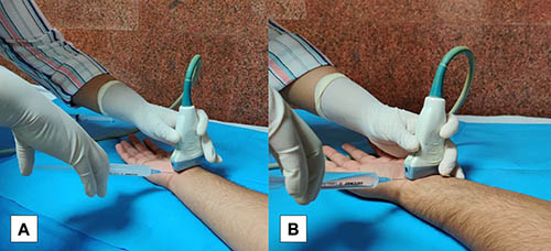
Figure 1. Photographs depict the in-plane technique of needle insertion for the US-guided hydrodissection procedure using the (A) ulnar approach in right wrist carpal tunnel syndrome (CTS) and the (B) radial approach in left wrist CTS.

A minimally invasive ultrasound-guided technique offers long-term relief for patients with carpal tunnel syndrome with fewer side effects than steroids, according to a new study published in Radiology, a journal of the Radiological Society of North America (RSNA).

Carpal tunnel syndrome occurs when the median nerve, a major nerve that runs from the forearm through a narrow space in the wrist called the carpal tunnel, becomes squeezed or compressed. It is associated with repetitive motions of the wrist and hands that lead to numbness, tingling, weakness and pain. Research has shown that the condition affects roughly 5% to 10% of the general population over their lifetimes, and incidence is rising.

Corticosteroids injection, a common treatment to reduce swelling and relieve pressure on the nerve, has limitations.

“Traditional steroid injection for carpal tunnel syndrome often provides only temporary relief, thus requiring repeats of the procedure,” said study co-author Anupama Tandon, MD, professor in the Department of Radiodiagnosis and Imaging at University College of Medical Sciences and Guru Teg Bahadur Hospital in Delhi, India. “Further, steroids carry risk of localised skin changes, fat thinning and tendon weakening.”

Nerve hydrodissection is a steroid-free alternative that involves careful injection of fluid around the affected nerve under ultrasound guidance to release the nerve from surrounding adhesions and improve its mobility. Hydrodissection is generally used in conjunction with steroid treatment, but little is known about its effectiveness as a standalone treatment.

To learn more, Dr Tandon, study lead author Anindita Bose, MD., assistant professor in the Department of Radiology at Mahatma Gandhi Medical College and Hospital in Jaipur, India, and colleagues performed a randomised control trial of 63 patients with confirmed carpal tunnel syndrome who had not responded to conventional treatment.

The researchers divided participants into three equal groups. Two groups underwent ultrasound-guided hydrodissection of the median nerve, with one group receiving normal saline alone and the other getting a combination of normal saline and corticosteroid. The third group received an ultrasound-guided corticosteroid injection alone.

The researchers assessed the patients clinically using standardised symptom, functional and pain scores. They also assessed the median nerve area on ultrasound at four weeks, 12 weeks and 24 weeks after treatment.

All three groups experienced significant improvement at four weeks post-treatment. However, the benefits of hydrodissection were sustained and continued to improve over the subsequent follow-up periods.

“At 12 weeks and 6 months, both hydrodissection groups demonstrated further improvement in clinical symptoms and function, whereas patients who received corticosteroid injection alone experienced a recurrence of the symptoms, suggesting that the initial benefit was short-lived,” Dr Tandon said.

The addition of corticosteroids to the hydrodissection injectate did not provide a significant additional long-term benefit compared with saline hydrodissection alone.

The ultrasound findings supported the clinical results. The cross-sectional area of the median nerve decreased by approximately 43% to 45% following hydrodissection, compared with approximately 15% following corticosteroid injection alone, indicating a greater reduction in nerve swelling.

“Most of our patients had been suffering from carpal tunnel syndrome for months to years and did not get much relief despite trying various treatments like pain medications, splints, lifestyle modification and corticosteroids,” Dr Tandon said. “It came as a pleasant surprise to find that a simple procedure of dissecting the median nerve under ultrasound guidance with normal saline provided them long-term relief.”

The procedure took only 15 minutes on average, and the patients tolerated it well. No significant immediate or delayed complications were observed. Questionnaire scores showed that the patients were highly satisfied with procedure. Its cost was low, no anaesthesia or hospitalisation was needed, and the patients were able to go back to work in an hour.

The findings point to a more prominent role for hydrodissection as a treatment for a condition that is increasing in prevalence around the world.

“Overall, the study suggests that ultrasound-guided saline hydrodissection is a safe, minimally invasive treatment that can provide sustained clinical and morphological improvement in patients with refractory carpal tunnel syndrome,” Dr Tandon said. “Thus, hydrodissection merits to be a minimally invasive option for mild to moderate carpal tunnel syndrome and can defer and even alleviate the need for surgery.”

Future multicenter studies with larger cohorts and longer follow-up are warranted, the researchers said. They have already launched a study comparing the long-term effectiveness of different injectates in hydrodissection for carpal tunnel syndrome, such as dextrose versus saline.“This trial features an extended follow-up period to track long-term patient outcomes and establish the true durability of the treatment,” Dr Tandon said.

Source: Radiological Society of North America

Impaired Relaxation Signal in Pre-eclampsia Vessels Points to New Treatment Targets

Source: Pixabay CC0

Researchers at the University of Manchester and Manchester University NHS Foundation Trust identified a major defect in microvascular regulation in women with pre-eclampsia. The breakthrough sheds new light on why blood pressure rises so dramatically in the condition and could point towards entirely new ways of treating it.

Pre-eclampsia affects 2–8% of pregnancies worldwide and is linked to serious complications including stroke, organ damage, premature birth and stillbirth. At present there is no cure other than delivering the baby and placenta, often forcing doctors to perform earlier deliveries.

But now the researchers have identified a key failure in the way small arteries regulate blood flow in women with the condition.

The team recruited patients at Saint Mary’s Hospital (SMH), part of MFT, and compared tiny arteries from pregnant women with and without preeclampsia. The researchers discovered that a vital natural relaxation mechanism was severely disrupted in those with the disease.

The vessels were taken from the omentum – a fold of fatty tissue inside the abdomen that helps protect and support the abdominal organs – and placed under pressure in a laboratory to study their functioning.

They found that microscopic calcium sparks (which normally signal arterial smooth muscle to relax) were reduced by up to 75% at physiological pressures. Calcium stores were depleted and less able to refill, impairing the pressure-balancing mechanism and contributing to vasoconstriction and hypertension.

The immune peptide HBD2 did not restore the sparks but still relaxed vessels via an alternative pathway component. This suggests potential therapies that bypass the defective mechanism to control maternal blood pressure, prolong pregnancy and reduce early delivery.

Study lead Dr Stephanie Worton (University of Manchester; Honorary Consultant in Obstetrics, Maternal and Fetal Health Research Centre, Saint Mary’s) stated this is the first demonstration of a profound impairment in this calcium signalling pathway, while vessels remain relaxable via alternative routes. While these are promising, there is further work needed on drug development for pregnancy.

Source: NHS Manchester University

Longer Life Exposure to Oestrogen Linked to Better Cerebrovascular Health

Photo by Ravi Patel on Unsplash

Women who had a longer reproductive period and had used or are using hormone replacement therapy showed fewer signs of damage to the small blood vessels in the brain later in life. This is shown by a study led by Karolinska Institute’s researchers, published in the journal Neurology.

In this new study, the researchers examined 352 women from Gothenburg. All were aged 70 with no signs of impaired cognitive functioning. The participants’ brains were scanned using magnetic resonance imaging (MRI) at the age of 70 and again five years later.

The researchers also had data on the age at first menstruation and the onset of the menopause, as well as previous use of the contraceptive pill and hormone replacement therapy during the menopause. This was compared with, amongst other things, signs of small-vessel disease – that is, damage to the brain’s tiniest blood vessels – the microstructure of the white matter, and the thickness of the cerebral cortex in areas vulnerable to Alzheimer’s disease.

A longer reproductive period – that is, the time between the first menstruation and the menopause – was associated with lower small-vessel disease burden in the brain. The use of hormone replacement therapy during the menopause was also associated with less small-vessel disease and a more favourable microstructure in the brain’s white matter. Women who had used both the contraceptive pill during their reproductive years and hormone replacement therapy during the menopause showed the most favourable profile for their vascular brain health.

May have cumulative effects

The researchers behind the study note that women not only are more likely than men to develop Alzheimer’s disease, a difference that may be partly related to oestrogen fluctuations across the lifespan. The new findings suggest that these hormonal exposures may also influence the health of the brain’s blood vessels.

”Our results suggest that hormonal exposure across different periods of a woman’s life may have cumulative effects also on the cerebrovascular health in later life. The findings suggest that the brain’s blood vessels may play an important role in the link between oestrogen and brain ageing,” says Anna Marseglia, assistant professor at the Department of Neurobiology, Care Sciences and Society, Karolinska Institutet, and senior author of the study.

One limitation of the study is that data on hormone use were self-reported, and there was a lack of detailed information on, for example, when treatment began, the dose and the type of treatment.

”Furthermore, the study is an observational study, and we cannot therefore determine cause and effect. The results cannot therefore be used as a basis for recommending hormone therapy for preventive purposes”, says first author Giulia Lorenzon, researcher at Yale University and former postdoctoral researcher at the Department of Neurobiology, Care Sciences and Society, Karolinska Institutet.

Source: Karolinska Institutet

Ultrasound Implant at Spinal Cord Injury Site Could Help Guide Recovery

A biomedical team has completed their first feasibility study to investigate the ability to place a temporary ultrasound device at the site of a spinal cord injury, which supports ongoing research to study its ability to monitor and modulate blood flow after surgery 

Researchers are studying how an ultrasonic neural interface device, which would be temporarily implanted at the site of injury following surgery, may be used to support recovery and personalise care for people recovering from spinal cord injuries.

A team of engineers and surgeons from Johns Hopkins Medicine has published its first proof-of-concept study that shows temporarily implanting a tiny ultrasonic device into the location of a spinal cord injury can be feasible, safe and potentially useful. These findings, which mark an early midpoint in a multitiered research journey to develop and study how the device could be used in clinical care, published today in Science Advances. 

“When the biology of spinal cord injury is local, our devices should also be able to track and treat local physiology,” says Amir Manbachi, PhD, the principal investigator and an associate professor of neurosurgery and biomedical engineering at the Johns Hopkins University School of Medicine. 

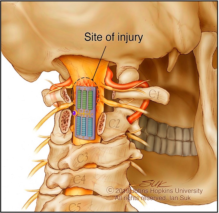
The device that Manbachi and researchers developed and studied, which is called Multimodal Ultrasound System for Injury Care (MUSIC), is designed to enhance feedback about what is happening at and around the site of injury following surgery and to support clinical decisions about care when a patient is recovering in the intensive care unit. 

Manbachi explains that the ultimate goal of developing and studying the device is to identify a way to support people while they are in the hospital and minimise risks for complications, such as stroke, blindness or losing additional control of movement, that they may face while recovering from a spinal cord injury.

To achieve this, the device was designed to double as a “mobile MRI” that could alleviate the need to move immobile patients in the days following their recovery from injury, while providing on-demand and as-needed feedback to medical teams, as opposed to single points in time. 

The device is designed to have dual capabilities of diagnostic ultrasound (sonography) and therapeutic ultrasound (physical therapy).

The diagnostic/imaging aspect of the device provides real-time insight about blood flow and changes surrounding the spinal cord injury, and it enables analysis of other measures, such as spinal fluids and excess fluid accumulation, how a person is responding to medication and what is happening outside of the injured area, which can help indicate how well a person is healing. This type of real-time detection in the days and weeks after surgery could provide instant feedback to medical teams, while helping to personalise treatment decisions. 

Therapeutic ultrasound (also known as focused ultrasound) is the more futuristic and exploratory aspect of the device that has been studied for its potential ability to identify and respond to changes in blood flow at and around the site of the injury (when blood flowing through the small vessels around the affected area drops below or above acceptable levels).

This ultrasound device, which received FDA Breakthrough Device Designation in 2023, is the size of a small Band-Aid and created to be placed over the spinal cord at the site of injury during the expected surgery. This publication reports the first proof-of-concept study in animals to show that MUSIC was able to be temporarily implanted and provide real-time feedback about blood flow and imaging around the site where it is located. 

Source: Johns Hopkin Medicine