

A biomedical team has completed their first feasibility study to investigate the ability to place a temporary ultrasound device at the site of a spinal cord injury, which supports ongoing research to study its ability to monitor and modulate blood flow after surgery
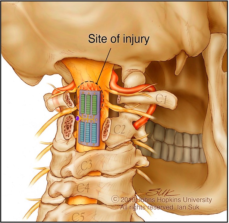
A team of engineers and surgeons from Johns Hopkins Medicine has published its first proof-of-concept study that shows temporarily implanting a tiny ultrasonic device into the location of a spinal cord injury can be feasible, safe and potentially useful. These findings, which mark an early midpoint in a multitiered research journey to develop and study how the device could be used in clinical care, published today in Science Advances.
“When the biology of spinal cord injury is local, our devices should also be able to track and treat local physiology,” says Amir Manbachi, PhD, the principal investigator and an associate professor of neurosurgery and biomedical engineering at the Johns Hopkins University School of Medicine.
The device that Manbachi and researchers developed and studied, which is called Multimodal Ultrasound System for Injury Care (MUSIC), is designed to enhance feedback about what is happening at and around the site of injury following surgery and to support clinical decisions about care when a patient is recovering in the intensive care unit.
Manbachi explains that the ultimate goal of developing and studying the device is to identify a way to support people while they are in the hospital and minimise risks for complications, such as stroke, blindness or losing additional control of movement, that they may face while recovering from a spinal cord injury.
To achieve this, the device was designed to double as a “mobile MRI” that could alleviate the need to move immobile patients in the days following their recovery from injury, while providing on-demand and as-needed feedback to medical teams, as opposed to single points in time.
The device is designed to have dual capabilities of diagnostic ultrasound (sonography) and therapeutic ultrasound (physical therapy).
The diagnostic/imaging aspect of the device provides real-time insight about blood flow and changes surrounding the spinal cord injury, and it enables analysis of other measures, such as spinal fluids and excess fluid accumulation, how a person is responding to medication and what is happening outside of the injured area, which can help indicate how well a person is healing. This type of real-time detection in the days and weeks after surgery could provide instant feedback to medical teams, while helping to personalise treatment decisions.
Therapeutic ultrasound (also known as focused ultrasound) is the more futuristic and exploratory aspect of the device that has been studied for its potential ability to identify and respond to changes in blood flow at and around the site of the injury (when blood flowing through the small vessels around the affected area drops below or above acceptable levels).
This ultrasound device, which received FDA Breakthrough Device Designation in 2023, is the size of a small Band-Aid and created to be placed over the spinal cord at the site of injury during the expected surgery. This publication reports the first proof-of-concept study in animals to show that MUSIC was able to be temporarily implanted and provide real-time feedback about blood flow and imaging around the site where it is located.
Source: Johns Hopkin Medicine
