Tag: diabetes

Neurons Cause Metabolic Havoc after Spinal Injury

Conditions such as diabetes, heart attack and vascular diseases commonly diagnosed in people with spinal cord injuries can be traced to abnormal post-injury neuronal activity that causes abdominal fat tissue compounds to leak and pool in the liver and other organs, a new animal study published in Cell Reports Medicine has found.

After discovering the connection between dysregulated neuron function and the breakdown of triglycerides in fat tissue in mice, researchers found that a short course of the drug gabapentin, commonly prescribed for nerve pain, prevented the damaging metabolic effects of the spinal cord injury – though not without side effects.

Gabapentin inhibits a neural protein that, after the nervous system is damaged, becomes overactive and causes communication problems – in this case, affecting sensory neurons and the abdominal fat tissue to which they’re sending signals.

“We believe there is maladaptive reorganisation of the sensory system that causes the fat to undergo changes, initiating a chain of reactions – triglycerides start breaking down into glycerol and free fatty acids that are released in circulation and taken up by the liver, the heart, the muscles, and accumulating, setting up conditions for insulin resistance,” said senior author Andrea Tedeschi, assistant professor of neuroscience in The Ohio State University College of Medicine.

“Through administration of gabapentin, we were able to normalise metabolic function.”

Previous research has found that cardiometabolic diseases are among the leading causes of death in people who have experienced a spinal cord injury. These often chronic disorders can be related to dysfunction in visceral white fat (or adipose tissue), which has a complex metabolic role of storing energy and releasing fatty acids as needed for fuel, but also helping keep blood sugar levels at an even keel.

Earlier investigations of these diseases in people with neuronal damage have focused on adipose tissue function and the role of the sympathetic nervous system, but also a regulator of adipose tissue that surrounds the abdominal organs.

Instead, Debasish Roy, a postdoctoral researcher in the Tedeschi lab and first author on the paper, decided to focus on sensory neurons in this context. Tedeschi and colleagues have previously shown that a neuronal receptor protein called alpha2delta1 is overexpressed after spinal cord injury, and its increased activation interferes with post-injury function of axons, the long, slender extensions of nerve cell bodies that transmit messages.

In this new work, researchers first observed how sensory neurons connect to adipose tissue under healthy conditions, and created a spinal cord injury mouse model that affected only those neurons – without interrupting the sympathetic nervous system.

Experiments revealed a cascade of abnormal activity within seven days after the injury in neurons – though only in their communication function, not their regrowth or structure – and in visceral fat tissue. Expression of the alpha2delta1 receptor in sensory neurons increased as they over-secreted a neuropeptide called CGRP, all while communicating through synaptic transmission to the fat tissue – which, in a state of dysregulation, drove up levels of a receptor protein that engaged with the CGRP.

“These are quite rapid changes. As soon as we disrupt sensory processing as a result of spinal cord injury, we see changes in the fat,” Tedeschi said. “A vicious cycle is established – it’s almost like you’re pressing the gas pedal so your car can run out of gas but someone else continues to refill the tank, so it never runs out.”

The result is the spillover of free fatty acids and glycerol from fat tissue, a process called lipolysis, that has gone out of control. Results also showed an increase in blood flow in fat tissue and recruitment of immune cells to the environment.

“The fat is responding to the presence of CGRP, and it’s activating lipolysis,” Tedeschi said. “CGRP is also a potent vasodilator, and we saw increased vascularisation of the fat – new blood vessels forming as a result of the spinal cord injury. And the recruitment of monocytes can help set up a chronic pro-inflammatory state.”

Silencing the genes that encode the alpha2delta1 receptor restored the fat tissue to normal function, indicating that gabapentin – which targets alpha2delta1 and its partner, alpha2delta2 – was a good treatment candidate. Tedeschi’s lab has previously shown in animal studies that gabapentin helped restore limb function after spinal cord injury and boosted functional recovery after stroke.

But in these experiments, Roy discovered something tricky about gabapentin: the drug prevented changes in abdominal fat tissue and lowered CGRP in the blood, in turn preventing spillover of fatty acids into the liver a month later, establishing normal metabolic conditions. But paradoxically, the mice developed insulin resistance, a known side effect of gabapentin.

The team instead tried starting with a high dose, tapering off and stopping after four weeks.

“This way, we were able to normalise metabolism to a condition much more similar to control mice,” Roy said. “This suggests that as we discontinue administration of the drug, we retain beneficial action and prevent spillover of lipids in the liver. That was really exciting.”

Finally, researchers examined how genes known to regulate white fat tissue were affected by targeting alpha2delta1 genetically or with gabapentin, and found both of these interventions after spinal cord injury suppress genes responsible for disrupting metabolic functions.

Tedeschi said the combined findings suggest starting gabapentin treatment early after a spinal cord injury may protect against detrimental conditions involving fat tissue that lead to cardiometabolic disease – and could enable discontinuing the drug while retaining its benefits and lowering the risk for side effects.

Source: Ohio State University

Metformin’s Weight Loss Tied to “Anti-hunger” Molecule

A new study finds that the modest weight loss from taking metformin is attributable to an appetite-suppressing molecule that is abundant after exercise

Photo by I Yunmai on Unsplash

An “anti-hunger” molecule produced after vigorous exercise is responsible for the moderate weight loss caused by the diabetes medication metformin, according to a new study in mice and humans. The anti-hunger molecule, lac-phe, was discovered by Stanford Medicine researchers in 2022.

The finding, made jointly by researchers at Stanford Medicine and at Harvard Medical School and published in Nature Metabolism, further cements the critical role the molecule, called lac-phe, plays in metabolism, exercise and appetite. It may pave the way to a new class of weight loss drugs.

“Until now, the way metformin, which is prescribed to control blood sugar levels, also brings about weight loss has been unclear,” said Jonathan Long, PhD, an assistant professor of pathology. “Now we know that it is acting through the same pathway as vigorous exercise to reduce hunger. Understanding how these pathways are controlled may lead to viable strategies to lower body mass and improve health in millions of people.”

Many people with diabetes who are prescribed metformin lose around 2% to 3% of their body weight within the first year of starting the drug. Although this amount of weight loss is modest when compared with the 15% or more often seen by people taking semaglutide, the discoveries that led to those drugs also grew from observations of relatively minor, but reproducible, weight loss in people taking first-generation versions of the medications.

Post-workout appetite loss

When Long and colleagues at Baylor University discovered lac-phe in 2022, they were on the hunt for small molecules responsible for curtailing hunger after vigorous exercise. What they found was a mishmash of lactate and an amino acid called phenylalanine. They dubbed the hybrid molecule lac-phe and went on to show that it’s not only more abundant after exercise but it also causes people (as well as mice and even racehorses) to feel less hungry immediately after a hard workout.

“There is an intimate connection between lac-phe production and lactate generation,” Long said. “Once we understood this relationship, we started to think about other aspects of lactate metabolism.”

Metformin was an obvious candidate because as it stimulates the breakdown of glucose (thus reducing blood sugar levels) it can trigger the generation of lactate.

The researchers found that obese laboratory mice given metformin had increased levels of lac-phe in their blood. They ate less than their peers and lost about 2 grams of body weight during the nine-day experiment.

Long and his colleagues also analysed stored blood plasma samples from people with Type 2 diabetes before and 12 weeks after they had begun taking metformin to control their blood sugar. They saw significant increases in the levels of lac-phe in people after metformin compared with their levels before treatment. Finally, 79 participants in a large, multi-ethnic study of atherosclerosis who were also taking metformin had significantly higher levels of lac-phe circulating in their blood than those who were not taking the drug.

“It was nice to confirm our hunch experimentally,” Long said. “The magnitude of effect of metformin on lac-phe production in mice was as great as or greater than what we previously observed with exercise. If you give a mouse metformin at levels comparable to what we prescribe for humans, their lac-phe levels go through the roof and stay high for many hours.”

Further research revealed that lac-phe is produced by intestinal epithelial cells in the animals; blocking the ability of mice to make lac-phe erased the appetite suppression and weight loss previously observed.

Finally, a statistical analysis of the people in the atherosclerosis study who lost weight during the several-year study and follow-up period found a meaningful association between metformin use, lac-phe production and weight loss.

“The fact that metformin and sprint exercise affect your body weight through the same pathway is both weird and interesting,” Long said. “And the involvement of the intestinal epithelial cells suggests a layer of gut-to-brain communication that deserves further exploration. Are there other signals involved?”

Long noted that, while semaglutide drugs are injected into the bloodstream, metformin is an oral drug that is already prescribed to millions of people. “These findings suggest there may be a way to optimize oral medications to affect these hunger and energy balance pathways to control body weight, cholesterol and blood pressure. I think what we’re seeing now is just the beginning of new types of weight loss drugs.”

Source: Stanford Medicine

Smart Moo-ve for Diabetes Treatment: Insulin Produced in Cow’s Milk

Photo by Pixabay on Pexels

An unassuming brown bovine from the south of Brazil has made history as the first transgenic cow capable of producing human insulin in her milk. The advancement, led by researchers from the University of Illinois Urbana-Champaign and the Universidade de São Paulo, could herald a new era in insulin production, one day eliminating drug scarcity and high costs for people living with diabetes.

“Mother Nature designed the mammary gland as a factory to make protein really, really efficiently. We can take advantage of that system to produce a protein that can help hundreds of millions of people worldwide,” said Matt Wheeler, professor in the Department of Animal Sciences, part of the College of Agricultural, Consumer and Environmental Sciences (ACES) at U. of I.

Wheeler is lead author on a new Biotechnology Journal study describing the development of the insulin-producing cow, a proof-of-concept achievement that could be scaled up after additional testing and FDA approval.

Precise insertion of DNA

Wheeler’s colleagues in Brazil inserted a segment of human DNA coding for proinsulin – the protein precursor of the active form of insulin – into cell nuclei of 10 cow embryos. These were implanted in the uteruses of normal cows in Brazil, and one transgenic calf was born. Thanks to updated genetic engineering technology, the human DNA was targeted for expression – the process whereby gene sequences are read and translated into protein products – in mammary tissue only.

“In the old days, we used to just slam DNA in and hope it got expressed where you wanted it to,” Wheeler said. “We can be much more strategic and targeted these days. Using a DNA construct specific to mammary tissue means there’s no human insulin circulating in the cow’s blood or other tissues. It also takes advantage of the mammary gland’s capabilities for producing large quantities of protein.”

When the cow reached maturity, the team unsuccessfully attempted to impregnate her using standard artificial insemination techniques. Instead, they stimulated her first lactation using hormones. The lactation yielded milk, but a smaller quantity than would occur after a successful pregnancy. Still, human proinsulin and, surprisingly, insulin were detectable in the milk.

“Our goal was to make proinsulin, purify it out to insulin, and go from there. But the cow basically processed it herself. She makes about three to one biologically active insulin to proinsulin,” Wheeler said. “The mammary gland is a magical thing.”

The insulin and proinsulin, which would need to be extracted and purified for use, were expressed at a few grams per liter in the milk. But because the lactation was induced hormonally and the milk volume was smaller than expected, the team can’t say exactly how much insulin would be made in a typical lactation.

Conservatively, Wheeler says if a cow could make 1 gram of insulin per liter and a typical Holstein makes 40 to 50 litres per day, that’s a lot of insulin. Especially since the typical unit of insulin equals 0.0347 milligrams.

“That means each gram is equivalent to 28,818 units of insulin,” Wheeler said. “And that’s just one liter; Holsteins can produce 50 liters per day. You can do the math.”

The team plans to re-clone the cow, and is optimistic they’ll achieve greater success with pregnancy and full lactation cycles in the next generation. Eventually, they hope to create transgenic bulls to mate with the females, creating transgenic offspring that can be used to establish a purpose-built herd. Wheeler says even a small herd could quickly outcompete existing methods – transgenic yeast and bacteria – for producing insulin, and could do so without having to create highly technical facilities or infrastructure.

“With regard to mass-producing insulin in milk, you’d need specialized, high-health-status facilities for the cattle, but it’s nothing too out of the ordinary for our well-established dairy industry,” Wheeler said. “We know what we’re doing with cows.”

An efficient system to collect and purify insulin products would be needed, as well as FDA approval, before transgenic cows could supply insulin for the world’s diabetics. But Wheeler is confident that day is coming.

“I could see a future where a 100-head herd, equivalent to a small Illinois or Wisconsin dairy, could produce all the insulin needed for the country,” he said. “And a larger herd? You could make the whole world’s supply in a year.

Source: University of Illinois College of Agricultural, Consumer and Environmental Sciences

Why People with Diabetes are More Vulnerable to Respiratory Infection

Credit: Scientific Animations CC4.0

It has long been known that people with diabetes are at a substantially increased risk of developing severe lung disease if they become infected with viruses such as influenza, as well as other pathogens. When the COVID-19 pandemic started in early 2020, it became even more important to understand this mysterious phenomenon. It became clear that people with diabetes were at a significantly higher risk of coming down with severe, even fatal, lung disease after developing severe COVID, but no one understood why. In fact, some 35% of the pandemic’s COVID mortalities had diabetes.

Now, research conducted at the Weizmann Institute of Science and published in Nature has revealed how, in diabetics, high levels of blood sugar disrupt the function of key cell subsets in the lungs that regulate the immune response. It also identifies a potential strategy for reversing this susceptibility and saving lives.

Prof. Eran Elinav‘s team in his lab at Weizmann, headed by Drs. Samuel Nobs, Aleksandra Kolodziejczyk and Suhaib K. Abdeen, subjected multiple mouse models of types 1 and 2 diabetes to a variety of viral lung infections. Just as in diabetic humans, in all these models the diabetic mice developed a severe, fatal lung infection following exposure to lung pathogens such as influenza. The immune reaction, which in nondiabetics eliminates the infection and drives tissue healing, was severely impaired in the diabetic mice, leading to uncontrolled infection, lung damage and eventual death.

Next, to decode the basis of this heightened risk, the team performed an evaluation of gene expression on the level of individual cells, in more than 150 000 single lung cells of infected diabetic and nondiabetic mice. The researchers also performed an extensive array of experiments involving immune and metabolic mechanisms, as well as an in-depth assessment of immune cell gene expression in infected diabetic mice. In the diabetic mice they identified a dysfunction of certain lung dendritic cells, the immune cells that orchestrate a targeted immune response against pathogenic infection. “High blood sugar levels severely disrupt certain subsets of dendritic cells in the lung, preventing these gatekeepers from sending the molecular messages that activate the critically important immune response,” says Nobs, postdoctoral fellow and study first author. “As a result, the infection rages on, uncontrolled.”

Next, they explored ways to prevent the harmful effects of hyperglycaemia in lung dendritic cells, as a means of lowering the infection’s risk in diabetic animals. Indeed, tight control of glycaemic levels by insulin supplementation prompted the dendritic cells to regain their capacity to generate a protective immune response that could prevent the cascade of events leading to a severe, life-threatening viral lung infection. Alternatively, administration of small molecules reversing the sugar-induced regulatory impairment corrected the dendritic cells’ dysfunction and enabled them to generate a protective immune response despite the presence of hyperglycaemia.

“Correcting blood sugar levels, or using drugs to reverse the gene regulatory impairment induced by high sugar, enabled our team to get the dendritic cells’ function back to normal,” says Abdeen, a senior intern who co-supervised the study. “This was very exciting because it means that it might be possible to block diabetes-induced susceptibility to viral lung infections and their devastating consequences.”

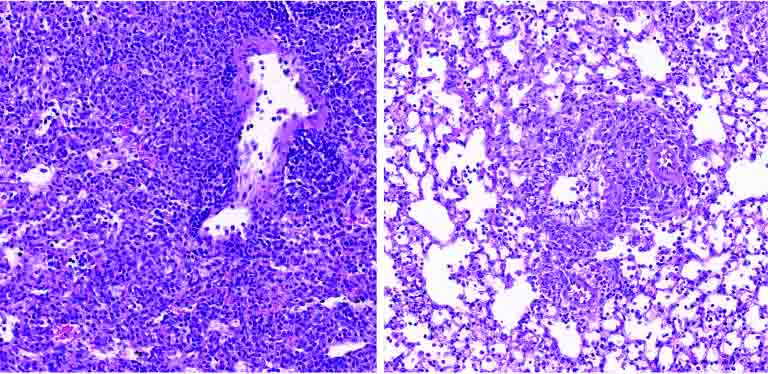
Lung tissue of a diabetic mouse (right) contains fewer immune cells (small purple dots) than that of a non-diabetic animal (left)

With over 500 million people around the world affected by diabetes, and with diabetes incidence expected to rise over the next decades, the new research has significant, promising clinical implications.

“Our findings provide, for the first time, an explanation as to why diabetics are more susceptible to respiratory infection,” Elinav says. “Controlling sugar levels may make it possible to reduce this pronounced diabetes-associated risk. In diabetic patients whose sugar levels are not easily normalized, small molecule drugs may correct the gene alterations caused by high sugar levels, potentially alleviating or even preventing severe lung infection. Local administration of such treatments by inhalation may minimize adverse effects while enhancing effectiveness, and merits future human clinical testing.”

Source: Weizmann Institute of Science

Scientists Identify New Cause of Diabetes – and Potential Treatment Target

Photo by Photomix Company on Pexels

Researchers have identified an enzyme that blocks insulin produced in the body – a discovery that could provide a new target to treat diabetes. Their study, published the journal Cellfocuses on nitric oxide, which dilates blood vessels, improves memory, fights infection and stimulates the release of hormones, among other functions.

How nitric oxide performs these activities had long been a mystery.

The researchers at Case Western Reserve University and University Hospitals discovered a novel “carrier” enzyme (called SNO-CoA-assisted nitrosylase, or SCAN) that attaches nitric oxide to proteins, including the receptor for insulin action.

They found that the SCAN enzyme was essential for normal insulin action, but also discovered heightened SCAN activity in diabetic patients and mice with diabetes.

Mouse models without the SCAN enzyme appeared to be shielded from diabetes, suggesting that too much nitric oxide on proteins may be a cause of such diseases.

“We show that blocking this enzyme protects from diabetes, but the implications extend to many diseases likely caused by novel enzymes that add nitric oxide,” said the study’s lead researcher Jonathan Stamler, professor at the Case Western Reserve School of Medicine.

“Blocking this enzyme may offer a new treatment.”

Given the discovery, next steps could be to develop medications against the enzyme, he said.

Many human diseases, including Alzheimer’s, cancer, heart failure and diabetes, are thought to be caused or accelerated by nitric oxide binding excessively to key proteins.

With this discovery, Stamler said, enzymes that attach the nitric oxide become a focus.

With diabetes, the body often stops responding normally to insulin.

The resulting increased blood sugar stays in the bloodstream and, over time, can cause serious health problems.

Individuals with diabetes, the Centers for Disease Control reports, are more likely to suffer such conditions as heart disease, vision loss and kidney disease.

But the reason that insulin stops working isn’t well understood.

Excessive nitric oxide has been implicated in many diseases, but the ability to treat has been limited because the molecule is reactive and can’t be targeted specifically, Stamler said.

“This paper shows that dedicated enzymes mediate the many effects of nitric oxide,” he said. “Here, we discover an enzyme that puts nitric oxide on the insulin receptor to control insulin. Too much enzyme activity causes diabetes. But a case is made for many enzymes putting nitric oxide on many proteins, and, thus, new treatments for many diseases.”

Source: Case Western Reserve University

Macrophages ‘Eat’ Pancreatic β Cells to Regulate Insulin Post Partum

A 3D map of the islet density routes throughout the healthy human pancreas. Source: Wikimedia CC0

Scientists have long known that pancreatic β cells increase during pregnancy and promptly return to their original number following birth. But the underlying mechanisms that cause the cells to go back to their original number are still not well understood.

In a significant breakthrough, a research group using mouse models, has discovered that macrophages ‘eat’ (phagocytose) the pancreatic β cells, thereby revealing the process behind their return to previous levels after pregnancy.

The research group, which was led by Associate Professor Junta Imai, Assistant Professor Akira Endo, and Professor Hideki Katagiri from Tohoku University’s Graduate School of Medicine, published the results in the journal Development Cell.

Initially, the group examined the number of pancreatic β cells in the islets of Langerhans in a mouse model of pregnancy.

They confirmed the cell number was double at the end of the pregnancy when compared to non-pregnant mice, but that it then gradually decreased, returning to the original amount after delivery.

“After we observed the islets of Langerhans before and after delivery, we noticed an increase in macrophages, which protect the body from infections by engulfing bacteria, foreign substances and dead cells, after delivery,” says Imai.

“When we applied treatment to inhibit this process, the blood glucose levels became too low (hypoglycaemia).”

Additional microscopic observation of the islets of Langerhans after birth revealed β cells to be phagocytosed by macrophages.

This mechanism appeared to keep the mother’s blood glucose levels from decreasing excessively after delivery by rapidly reducing pancreatic β cells to their normal pre-pregnancy number.

Next, the group identified the protein responsible for attracting the macrophages into the islets of Langerhans: cytokine CXCL10.

Accordingly, the inhibition of CXCL10 function suppressed the decrease in pancreatic β cells after birth.

“We hope our results will contribute to clarifying the means by which normal blood glucose levels are maintained as well as the development of methods to prevent and treat diabetes,” adds Imai.

Source: Tohoku University

Collaboration Key to Address SA’s Fatal, Diabetes-linked Cardiovascular Disease Burden

Photo by Hush Naidoo on Unsplash

Only concerted multi-disciplinary collaboration and research will stem the tide of diabetes and diabetes-linked cardiovascular disease (CVD), the latter currently the leading cause of death locally and worldwide, claiming 17.9 million lives annually1.

This was the consensus among some of the world’s leading cardiologists and researchers gathered at the SA Heart Association’s annual congress aptly themed: ‘The Cardiac Collaboration,’ which took place at the Sandton Convention Centre in Johannesburg from 26-29 October this year.

Globally, CVD takes more lives than TB, HIV and malaria combined, while 215 South Africans are killed by CVD every day – with 80% of CVD and strokes being preventable.1,2 The prevalence of diabetes has also increased in South Africa, from 4.5% in 2010 to 12.7% in 2019. Of the 4.58 million people aged 20-79 years who were estimated to have diabetes in 2019, 52.4% were undiagnosed.3

With diabetes being a key driver of CVD – especially in Africa (with limited access to novel drugs and the prevalence of sugar-rich, poverty-driven lifestyles), the mutual consensus at this year’s congress was that collaboration is key.

Dr Zaheer Bayat, Chairperson of the Society for Endocrinology, Metabolism and Diabetes of South Africa (SEMDSA), told delegates that endocrinologists and cardiologists would have to work together to improve outcomes for diabetic patients, 30% of whom suffered cardiovascular events. He warned that a 134% increase of people living with diabetes was predicted over the next two decades, translating into a dramatic surge in chronic kidney disease, cardiovascular disease, blindness, and amputations.

Dr Bayat said he intends appealing for mass diabetes screening to find the 52% of people whom researchers estimate are undiagnosed. Ideally, this should be followed by access to cheaply acquired, effective new glucose-lowering drugs.

“The reality is that this country cannot afford all the new treatments for everyone – not private funders, not government. So, drugs are not really a solution – the best solution is to change lifestyle and prevent disease in the first place,” said Dr Bayat.

“We’re here to fight for our patients, not our pockets. Can we afford to have 52% of our patients not knowing they’re diabetic? People who should be contributing to our economy are living with diabetes and eventually dying,” he asserted.

Dr Bayat also said that globally, First World countries such as the USA and Sweden are reducing myocardial infarctions, strokes, and amputations, because they’re doing all the right things together. This included adopting a healthy lifestyle, effective management of sugar, blood pressure and cholesterol and smoking cessation.

“However, here in South Africa with private healthcare representing 15% of healthcare delivery but consuming 50% of the spend and the public sector representing 85% of the population and consuming the other half – we’re not doing nearly as well. With only 200 cardiologists in the country (one per 190 000 population), and even less nephrologists, we need to join together and change the trajectory of diabetes. We must work together to reduce morbidity and mortality,” said Dr Bayat.

According to the SA Heart Association, this graphically illustrates the importance of a multi-disciplinary approach, the very reason why the conference was called ‘The Cardiac Collaboration.’

The SA Heart Association has already begun forging formal ties with other academic societies and next year, it hopes to join and host joint sessions with collaborative meetings to connect a multidisciplinary team in order to achieve a well-rounded balance of care.

References:

  1. https://www.heartfoundation.co.za/wp-content/uploads/2017/10/CVD-Stats-Reference-Document-2016-FOR-MEDIA-1.pdf.
  2. https://world-heart-federation.org/what-we-do/prevention/#:~:text=An%20estimated%2080%25%20of%20cardiovascular,and%20%E2%80%9Cknowing%20your%20numbers%E2%80%9D.
  3. International Diabetes Federation. IDF Diabetes Atlas.10th ed. International Diabetes Federation; Brussels, Belgium: 2021. [Google Scholar] (primary). https://www.ncbi.nlm.nih.gov/pmc/articles/PMC10218408/#:~:text=The%20prevalence%20of%20diabetes%20mellitus,%25%20were%20undiagnosed%20%5B5%5D. (secondary).

Diabetes Worsens Colorectal Cancer Survival Odds by 41%

Photo by Nataliya Vaitkevich on Pexels

In an analysis of information on adults with colorectal cancer, patients who also had diabetes, particularly those with diabetic complications, faced a higher risk of early death. The results are published in CANCER, a peer-reviewed journal of the American Cancer Society.

For the study, Kuo‐Liong Chien, MD, PhD, of National Taiwan University, and his colleagues examined data registered between 2007 and 2015 in the Taiwan Cancer Registry Database, which is linked to health insurance and death records. Their analysis included 59 202 individuals with stage I–III colorectal cancer who underwent potentially curative surgery to remove their tumours. Among these patients, 9448 experienced a cancer recurrence and 21 031 died from any cause during the study period.

Compared with individuals without diabetes, those with uncomplicated diabetes were at a minimally or insignificantly higher risk of all‐cause and cancer‐specific death, whereas those with complicated diabetes had 85% higher odds of death from any cause and 41% higher odds of death from cancer. These associations were more pronounced in women and in patients with early‐stage colorectal cancer.

Also, compared to patients without diabetes, patients with uncomplicated or complicated diabetes had a 10–11% higher risk of colorectal cancer recurrence.

The mechanisms behind the relationship between diabetic severity and poor colorectal cancer prognosis could involve various pathways and responses triggered by high insulin and glucose levels in the blood, as well as elevated inflammatory states, which are characteristic of type 2 diabetes.

“While a higher diabetes prevalence was noted in patients with colorectal cancer, the study suggests that coordinated medical care involving multiple specialists can help prevent diabetes complications, potentially improving long-term colorectal cancer oncological outcomes, particularly in women and patients with early-stage cancer,” said Dr Chien.

Source: Wiley

Don’t Overlook Latent Autoimmune Diabetes in Adults, Researchers Caution

Photo by Photomix Company on Pexels

To reduce the risk of complications, it is important to measure antibodies those with adult onset diabetes, while also considering the levels of these antibodies.

In a study published in the journal Diabetes Care, researchers demonstrate that individuals with Latent Autoimmune Diabetes in Adults (LADA) have an equally high risk of developing cardiovascular disease as people with type 2 diabetes, but a higher risk of developing retinopathy and poorer glucose control. Many also lack adequate treatment.

LADA is a common but relatively unknown form of diabetes. Similar to type 1 diabetes, it is an autoimmune disease characterised by antibodies against insulin-producing cells. It develops in adulthood, and the autoimmune process progresses more slowly than in type 1 diabetes. LADA also shares features with type 2 diabetes, which means those affected risk getting the wrong diagnosis if antibodies are not measured. Incorrect diagnosis can result in inadequate treatment. Previous studies suggest that between five and ten percent of all individuals initially diagnosed with type 2 diabetes actually have LADA. Researchers at Karolinska Institutet, and the Universities of Lund and Helsinki set out to examine the risk of complications in LADA.

Our results emphasise the importance of diagnosing LADA correctly and careful monitoring of glucose control in these individuals, so that treatment can be intensified if needed, thereby reducing the risk of complications.

Yuxia Wei, PhD-student and Sofia Carlsson, senior lecturer, Institute of Environmental Medicine, Karolinska Institutet

According to the study LADA was characterised by fewer metabolic risk factors than type 2 diabetes, such as high blood pressure and high blood lipids. However, a lower proportion of individuals with LADA achieved good glucose control. The lack of glucose control was most evident in LADA patients with high levels of the antibody GADA (glutamic acid decarboxylase antibody). A significant portion of individuals with LADA lacked any glucose-lowering treatment.

The results of the new study are based on the ESTRID study, where researchers followed over 4000 individuals with diabetes, of whom 550 had LADA, for up to 12 years after diagnosis. According to the researchers, it is the most comprehensive study to date regarding the risk of complications in LADA.

Source: Karolinska Institutet

‘We Will Rock You’: The Special Cells that Secrete Insulin to Music

Freddie Mercury performing with Queen in 1977. Source: Wikimedia Commons

Music has often been touted as a soothing treatment to aid healing. Now, researchers at ETH Zurich in Basel have come up with another medical approach. They have developed a novel method to get music to make specially designed cells secrete insulin. They found that this works especially well with the bass rhythm “We Will Rock You,” a global hit by British rock band, Queen.

Diabetics depend on an external supply of insulin via injection or pump. Researchers led by Martin Fussenegger from the Department of Biosystems Science and Engineering at ETH Zurich in Basel want to make the lives of these people easier and are looking for solutions to produce and administer insulin directly in the body. Any alternatives must be able to release insulin in controlled quantities on command.

One such solution the scientists are pursuing is enclosing insulin-producing designer cells in capsules that can be implanted in the body. To be able to control from the outside when and how much insulin the cells release into the blood, researchers have studied and applied different triggers in recent years: light, temperature and electric fields.

Equipping cells to receive sound waves

To make the insulin-producing cells receptive to sound waves, the researchers used a protein from the bacterium E. coli. Such proteins respond to mechanical stimuli and are common in animals and bacteria. The protein is located in the membrane of the bacterium and regulates the influx of calcium ions into the cell interior. The researchers incorporated the blueprint of this bacterial ion channel into human insulin-producing cells, letting these cells create the ion channel themselves and embed it in their membrane.

As the scientists have been able to show, the channel in these cells opens in response to sound, allowing positively charged calcium ions to flow into the cell. This leads to a charge reversal in the cell membrane, which in turn causes the tiny insulin-filled vesicles inside the cell to fuse with the cell membrane and release the insulin to the outside.

Turn up the bass

In cell cultures, the researchers first determined which frequencies and volume levels activated the ion channels most strongly. They found that volume levels around 60 decibels (dB) and bass frequencies of 50 hertz were the most effective in triggering the ion channels. To trigger maximum insulin release, the sound or the music had to continue for a minimum of three seconds and pause for a maximum of five seconds. If the intervals were too far apart, substantially less insulin was released.

Finally, the researchers looked into which music genres caused the strongest insulin response at a volume of 85dB. Rock music with booming bass like the song “We Will Rock You,” from Queen, came out on top, followed by the soundtrack to the action movie The Avengers. The insulin response to classical music and guitar music was rather weak by comparison.

“We Will Rock You” triggered roughly 70% of the insulin response within five minutes, and all of it within 15 minutes. This is comparable to the natural glucose-induced insulin response of healthy individuals, Fussenegger says.

Sound source must be directly above the implant

To test the system as a whole, the researchers implanted the insulin-producing cells into mice and placed the animals so that their bellies were directly on the loudspeaker. This was the only way the researchers could observe an insulin response. If, however, the animals were able to move freely in a “mouse disco,” the music failed to trigger insulin release.

“Our designer cells release insulin only when the sound source with the right sound is played directly on the skin above the implant,” Fussenegger explains. The release of the hormone was not triggered by ambient noise such as aircraft noise, lawnmowers, fire brigade sirens or conversations.

Ambient noise won’t do

As far as he can tell from tests on cell cultures and mice, Fussenegger sees little risk that the implanted cells in humans would release insulin constantly and at the slightest noise.

Another safety buffer is that insulin depots need four hours to fully replenish after they have been depleted. So even if the cells were exposed to sound at hourly intervals, they would not be able to release a full load of insulin each time and thereby cause life-threatening hypoglycaemia. “It could, however, cover the typical needs of a diabetes patient who eats three meals a day,” Fussenegger says. He explains that insulin remains in the vesicles for a long time, even if a person doesn’t eat for more than four hours. “There’s no depletion or unintentional discharge taking place.”

As a proof of concept only, clinical application is a long way off, but it shows that genetic networks can be controlled by mechanical stimuli such as sound waves. Whether this principle will ever be put to practical use depends on whether a pharmaceutical company is interested in doing so. It could, after all, be applied broadly: the system works not only with insulin, but with any protein that lends itself to therapeutic use.

Source: ETH Zurich