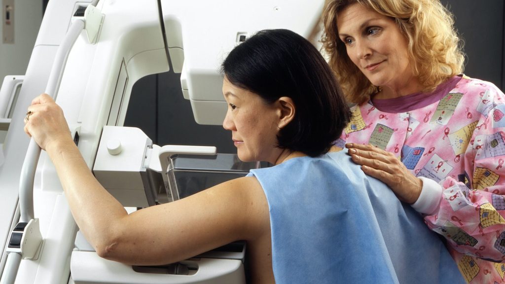
A new study reported in the journal Cancer reconsiders guidelines for when to start screening with mammograms if a woman has a first degree relative who was diagnosed with breast cancer.
Women with a first-degree family relative diagnosed with breast cancer, who are otherwise at average risk, are often advised to get screened 10 years earlier than the relative’s diagnosis age. However, there is little evidence to support the long-standing recommendation.
UC Davis Comprehensive Cancer Center researcher Diana Miglioretti joined Danielle Durham, with the Department of Radiology at University of North Carolina at Chapel Hill, and five other researchers on the study. They analysed data from the Breast Cancer Surveillance Consortium on screening mammograms conducted from 1996–2016 to evaluate when screenings should begin for women with a family history of breast cancer.
More than 300 000 women were included in the national study. Researchers compared cumulative 5-year breast cancer incidence among women with and without a first-degree family history of breast cancer by relative’s age at diagnosis and screening age.
“The study concluded that a woman with a relative diagnosed at or before age 45 may wish to consider, in consultation with her doctor, initiating screening 5–8 years earlier than their relative’s diagnosis age, rather than a decade earlier. That puts them at a risk that is equal to that of an average-risk woman who is age 50, which is the most recommended age for starting mammograms,” said Durham.
BRCA gene mutation carriers may benefit from starting screenings earlier. Women ages 30–39 with more than one first-degree relative diagnosed with breast cancer may wish to consider genetic counselling.
Increasing the age for initiating screening could reduce the potential harms of starting breast cancer screenings too early. These include increased radiation exposure and false positive results that require women to return to the clinic for diagnostic imaging and possibly invasive procedures, but do not result in a breast cancer diagnosis. The earlier a woman starts receiving mammograms, the more screenings they will undergo over their lifetime – and that increases the chances of experiencing these harms.
“Mammography also may not perform as well in younger women because they are more likely to have dense breasts which increase the difficulty of finding cancer on the images and results in more false-positives,” Miglioretti said.
The prognosis for breast cancer has improved, allowing more and more women to be cured with a combination of surgery, radiotherapy and medical treatment. A new trial led by Karolinska Institutet will investigate whether combining neoadjuvant chemotherapy with exercise will improve the outcomes of breast cancer patients.
Neoadjuvant chemotherapy (NACT) is increasingly used in breast cancer. The main benefit of NACT is its ability to downstage large tumours with a view to treatment by breast-conserving surgery, although there is a non-significant increase in the local recurrence rate. The best proof of NACT efficacy is pathological complete response (pCR), ie the absence of invasive tumour on post-NACT on surgical histopathology.
“While it is known that physical exercise can help patients to better tolerate often harsh cancer treatments, it is an emerging area of research to understand if and how exercise exerts anti-tumour effects and improves oncological outcomes”, explained Jana de Boniface, principal investigator of the trial and associate professor in the Breast Surgery Group, Department of Molecular Medicine and Surgery.
The Neo-ACT trial opened for recruitment in September 2022, and it is estimated that inclusion may be completed in December 2025.
Higher rates of brain metastases in patients with inflammatory breast cancer, a rare subtype of breast cancer, have been observed in studies, but detailed information is lacking. Now, a new study published in CANCER indicates that patients inflammatory breast cancer face a higher risk that their cancer will metastasise to the brain.
To provide insights into the incidence and risk factors for brain metastases in this patient population, Laura E.G. Warren, MD, and colleagues analysed data on 372 patients with stage III inflammatory breast cancer and 159 with stage IV inflammatory breast cancer.
Over a median follow-up of five years, the incidence of brain metastases at one, two, and five years was 5%, 9%, and 18% among patients who presented with stage III disease, and 17%, 30%, and 42% among those with stage IV disease. Patients with triple-negative breast cancer faced a particularly high risk, and when they did experience brain metastases, their survival time was shorter than those with hormone receptor–positive or HER2-positive breast cancer who experienced brain metastases. Higher risks of brain metastases were also seen in patients whose cancer had metastasised to other parts of the body besides the brain, especially when this occurred at a young age.
“The relatively high incidence of brain metastases seen in the study population highlights the need for future research on the potential role for surveillance brain imaging for high-risk patients. There is an open, phase II, single arm study at Dana-Farber Cancer Institute examining this question,” said Dr. Warren. “It also emphasises the need to obtain brain imaging in patients with inflammatory breast cancer presenting with neurologic symptoms given the high incidence of brain metastases in this population.” Most patients in this study who were diagnosed with brain metastases had neurologic symptoms, but because some patients may have undetected, asymptomatic brain metastases, the true incidence in patients with inflammatory breast cancer is likely even higher than what Dr Warren and her colleagues observed.
An accompanying editorial notes that when considering whether to implement routine brain imaging tests in patients with inflammatory breast cancer, it will be important to determine whether earlier detection of brain metastases leads to improvements in both survival and quality of life.
Why breast cancer survivors experience troubling cognitive problems is a long-standing mystery, and for which inflammation is one possible culprit. A new long-term study of older breast cancer survivors published in the Journal of Clinical Oncology adds evidence to this link.
Higher levels of the inflammatory marker C-reactive protein (CRP) were related to older breast cancer survivors reporting cognitive problems in the new study.
“Blood tests for CRP are used routinely in the clinic to determine risk of heart disease. Our study suggests this common test for inflammation might also be an indicator of risk for cognitive problems reported by breast cancer survivors,” said study lead author Judith Carroll, an associate professor at UCLA.
The Thinking and Living with Cancer (TLC) Study is one of the first long-term efforts to examine the potential link between chronic inflammation and cognition in breast cancer survivors 60 and older, who make up a majority of the nearly 4 million breast cancer survivors in the United States. Previous research has focused largely on younger women and women immediately after therapy, making it difficult to draw conclusions about CRP’s role in long-term cognitive problems among older breast cancer survivors.
In TLC, teams of researchers from around the country talked to, and obtained blood samples from, hundreds of breast cancer survivors and women without cancer up to six times over the course of five years. The study was motivated by hearing from survivors and advocates that cognitive problems are one of their major worries.
“Cognitive issues affect women’s daily lives years after completing treatment, and their reports of their own ability to complete tasks and remember things was the strongest indicator of problems in this study,” said co-senior study author Dr Jeanne Mandelblatt, a professor of oncology at Georgetown University who is the lead of the TLC study.
“Being able to test for levels of inflammation at the same time that cognition was being rigorously evaluated gave the TLC team a potential window into the biology underlying cognitive concerns,” said Elizabeth C. Breen, a professor emerita of psychiatry and biobehavioral sciences at the Cousins Center for Psychoneuroimmunology at UCLA, who also served as co-senior study author.
The women’s cognition was evaluated through a commonly used questionnaire. The study found higher CRP levels among survivors were predictive of lower reported cognitive function among breast cancer survivors. There was no similar relationship between CRP levels and reported cognition in the women without cancer.
Cognitive performance, as measured by standardised neuropsychological tests, failed to show a link between CRP and cognition. The authors say this may indicate women are more sensitive to differences in their everyday cognitive function, self-reporting changes that other tests miss.
The authors said their study supports the need for research on whether interventions that can lower inflammation – including increased physical activity, better sleep, and anti-inflammatory medications – may prevent or reduce cognitive concerns in older breast cancer survivors.
Giving standard chemotherapy drugs in a specific sequence for certain types of metastatic breast cancer can cut costs while preserving quality of life, according to a study in theJournal of Clinical Oncology.
The study, led by researchers from UNC Lineberger Comprehensive Cancer Center and UNC Gillings School of Global Public Health, developed three different computer models to predict how a hypothetical set of 10 000 patients with specific types of metastatic breast cancer would respond to different sequences and types of chemotherapy. For this study, the patient’s cancer was either endocrine resistant or was triple-negative breast cancer.
Many chemotherapy choices are available to treat metastatic breast cancer. While oncologists may prefer certain drugs to use early in treatment, the best order in which to give the drugs is unclear. The researchers consulted oncologists and experts in the field to choose which chemotherapy drugs were preferred choices to include in the study.
Mimicking clinical practice, and based upon existing data, the researchers then assumed that if a person started treatment with one drug, they would change to a second-choice treatment after their cancer stopped responding to the first drug, or if the side effects weren’t tolerable. The purpose of the study was to test whether putting the drugs in one sequence compared to another could keep the patient on treatment for similar times while decreasing their side effect and/or cost burden.
“The cost of cancer drugs in the US has rapidly increased, even for generics. As a society, we urgently need more strategies to reduce cancer drug costs without compromising outcomes, and our analysis provides quantifiable evidence to help providers choose lower priced, but equally effective sequences of drugs,” said Stephanie B. Wheeler, PhD, MPH, professor of health policy & management at UNC Gillings and associate director of community outreach and engagement at UNC Lineberger and corresponding author of the article. “More spending on cancer care does not necessarily confer greater health benefits.”
The costs calculated in this study were inclusive of medical and nonmedical costs borne by patients, including lost productivity. In this simulation, after two years, nearly all women would have completed the first three sets of treatment, but the cancer would cause the death of about one-third of the women. Productivity days lost due to sickness were similar across chemotherapy sequences, so most of the cost difference was due to drug savings. In the simulation, patients were placed in three groups, depending on what treatments they had already received for earlier episodes of breast cancer.
Outcomes in the three groups were:
For people who had not previously received the common chemotherapy drug categories, including a taxane (e.g., paclitaxel) or an anthracycline (e.g., capecitabine), treatment with paclitaxel then capecitabine followed by doxorubicin corresponded to the highest expected gains in quality of life and lowest costs.
For people who had previously received a taxane and an anthracycline drug, treatment with carboplatin, followed by capecitabine, followed by eribulin, corresponded to the highest expected gains in quality of life and lowest costs.
For people who had previously received a taxane but not an anthracycline, treatment sequences beginning with capecitabine or doxorubicin, followed by eribulin, were most cost-effective.
“The drugs we studied are already recommended and reimbursed for the treatment of metastatic breast cancer, but the optimal sequencing of them has been unclear, which has led to considerable variation in physician preference and practice. Our study suggests that treatment sequencing approaches that minimise costs early may improve the value of care,” Wheeler said. “The implications of this study are fairly straightforward for medical oncologists and those developing value-based clinical pathways to implement in practice now.”
Associate professor Katherine E. Reeder-Hayes, one of the study’s authors, said the treatment choices for metastatic breast cancer are constantly changing, and new options for targeted therapy have emerged even since this study was conducted. “Many oncologists and patients find that there aren’t any more targeted therapies that fit the cancer’s molecular profiles, so they are left with the choice of a number of chemotherapy drugs that may feel pretty similar or have an unclear balance of pros and cons.
“In that scenario, I hope our study will help expand the framework that we use to make these decisions from one where we just think about the biologic action of the drug to one where we also consider the bigger picture of what the treatment experience is like for the patient, including their financial burden, investment of time, and side effects,” Reeder-Hayes added. “The most potent drug isn’t always the next best choice depending on what the patient values and wants to accomplish with their treatment.”
Researchers have developed a two-pronged approach to imaging breast density in mice, resulted in better detection of changes in breast tissue, including spotting early signs of cancer. If applied in humans, the technology may also help with prognosis of disease as density can be linked to specific patterns of mammary gland growth, including signs of cancer development. The findings appeared in the American Journal of Pathology.
“Having a means to accurately assess mammary gland density in mice, just as is done clinically for women using mammograms, is an important research advance,” said Priscilla A. Furth, MD, professor of oncology and medicine at Georgetown Lombardi Comprehensive Cancer Center, and corresponding author of the study. “This method has the benefit of being applicable across all ages of mice and mammary gland shapes, unlike some methods used in earlier studies.”
While working as an undergraduate in Furth’s lab, Brendan Rooney developed an innovative analytic computer program (C’20), which allowed for sorting of mammary gland tissue to one of two imaging assessments. At first, Rooney looked at younger mouse glands and found that a program that removed background ‘noise’ in those images helped boost detection of abnormalities in what are typically rounder, more lobular tissues. But as aging occurs and the chances of developing cancer increase, lobules diminish and ridges become more apparent, just as falling autumn leaves expose tree branches. The mammary ridges represent ducts that carry milk and other fluids. When the de-noising technique was applied to the images from the older mice, it was found to be less reliable in detecting ridges. Therefore Rooney and the team turned to a different imaging program, which has primarily been used to detect blood vessel changes in the eye’s retina.
“The idea for the analytic program came from routine visual observations of tissue samples and the challenges inherent in observing differences in breast tissue with just a microscope. We found that visual human observations are important but having another read on abnormalities from optimal imaging programs added validity and rigor to our assessments,” says Rooney, the lead author of the study. “Not only does our program result in a high degree of diagnostic accuracy, it is freely available and easy to use.”
Now that the broad strokes of the research have been laid down and proof-of-principle has been established, Rooney has started medical school with a possible eye toward specializing in oncology. Both Furth and Rooney believe that future studies will need to refine and streamline their research approach in mice, including better density measurements that could enable sorting of samples into higher and lower probabilities of cancer.
Breast cancer cells. Image source: National Cancer Institute on Unsplash
Type XII collagen plays a key role in regulating the organisation of the tumour matrix, according to research published in the journal Nature Communications. The study investigators also discovered that high levels of collagen XII can trigger metastasis.
Cancer cells continually interact with the tumour microenvironment one component of which is the extracellular matrix. Collagen is an important part of this tumour microenvironment, but just how it influences tumours has not been understood.
“There’s still a lot we don’t know about the role of the extracellular matrix in cancer metastasis. Our study shows that collagen XII plays an important role in breast cancer progression and metastasis,” said Associate Professor Thomas Cox, senior author of the study.
“Imagine cancer cells as seeds, and the tumour microenvironment as the soil. By studying the soil – the extracellular matrix – we can begin to understand what makes some tumours more aggressive than others, and by extension, begin to develop new ways to treat cancer,” he explained.
The research also suggests that measuring the level of collagen XII in a patient’s tumour biopsy could potentially be used as an additional screening tool to identify aggressive breast cancers with higher rates of metastasis, such as in the triple-negative type of breast cancer. Furthermore, collagen XII might be a possible target for future treatments.
The extracellular matrix is a 3D meshwork of around 300–400 core molecules, including several collagen proteins. This matrix provides structural and functional support to cells and tissues in all parts of the body.
In this study, the researchers catalogued how the tumour matrix changes over time and have generated a comprehensive database of these changes, which has been made freely available to researchers.
The team focused on collagen XII, one of 28 types of collagen in the body. Collagen XII plays an important role in organising other collagens and can have profound effects on the 3D structure of the extracellular matrix.
The researchers studied tumours in mouse models from the earliest pre-clinical stages of cancer, right through to late-stage tumours. They found that as the tumours developed, many matrix molecules changed, and importantly the level of collagen XII was also increased.
“Collagen XII seems to be altering the properties of the tumour and makes it more aggressive,” said first author Michael Papanicolaou. “It changes how collagens are organised to support cancer cells escaping from the tumour and moving to other sites like the lungs.”
The team then genetically manipulated collagen XII production, looking at the effects of metastasis to other organs. They found that as levels of collagen XII increased, so did metastasis. These findings were then confirmed in human tumour biopsies, which showed that high levels of collagen XII are associated with higher metastasis and poorer overall survival rates.
Further research will focus on studying more human samples, and investigating possible therapeutic pathways.
Changing the order of treatments given to breast cancer patients could reduce side effects resulting from mastectomy and improve outcomes, according to a clinical feasibility trial, published in The Lancet Oncology.
In the study, researchers found that switching the sequence of treatments given to breast cancer patients was safe, without any increase in complications and could lead to patients receiving faster and more effective care compared to current methods.
Thirty-three women with breast cancer requiring a mastectomy and post-mastectomy radiotherapy, were recruited to the primary radiotherapy and deep inferior epigastric perforator flap reconstruction for patients with breast cancer (PRADA) trial between January 2016 and December 2017. They were also eligible for a breast reconstruction using tissue from another part of their body.
They were given chemotherapy followed by radiotherapy before having a mastectomy and a breast reconstruction. The team found that this approach was feasible and safe. They also found that side effects were low and that 12 months after surgery patients reported high levels of satisfaction with their breast reconstruction.
Lead author Daniel Leff said: “We believe that, in the long term, this approach will improve patients’ mental and physical wellbeing with higher quality of life scores and satisfaction with their reconstructed breasts compared to current care. It also means that many patients who are currently denied reconstruction due to concerns of further complications due to radiotherapy may be able to get access to this treatment in future.”
Research published in the Journal of the National Cancer Institute found that menopausal hormone therapy for breast cancer survivors is not associated with breast cancer reoccurrence, despite worries among some researchers and physicians.
Hot flashes and night sweats, as well as vaginal dryness and urinary tract infections, are common in breast cancer survivors, worsening quality of life and can lead patients to discontinue therapy. These symptoms may be alleviated by vaginal oestrogen therapy or menopausal hormone therapy (MHT). However, the safety of systemic and vaginal oestrogen use among breast cancer survivors, particularly those with oestrogen receptor-positive disease, has been unclear.
Many doctors caution breast cancer survivors against using MHT following the demonstration of an increased risk of breast cancer recurrence in two trials in the 1990s. Though later studies have not shown increased recurrence, they were seriously limited, with small sample sizes and short follow-up periods.
This study compared hormonal treatment with the risk of breast cancer recurrence and mortality in a large cohort of Danish postmenopausal women treated for early-stage oestrogen receptor-positive breast cancer.
Participants were diagnosed between 1997 and 2004 with early-stage breast cancer who received no treatment or five years of hormone therapy.
Among 8461 women, 1957 and 133 used vaginal oestrogen therapy or MHT, respectively, after diagnosis. No increase was seen in the risk of recurrence or mortality for those who received either vaginal oestrogen therapy or MHT.
“This large cohort study helps to inform the nuanced discussions between clinicians and breast cancer survivors about the safety of vaginal oestrogen therapy,” said Elizabeth Cathcart-Rake, writing in an accompanying editorial. “These results suggest that breast cancer survivors on tamoxifen with severe genitourinary symptoms can take vaginal estrogen therapy without experiencing an increase in their risk for breast cancer recurrence. However, caution is still advised when considering vaginal oestrogen for breast cancer survivors on aromatase inhibitors, or when considering menopausal hormonal therapy.”
Researchers previously assumed that metastasising tumours release cells continuously. However, a new study has reached a surprising conclusion: circulating cancer cells that later form metastases mainly arise during the sleep phase of the affected individuals. This may have implications for oncologists, as timing of samples may affect their results. The study findings have just been published in Nature.
Circadian rhythm-regulated hormones control metastasis
“When the affected person is asleep, the tumour awakens,” said study leader Professor Nicola Aceto at ETH Zurich. During their study, which included 30 female cancer patients and mouse models, the researchers found that the tumour generates more circulating cells when the organism is asleep. Cells that leave the tumour at night also divide more quickly and therefore have a higher potential to form metastases, compared to circulating cells that leave the tumour during the day.
“Our research shows that the escape of circulating cancer cells from the original tumour is controlled by hormones such as melatonin, which determine our rhythms of day and night,” said Zoi Diamantopoulou, the study’s lead author and a postdoctoral researcher at ETH Zurich.
An accidental discovery led to the study
In addition, the study indicates that the time in which tumour or blood samples are taken for diagnosis may influence the findings of oncologists. It was an accidental finding along these lines that first put the researchers on the right track, “Some of my colleagues work early in the morning or late in the evening; sometimes they’ll also analyse blood at unusual hours,” Prof Aceto said with a smile. The scientists were surprised to find that samples taken at different times of the day had very different levels of circulating cancer cells.
Another clue was the surprisingly high number of cancer cells found per unit of blood in mice compared to humans. The reason was that as nocturnal animals, mice sleep during the day, which is when scientists collect most of their samples.
“In our view, these findings may indicate the need for healthcare professionals to systematically record the time at which they perform biopsies,” Prof Aceto said. “It may help to make the data truly comparable.”
The researchers’ next step will be to figure out how these findings can be incorporated into existing cancer treatments to optimise therapies. As part of further studies with patients, Prof Aceto wants to investigate whether different types of cancer behave similarly to breast cancer and whether existing therapies can be made more successful if patients are treated at different times.