Tag: Alzheimer's disease

Adults Get the Least Sleep From Their 30s to 50s

Photo by Ketut Subiyanto on Pexels

People sleep less in mid-adulthood than they do in early and late adulthood, according to a large study published in Nature Communications. The study investigators found that sleep duration declines in early adulthood until age 33, and then picks up again at age 53.

The study, involving 730 187 participants spread over 63 countries, revealed how sleep patterns change across the lifespan, and how they were largely the same across countries.

Study participants were playing the Sea Hero Quest mobile game, a citizen science venture designed for neuroscience research, which was designed to aid Alzheimer’s research by shedding light on differences in spatial navigational abilities. Thus far, over four million people have played Sea Hero Quest, contributing to numerous studies across the project as a whole.

In addition to completing tasks testing navigational ability, anyone playing the game is asked to answer questions about demographic characteristics as well as other questions that can be useful to neuroscience research, such as on sleep patterns.

The researchers, led by Professor Hugo Spiers (UCL Psychology & Language Sciences) and Dr Antoine Coutrot (CNRS, University of Lyon) found that across the study sample, people sleep an average of 7.01 hours per night, with women sleeping 7.5 minutes longer than men on average. They found that the youngest participants in the sample (age 19) slept the most, and sleep duration declined throughout people’s 20s and early 30s before plateauing until their early 50s and increasing again. The pattern, including the newly-identified key time points of age 33 when declining sleep plateaus and 53 for sleep to increase again, was the same for men and women, and across countries and education levels.

The researchers suggest the decline in sleep during mid-life may be from the demands of childcare and working life.

Professor Spiers said: “Previous studies have found associations between age and sleep duration, but ours is the first large study to identify these three distinct phases across the life course. We found that across the globe, people sleep less during mid-adulthood, but average sleep duration varies between regions and between countries.”

People who report sleeping the most are in Eastern European countries such as Albania, Slovakia, Romania and the Czech Republic, reporting 20–40 minutes extra sleep per night and the least in South East Asian countries including the Philippines, Malaysia and Indonesia. People in the United Kingdom reported sleeping slightly less than the average. People tended to sleep a bit less in countries closer to the equator.

The researchers found that navigational ability was unaffected by sleep duration for most of the sample, except for among older adults (aged 54–70) whose optimal sleep duration was seven hours, although they caution that the findings among older adults might be impacted by underlying health conditions.

Source: University College London

Looking Back at 2022: Pandemic Fades but Other Challenges Remain

Photo by Joshua Hoehne on Unsplash

The year 2022 finally saw the COVID pandemic petering out, largely through the less-lethal but still highly contagious Omicron variant. Significant strides were made in cancer and Alzheimer’s research, although not without controversy. Amid growing public healthcare challenges in South Africa, the NHI Bill advanced closer to reality.

As Omicron displayed greatly reduced severity compared to prior strains, South African medical experts were some of the first to justify no longer being at ‘code red’. This brought an end to the cycles of lockdowns and travel restrictions characterised by the two previous years.

It even saw the lifting of some aspects of China’s ultra-strict ‘zero-COVID’ policy, with citizens paying online tribute to the memory of the heroic doctor who defied government censorship to warn the world. However, the pandemic’s true cost became apparent as the World Health Organization put global excess deaths for the pandemic at almost 15 million.

A number of key medical advances were made during the year for a variety of conditions. Studies showed that administering steroids after COVID hospitalisation with severe inflammation reduced mortality up to one year post-infection.

COVID was found to be linked to a spate of new-onset Type 1 diabetes, but this may just have been due to medical checkups as a result of developing COVID. The rheumatoid arthritis drug auranofin was found to relieve diabetes symptoms. And research suggested a possible way to deliver insulin and cancer drugs orally, by adding a ‘tag’ that lets them enter the bloodstream through the intestines.

The fields of cancer and Alzheimer’s research was rocked by findings of numerous red flags. This controversy did not stop real progress: the first new drug that had any real effectiveness against Alzheimer’s disease was confirmed in a historic trial. Fortunately, the flu jab also seems to protect against developing the disease. Indeed, serious infections appear to increase the risk of both Alzheimer’s and Parkinson’s.

In advanced ER-positive, HER-2 negative breast cancers, the new drug capivasertib halved the rate of progression.

It was also revealed that humans are paying through the nose for common medications compared to those that animals receive. Antimicrobial resistance also remains a growing problem, causing an estimated 1.2 million deaths in 2019.

A major South African Medical Research Council (SMARC) study told a familiar story: unsafe sex, interpersonal violence, obesity, hypertension, and alcohol consumption are the top risk factors for disease and death in South Africa.

Despite lessons learned in the COVID pandemic, South Africa saw the progression of systemic problems in healthcare such as a critical shortage of nurses. Dr Tim de Maayer’s open letter on appalling conditions at Rahima Moosa exposed deep-seated problems in Gauteng’s public healthcare system. This was followed by the shock resignation of top cancer surgeon Professor Carol-Ann Benn. The appointment of Nomantu Nkomo-Ralehoko as Gauteng Health MEC should hopefully change the province’s situation.

As for the National Health Insurance (NHI) Bill, medical aids have aimed to reposition themselves in the new uncertain paradigm while the threat of a mass exodus of healthcare professionals from the country still hangs in the air. A slew of legal challenges now await the Bill, which still has no details on how it will be financed.

‘Thor’ Actor Takes a Break from Acting after Alzheimer’s Gene Discovery

Actor Chris Hemsworth. Credit: Gage Skidmore / Wikimedia Commons

Actor Chris Hemsworth has announced that he is stepping back from acting in order to focus on preventative measures for Alzheimer’s disease.

The 39-year-old star of ‘Thor’ told Vanity Fair that genetic testing had confirmed that he had two pairs of a gene, APOE4. which is highly predictive of developing Alzheimer’s. About one in four have a single copy while 2–3% carry two copies of the gene.

The reason APOE4 increases Alzheimer’s risk isn’t not well understood. The APOE protein helps carry cholesterol and other types of fat in the bloodstream. Recent studies suggest that problems with brain cells’ ability to process lipids may play a key role in Alzheimer’s and related diseases.

Lipid imbalances can impair many of a cell’s essential processes. This includes creating cell membranes, moving molecules within the cell, and generating energy.

Hemsworth had made the discovery while making the TV series ‘Limitless‘, in which he engages in a variety of activities to push the limits of his own body and mind and explores ways of extending the lifespan.

“My concern was I just didn’t want to manipulate it and overdramatise it, and make it into some sort of hokey grab at empathy, or whatever, for entertainment,” said Hemsworth. “It’s not like I’ve been handed my resignation.”

He emphasises that he is thankful at having made the discovery, as it has made him more appreciative of his life, and it now means he can now take steps to protect his health.

Fortunately, research suggests that there are lifestyle changes that may offer preventative effects for APOE4 carriers, such as reducing stress and getting regular exercise – though the latter is unlikely to be a problem for the already athletic actor. Dietary measures include various low-carbohydrate diets (including ketogenic diets), regular portions of oily fish, cruciferous vegetables and abstaining from alcohol.

Supplements with potential benefits include DHA, quercetin, resveratrol, vitamin D3, vitamin K2, B-vitamin complex and possibly lithium.

Alzheimer’s Prions also Appear in Down Syndrome

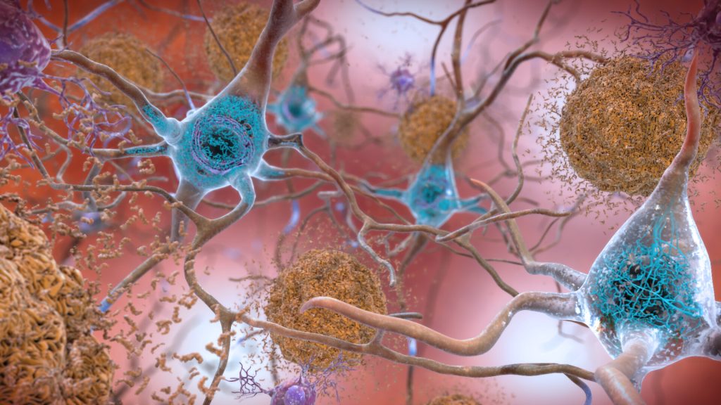
Plaques and neurons. Source: NIAH

The brains of people with Down syndrome develop the same neurodegenerative tangles and plaques associated with Alzheimer’s disease and they frequently demonstrate signs of the neurodegenerative disorder in their 40s or 50s. A new study in the journal PNAS shows that these tangles and plaques are driven by the same amyloid beta (Aß) and tau prions that they showed are behind Alzheimer’s disease.

Prions begin as normal proteins that become misshapen and self-propagate. They spread through tissue like an infection by forcing normal proteins to adopt the same misfolded shape. In both Alzheimer’s and Down syndrome, as Aß and tau prions accumulate in the brain, they cause neurological dysfunction that often manifests as dementia.

Tau tangles and Aß plaques are evident in most people with Down syndrome by age 40, according to the National Institute on Aging, with at least 50% of this population developing Alzheimer’s as they age.

The new study highlights how a better understanding of Down syndrome can lead to new insights about Alzheimer’s, as well.

“Here you have two diseases – Down syndrome and Alzheimer’s disease – that have entirely different causes, and yet we see the same disease biology. It’s really surprising,” said Stanley Prusiner, MD, the study’s senior author, who was awarded the Nobel Prize in 1997 for his discovery of prions.

Down syndrome is the most common neurodegenerative disease among younger people in the United States, while Alzheimer’s is the most common among adults.

Down syndrome occurs because of an extra copy of chromosome 21. Among the many genes on that chromosome is one called APP, which codes for one of the major components of amyloid beta. With an extra copy of the gene, people with Down syndrome produce excess APP, which may explain why they develop amyloid plaques early in life.

A clearer picture in young brain

It’s been known for some time that Aß plaques and tau tangles are present in both Down syndrome and Alzheimer’s. Having shown earlier that these neurodegenerative features are provoked by prions in Alzheimer’s, the researchers wanted to know whether the same aberrant proteins were present in the brains of people with Down syndrome.

While these plaques and tangles in the brains of people with Alzheimer’s disease have been well-studied, it can be challenging to discern which changes in the brain are from old age and which are from prion activity, said Prusiner.

“Because we see the same plaques-and-tangles pathology at a much younger age in people with Down syndrome, studying their brains allows us to get a better picture of the early process of disease formation, before the brain has become complicated by all the changes that go on during aging,” he said. “And ideally, you want therapies that address these early stages.”

Employing a variation on the novel assay they used in the Alzheimer’s study, the team looked at donated tissue samples from deceased people with Down syndrome, which they obtained from biobanks around the world. Of the 28 samples from donors aged 19 to 65 years old, the researchers were able to isolate measurable amounts of both Aß and tau prions in almost all of them.

New insights could yield preventative measures

The results confirm not only that prions are involved in the neurodegeneration seen in Down syndrome, but that Aß drives the formation of tau tangles as well as amyloid plaques, a relationship that has been suspected but not proven.

“The field has long tried to understand what the intersection is between these two pathologies,” said lead author Carlo Condello, PhD, also a member of the UCSF Institute for Neurodegenerative Diseases. “The Down syndrome case corroborates the idea; now you have this extra chromosome that’s driving the Aß, and there’s no tau gene on the chromosome. So, it’s truly by increasing the expression of Aß that you kick off production of the tau.”

These and other insights gained from studying the brains of people with Down syndrome will lead to a much better picture of how prions begin to form in the first place, said Condello.

Whether the Down syndrome brain tissue will prove to be the ultimate model for developing treatments for Alzheimer’s remains to be seen, the researchers said. While the two disorders share many similarities in their prion pathobiology, there are some differences that may be limiting.

Still, the researchers said, studying the plaques and tangles in Down syndrome is a promising route to identifying the specific prions that arise at the very earliest stages of the disease process. That insight could open new vistas on not only treating but perhaps even fending off Alzheimer’s disease.

“If we can understand how this neurodegeneration begins, we are one big step closer to being able to intervene at a meaningful point and actually prevent these large brain lesions from forming,” Condello said.

Source: University of California – San Francisco

Serious Infections Increase Alzheimer’s and Parkinson’s Risk

Old man
Source: JD Mason on Unsplash

Infections needing hospitalisal treatment in early- and mid-life are associated with an increased subsequent risk of Alzheimer’s and Parkinson’s diseases, according to a population-based, case control study by published in PLOS Medicine. No such increase was seen for amyotrophic lateral sclerosis (ALS), however.

The study used Swedish data on individuals diagnosed with Alzheimer’s disease, Parkinson’s disease or ALS, from 1970–2016, as well as five matched controls per case. The analysis included more than 290 000 Alzheimer’s disease cases, 100 000 Parkinson’s disease cases and 10 000 ALS cases.

The results show that a hospital-treated infection five or more years before diagnosis was associated with a 16% increased risk of Alzheimer’s disease and a 4% higher risk of Parkinson’s disease. The associations only applied to individuals diagnosed before the age of 60, whereas no association was found for those diagnosed later in life.

Individuals with multiple hospital treatments for infections before age 40 was associated with the highest risk of disease, with more than doubled risk of Alzheimer’s disease and more than 40% increase in the risk of Parkinson’s disease.

No association was observed for ALS, regardless of age at diagnosis. Due to the observational nature of the study, no causal link could be established.

“These findings suggest that infectious events may be a trigger or amplifier of a pre-existing disease process, leading to clinical onset of neurodegenerative disease at a relatively early age,” said Jiangwei Sun, the study’s first author and postdoctoral researcher at Karolinska Institutet.

Source: Karolinska Institutet

Researchers Uncover Major Contributor to Alzheimer’s Disease

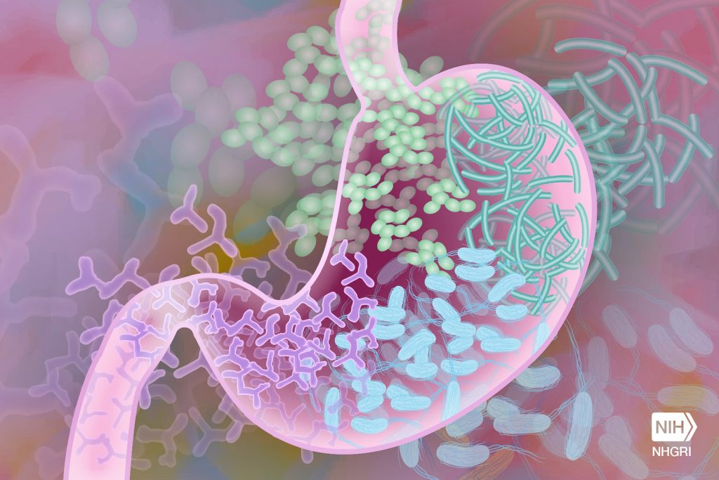
Gut microbiome. Credit: Darryl Leja, NIH

Research reports for the first time a pathway that begins in the gut and ends with a potent pro-inflammatory toxin in brain cells contributing to the development of Alzheimer’s disease (AD). Results are published in Frontiers in Neurology, where the researchers also report a simple way to counter the process.

The researchers, led by Drs Yuhai Zhao and Walter J Lukiw, found evidence that a molecule containing a very potent microbial-generated neurotoxin (lipopolysaccharide or LPS) derived from the Gram-negative bacteria Bacteroides fragilis in the human gastrointestinal (GI) tract generates a neurotoxin known as BF-LPS.

“LPSs in general are probably the most potent microbial-derived pro-inflammatory neurotoxic glycolipids known,” explained Dr Lukiw. “Many laboratories, including our own, have detected different forms of LPS within neurons of the Alzheimer’s disease-affected human brain.”

The researchers detailed the pathway of BF-LPS from the gut to the brain and its mechanisms of action once there. BF-LPS leaks out of the GI tract, crosses the blood brain barrier via the circulatory system, and accesses brain compartments. Then it increases inflammation in brain cells and inhibits neuron-specific neurofilament light (NF-L,) a protein that supports cell integrity. A deficit of this protein leads to progressive neuronal cell atrophy, and ultimately cell death, as is observed in AD-affected neurons. They also report that adequate intake of dietary fibber can head off the process.

The novel features of this newly described pathological pathway are threefold. The AD-stimulating pathway begins in the gut microbiome and therefore is very “locally sourced” and active throughout our lives. The highly potent neurotoxin BF-LPS is a natural by-product of gut-based microbial metabolism. Bacteroides fragilis abundance in the microbiome, which is the source of the neurotoxin BF-LPS, can be regulated by dietary fiber intake.

“Put another way, dietary-based approaches to balance the microorganisms in the microbiome may be an attractive means to modify the abundance, speciation, and complexity of enterotoxigenic forms of AD-relevant microbes and their potential for the pathological discharge of highly neurotoxic microbial-derived secretions that include BF-LPS and other forms of LPS,” Dr Lukiw explained.

The researchers conclude that an improved understanding of the interaction between the Gut–Brain axis and the gut microbiome and Alzheimer’s disease has considerable potential to lead to new diagnostic and therapeutic strategies in the clinical management of Alzheimer’s disease and other lethal, progressive, and age-related neurodegenerative disorders.

Source: Louisiana State University Health Sciences Center

‘Red Flags’ Uncovered in Alzheimer’s and Cancer Research

Image source: National Cancer Institute

The fields of Alzheimer’s disease and cancer research have both been shaken by recent investigations which have revealed image falsification and plagiarism. These findings call into question specific avenues of research which have received considerable funding.

Neuroscientist Matthew Schrag, a junior professor studying Alzheimer’s, had already ruffled some feathers criticising the FDA approval of the Alzheimer’s drug Aduhelm when he was approached by an attorney to investigate Simufilam, another Alzheimer’s drug under development.

According to Science, he used funding given to him by the attorney to investigate the data behind the drug’s development. The research focuses on amyloid beta (Aβ) plaques, long thought to be the culprit behind Alzheimer’s.

Schrag identified apparently altered or duplicated images in dozens of journal articles, and sent them to the National Institutes of Health (NIH), which had funded tens of millions of dollars. 

The investigation drew him towards a bedrock of modern Alzheimer’s research, a 2006 Nature study by Sylvain Lesné of the University of Minnesota in the laboratory of Karen Ashe, which identified an amyloid beta protein.

Schrag avoids describing the suspicious data as fraud, since that would require unfettered access to the original material. “I focus on what we can see in the published images, and describe them as red flags, not final conclusions,” he said. “The data should speak for itself.”

The work focused on ‘toxic oligomers’, subtypes of Aβ that dissolve in some bodily fluids, a potential Alzheimer’s cause that had gained traction in the early 2000s partly due to their discovery in autopsied Alzheimer’s patients.

Using transgenic mice, the UMN team discovered a previously unknown oligomer species, Aβ*56. They isolated Aβ*56 and injected it into young rats, causing a reduction in cognitive ability.

This discovery, the first to show a direct cause, resulted in an explosion in related research, with related studies receiving $287 million in National Institutes of Health funding in 2021, compared to no funding in 2006.

In concert with molecular biologist Elisabeth Bik, no less than 20 of Lesné’s papers were flagged, 10 of which related to Aβ*56. A finding which some Alzheimer’s experts say calls into question 16 years of amyloid beta research. Some had been suspicious and had failed to replicate the findings, but journals are reluctant to publish research which proves a negative or which contradicts a prominent researcher’s findings.

Cancer research has been dogged by its own crisis with fabricated data, according to an investigative report by Nature. For years, a prominent US cancer-research laboratory run by leading cancer geneticist Carlo Croce at the Ohio State University (OSU) had been dogged by allegations of plagiarism and falsified images. To date 11 papers he has co-authored have been retracted, and 21 have needed corrections.

In 2017, The New York Times first reported on allegations of research misconduct against Croce, when OSU opened inquiries into papers from Croce’s lab. These proceeded to formal investigations, Nature learnt, two of which found multiple instances of research misconduct, including data falsification and plagiarism, by scientists Michela Garofalo and Flavia Pichiorri, in papers they’d authored while in Croce’s laboratory.

Garofalo claimed she did not received proper image manipulation training and Pichiorri said she did not understand plagiarism at the time. They have since moved on from OSU.

OSU declined to charge Croce with misconduct as his involvement did not relate to direct plagiarism or image fabrication, but did note that these cases resulted out of his “poor mentorship and lack of oversight.”

Croce was removed from a number of his positions – for which he attempted to sue – but is still employed by OSU, and many of the papers identified by OSU have not been retracted by their journals.

Flu Jab May Protect Against Developing Alzheimer’s

Old man
Source: JD Mason on Unsplash

In a study with nearly 2 million older adults, those who received at least one influenza vaccine were 40% less likely than their non-vaccinated peers to develop Alzheimer’s disease over four years of follow-up, according to a new study from UTHealth Houston.

An early online version of the paper detailing the findings is available in advance of its publication in the Journal of Alzheimer’s Disease in August.

“We found that flu vaccination in older adults reduces the risk of developing Alzheimer’s disease for several years. The strength of this protective effect increased with the number of years that a person received an annual flu vaccine – in other words, the rate of developing Alzheimer’s was lowest among those who consistently received the flu vaccine every year,” said first author Avram S. Bukhbinder, MD. “Future research should assess whether flu vaccination is also associated with the rate of symptom progression in patients who already have Alzheimer’s dementia.”

The study comes two years after UTHealth Houston researchers found a possible link between the flu vaccine and reduced risk of Alzheimer’s disease. This new study analysed a much larger sample than previous research, including 935 887 flu-vaccinated patients and 935 887 non-vaccinated patients.

During four-year follow-up appointments, about 5.1% of flu-vaccinated patients were found to have developed Alzheimer’s disease. Meanwhile, 8.5% of non-vaccinated patients had developed Alzheimer’s disease during follow-up.

These results underscore the strong protective effect of the flu vaccine against Alzheimer’s disease, according to Bukhbinder and Schulz. However, the underlying mechanisms behind this process require further study.

“Since there is evidence that several vaccines may protect from Alzheimer’s disease, we are thinking that it isn’t a specific effect of the flu vaccine,” said Professor Paul. E. Schulz, MD, senior author of the study. “Instead, we believe that the immune system is complex, and some alterations, such as pneumonia, may activate it in a way that makes Alzheimer’s disease worse. But other things that activate the immune system may do so in a different way – one that protects from Alzheimer’s disease. Clearly, we have more to learn about how the immune system worsens or improves outcomes in this disease.”

Past research has uncovered a decreased risk of dementia associated with prior exposure to various adulthood vaccinations, including those for tetanus, polio, and herpes, in addition to the flu vaccine and others.

Additionally, as more time passes since the introduction of the COVID vaccine and longer follow-up data becomes available, Dr Bukhbinder said it seeing if there is a similar link between COVID vaccination and the risk of Alzheimer’s disease.

Source: The University of Texas Health Science Center at Houston

Newly Discovered Neuron Type may Help Explain Memory Formation

A healthy neuron.
A healthy neuron. Credit: NIH

Scientists publishing in Neuron have described how a newly discovered neuron type may be involved with the formation of memory in the hippocampus, which is marked by high-frequency electrical events.

It is known that memory is represented by changes in the hippocampus. One of the well-established changes in the hippocampus that has been associated with memory is the presence of so-called sharp wave ripples (SWR). These are brief, high-frequency electrical events generated in the hippocampus, and they are believed to represent a major event occurring in the brain in the so-called episodic memory, such as recalling a life event or a friend’s phone number.

However, what happens in the hippocampus when SRWs are generated has not been well understood.

Now a new study sheds light on the existence of a neuron type in the mouse hippocampus that might be a key to better understanding of episodic memory.

Professor Marco Capogna and Assistant professor Wen-Hsien Hou have contributed to the discovery of the novel neuron that is associated with sharp wave ripples and memory.

Possible disruption in dementia and Alzheimer’s

The study describes the novel neuron type in the hippocampus.

“We have found that this new type of neuron is maximally active during SWRs when the animal is awake – but quiet – or deeply asleep. In contrast, the neuron is not active at all when there is a slow, synchronized neuronal population activity called “theta” that can occur when an animal is awake and moves or in a particular type of sleep when we usually dream,” Prof Capogna said.

Because of this dichotomic activity, this novel type of neuron is named theta off-ripples on (TORO).

“How come, TORO-neurons are so sensitive to SWRs? The paper tries to answer this question by describing the functional connectivity of TORO-neurons with other neurons and brain areas, an approach called circuit mapping. We find that TOROs are activated by other types of neurons in the hippocampus, namely CA3 pyramidal-neurons and are inhibited by inputs coming from other brain areas, such as the septum,” Prof Capogna explained.

“Furthermore, the study finds that TOROs are inhibitory neurons that release the neurotransmitter GABA. They send their output locally – as most GABAergic neurons do – within the hippocampus, but also project and inhibit other brain areas outside the hippocampus, such as the septum and the cortex. In this way, TORO-neurons propagate the SWR information broadly in the brain and signal that a memory event occurred,” he concluded.

The team has monitored the activity of the neuron by using electrophysiology – a technique that detects activity of the neurons by measuring voltage versus time, and by using imaging that detects activity by measuring changes in calcium signalling inside the neurons.

Demonstrating a causal link between the activity of TORO-nerve cells and memory will be the next step, and exploring whether inhibition of TORO-neurons and sharp wave ripples occurs in dementia and Alzheimer’s diseases. 

Source: Aarhus University

For Alzheimer’s, Old Dogs Can Teach Humans New Tricks

Photo by Pauline Loroy on Unsplash

Researchers have found that quantifiable changes can be measured in dogs suspected of suffering from cognitive decline: an approach that could serve as a model for evaluating cognitive decline progression in, and treatments for, humans with Alzheimer’s disease.

In dogs there is a similar condition to similar to Alzheimer’s disease in humans, canine cognitive dysfunction syndrome (CCDS). In CCDS, cognitive decline is associated with the development of amyloid plaques as well as cortical atrophy. CCDS is also challenging to diagnose. Traditionally, CCDS is diagnosed based on ruling out any obvious physical conditions and an owner’s answers to a questionnaire.

“One problem with the current approach is that questionnaires only capture a constellation of home behaviours,” explained Professor Natasha Olby, co-senior author of the paper. “There can be other reasons for what an owner may perceive as cognitive decline – anything from an undiagnosed infection to a brain tumour.”

Olby and co-senior author Assistant Professor Margaret Gruen, wanted to see if cognitive function could be accurately quantified in dogs.

“Our goal was to bring together multiple tools in order to get a more complete picture of how CCDS presents in dogs,” A/Prof Gruen said.

To accomplish this, they recruited 39 dogs from 15 breeds. All of them were in the senior and geriatric age range, but in good health overall. A dog is considered ‘senior’ if it is in the last 25% of its expected life span based on breed and size, and geriatric beyond that.

The dogs underwent physical and orthopaedic exams, as well as lab work that included a blood test that is a marker of neuronal death. Their owners filled out two commonly used diagnostic questionnaires, and then the dogs participated in a series of cognitive tests designed to assess executive function, memory and attention.

“The approach we took isn’t necessarily designed to be diagnostic; instead, we want to use these tools to be able to identify dogs at an early stage and be able to follow them as the disease progresses, quantifying the changes,” Prof Olby said.

The team found that cognitive and blood test results correlated well with the questionnaire scores, suggesting that a multi-dimensional approach can be used to quantify cognitive decline in aging dogs.

“Being able to diagnose and quantify CCDS in a way that is clinically safe and relevant is a good first step toward being able to work with dogs as a model for Alzheimer’s disease in humans,” Prof Olby said. “Many of the current models of Alzheimers disease – in rodents, for example – are good for understanding physiological changes, but not for testing treatments.”

“Dogs live in our homes and develop naturally occurring disease just like we do,” A/Prof Gruen said. “These findings show promise for both dogs and humans in terms of improving our understanding of disease progression as well as for potentially testing treatments.”

Source: North Carolina State University