Tag: 27/10/25

New Laser System Measures Scalp and Brain Blood Flow

This optical measurement could offer an affordable and scalable way to diagnose stroke, brain injury and other conditions

Experimental arrangement of the SCOS system for measuring cerebral blood dynamics during superficial temporal artery (STA) occlusion. (a) 3D visualization of the SCOS device positioned over the temple region and the occlusion site near the ear bone. (b) Top and lateral views of the device, illustrating different detecting channels for sensing the scalp, skull, and brain layers. Credit: Liu et al., APL Bioengineering, 2025

Measuring blood flow in the brain is critical for responding to a range of neurological problems, including stroke, traumatic brain injury (TBI) and vascular dementia. But existing techniques, including magnetic resonance imaging and computed tomography, are expensive and therefore not widely available.

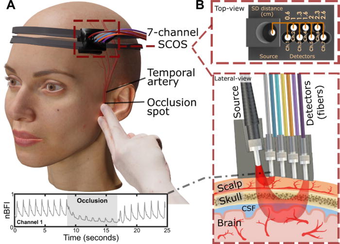
Researchers from the USC Neurorestoration Center and the California Institute of Technology (Caltech) have built a simple, noninvasive alternative. The device takes a technique currently used in animal studies known as speckle contrast optical spectroscopy (SCOS) and adapts it for potential clinical use in humans. It works by capturing images of scattered laser light with an affordable, high-resolution camera.

“It’s really that simple. Tiny blood cells pass through a laser beam, and the way the light scatters allows us to measure blood flow and volume in the brain,” said Charles Liu, MD, PhD, professor of clinical neurological surgery, urology and surgery at the Keck School of Medicine of USC, director of the USC Neurorestoration Center and co-senior author of the new research.

The device has already been tested with humans in small proof of concept studies demonstrating the tool’s utility for assessing stroke risk and detecting brain injury. In the current study, published in APL Bioengineering, Liu and his team sought to confirm that SCOS is truly measuring blood flow in the brain, rather than in the scalp, which also contains many blood vessels. The question has long plagued researchers who use light-based technology to visualize the brain.

Liu’s team took an innovative approach: By temporarily blocking blood flow to the scalp, they confirmed that SCOS readings were indeed measuring signals from blood vessels in the brain. Readings from 20 participants showed that positioning the detector at least 2.3cm away from the laser source provided the clearest measurement of brain blood flow. The study, funded in part by the National Institutes of Health, the Alfred Mann Foundation and the USC Neurorestoration Center, was just published in the journal APL Bioengineering.

“For the first time in humans, this experimental evidence shows that a laser speckle optical device can probe beyond the scalp layers to access cerebral signals,” said Simon Mahler, PhD, who is now an assistant professor in the Department of Biomedical Engineering at the Stevens Institute of Technology and one of the paper’s coauthors. “This is an important step toward using SCOS to non-invasively measure blood flow in the brain.”

Tracking brain blood flow

For years, researchers measuring brain signals with light-based technology, such as lasers and fibre optics, have used statistical simulations to estimate which signals originate in the brain versus the scalp. The USC Neurorestoration Center team found a direct way to test the difference, thanks to a collaboration between surgeons, engineers and neurologists.

“I perform surgeries to increase blood flow in the brain, and many of these involve temporarily stopping blood flow in the scalp,” said Jonathan Russin, MD, now professor and chief of neurosurgery at the University of Vermont, who continues to collaborate with the USC Neurorestoration Center. “That gave us a simple way to test the technology – by creating a change that affected only the scalp’s circulation while leaving the brain’s blood flow untouched.”

In 20 participants, the researchers temporarily stopped blood flow to the scalp, then collected a series of SCOS readings. By gradually moving the detector further from the head, they captured signals reaching progressively deeper towards the brain. They found that positioning the detector 2.3cm from the head allowed them to measure brain blood flow while minimising interference from the scalp.

The findings confirm the utility of SCOS for non-invasively detecting brain blood flow and provide important guidance for other researchers working with light-based technology, Liu said.

Bringing SCOS to patients

Beyond advancing research, the study helps confirm the clinical potential of SCOS for detecting and responding to stroke, brain injury and dementia. Because all of the team’s research has been done with humans, the tool is poised for rapid translation from the lab to the clinic.

“We look directly at humans in essentially the same way the tool will be applied, so there’s nothing lost in translation,” Liu said. “We are never more than one step away from the problem we’re trying to solve.”

The technique is already being used by some of the team’s collaborators to help diagnose stroke and TBI. Next, the researchers will continue to refine the technology and software, working to improve the resolution of images and the quality of data extracted from readings.

“With the knowledge that we’re now measuring exactly what we intend to measure, we’re also going to expand our testing of this technique with patients in clinical settings,” Liu said.

Source: Keck School of Medicine of USC

Fibroblasts Have Hidden Powers That Could Heal Brain Injuries

A mouse brain cortex seven days after a stroke that caused injury. Fibroblasts (green) have created collagen (pink) to form a protective scar layer around the injury. All images by Molofsky Lab, UCSF

Healing from any injury involves a delicate balance between scarring and inflammation – two processes that can wreak havoc as well as make repairs.

When the injury is to the brain, the balance is that much more important, yet scientists know almost nothing of how this process works.

Now, a study from UC San Francisco spotlights how a cell type called a fibroblast, that plays a healing role in other parts of the body also performs a similar function in the brain. The discovery is a step toward finding new ways to treat brain injuries, which are the nation’s leading cause of death and disability and for which there aren’t any drugs that can intervene.

Fibroblasts were only identified in the brain in the last decade. They reside mostly in the meninges, a set of protective membranes that surround the brain and spinal cord. Until now, scientists thought they mostly served to maintain the structure of the meninges and its network of blood vessels.

Ari Molofsky, MD, PhD, a professor of laboratory medicine, suspected the fibroblasts might be doing much more than that. He and Tom Arnold, MD, a professor of paediatrics, discovered that when the brain is injured – whether from a blow or a stroke – fibroblasts navigate from the meninges and surround the injured tissue where they create a protective barrier, or scar.

The same injury 14 days after the stroke. The scar now surrounds the whole injury, which is less swollen. Some fibroblasts have returned to their usual location in the meninges. Those that remain have switched roles and are now recruiting immune cells to moderate inflammation.

Then, about a week later, after the scar has formed, the fibroblasts adopt new roles. Some recruit immune cells that are required for healing; others ensure that the immune response doesn’t cause too much inflammation; and still others return to the meninges. Understanding these distinct stages could spur new interventions to help people with serious injuries.

Various views of a mouse brain cortex seven days after a stroke that caused injury. Green dots show fibroblast cells; pink areas show collagen produced by the fibroblasts to create a protective scar layer; and blue shows blood vessels with fibroblasts.

“Our study reveals opportunities to enhance the natural repair process,” said Molofsky, the senior author of the study, which appeared in Nature. “The goal is to give someone who’s experienced a traumatic brain injury or stroke the best outcome possible, based on the stage of healing they’re in.”

Therapies currently in clinical trials for lung and liver fibrosis target a molecule that prompts fibroblasts to create scarring. This suggests that other similar drugs could enhance healing in the early stages of a brain injury.

Molofsky’s study also offers an ideal venue for scientists to learn how fibroblasts are doing their work elsewhere in the body. Being largely devoid of immune cells, the brain offers a much clearer view than other organs like the lungs or liver, where immune cells may be too crowded around fibroblasts to see what they are doing.

“There’s a lot of potential here,” Molofsky said. “These overlooked cells seem adept at solving the common challenge of balancing healing and inflammation.”

Source: University of California – San Francisco

FundiHealth: Quality Healthcare Now Accessible for Students

Photo by Andrea Piacquadio on Pexels

Fundi, South Africa’s leading education finance and student solutions provider, has launched FundiHealth: a healthcare platform designed to meet the unique needs of students, young professionals and organisations committed to supporting the health and wellbeing of their people.

As part of its digital transformation and enablement strategy, Fundi has launched its own healthcare platform: FundiHealth.

Born out of Fundi’s deep understanding of the challenges students face and the recognition that health is a critical enabler of success, the platform offers affordable quality healthcare solutions for students and young professionals. “We’ve always believed that education unlocks potential, but we also see every day how poor health and the inability to afford care can derail even the most promising futures,” explains Benedict Johnson, Fundi Executive Head: EBS and New Initiatives. “Our platform was created with one clear purpose in mind: to remove healthcare as a barrier to education, productivity and meaningful participation in the economy.”

Fundi CEO Mala Suriah, moderator on the day ,Thembekile Mrototo and Fundi Executive Head Benedict Johnson at the FundiHealth launch.

The solution has been launched at a time when South Africa is grappling with a significant gap in healthcare coverage. According to the Council for Medical Schemes (2024), over 80% of South Africans do not belong to a medical aid scheme and rely entirely on the overstretched public health system. Young people are particularly vulnerable notes Charles Irumba, FundiHealth Executive: “Research from the Board of Healthcare Funders (2023) shows that fewer than 12% of students and young professionals under the age of 30 have adequate medical cover. This lack of access has serious implications not only for individual wellbeing but also for South Africa’s productivity and growth.” Data from Statistics South Africa (2024) further supports this; with absenteeism due to preventable illness costing the economy over R16 billion annually, with students and entry-level workers among the most affected.

“When students drop out of school because of untreated illnesses – including stress, depression or anxiety – or when young professionals miss work and lose income as a result, it has a detrimental effect on them as well as their families,” says Irumba. “FundiHealth ensures that young South Africans stay healthy in class and at work, contributing meaningfully to their futures and to our country’s growth and development.”

The new platform offers affordable, accessible medical cover that includes access to private doctors, medication, mental health support and preventative care services, all designed to fit the budgets of students, families and young professionals. “Critically, we’ve also created an option that allows organisations to offer their entry-level staff healthcare benefits at a fraction of the cost of traditional schemes. This is ideal for SMMEs and growing businesses, as it will assist them improve staff retention, morale and productivity,” adds Johnson. “Employers often underestimate the economic impact of poor health among their youngest and most vulnerable employees. By offering a healthcare solution like FundiHealth, they’re not just supporting their people, they’re strengthening their businesses too. Healthy employees are more engaged, more present and more productive. It’s a win-win.”

Beyond immediate productivity benefits, FundiHealth also has the potential to alleviate pressure on South Africa’s public health system by shifting some demand into the private sector and by promoting preventative care among young people. “If we want to grow as a nation, we have to keep our young people healthy in body, mind and spirit,” notes Irumba. “That’s what FundiHealth is all about. Good health is not a ‘nice-to-have’. It is a fundamental part of what it takes to succeed.”

FundiHealth is available to students, their families, young professionals and organisations at fundihealth.co.za. Customers can both explore different plan options and sign-up online.

“With FundiHealth, we’re tackling one of the most overlooked challenges to achieving one’s full potential: health. Good health underpins everything. By keeping our students and young professionals healthy, we’re keeping their dreams alive and South Africa moving forward,” Johnson concludes.

SAHPRA Registers Lenacapavir

Photo by Elen Sher on Unsplash

Pretoria, 27 October 2025 – The South African Health Products Regulatory Authority (SAHPRA) is pleased to announce the registration of Lenacapavir. Lenacapavir is an antiviral medicine that is recommended, in combination with safer sex practices, for pre-exposure prophylaxis (PrEP) to prevent HIV-1 infection in adults and adolescents weighing at least 35kg.

An application by Gilead was submitted to SAHPRA in March 2025. The SAHPRA review process was done in collaboration with the European Medicines for All Procedure (EU-M4all).  This procedure enables the European Medicines Agency (EMA), together with the participating regulatory authorities, to provide scientific opinions on high-priority medicines, such as Lenacapavir, intended for markets outside the European Union. The benefits of this pathway are to strengthen regulatory systems and accelerate access to essential medicines.

Lenacapavir mechanism. Credit: Dzinamarira, T et al., 2023. CC 4.0

Dosage

This product, developed to prevent new HIV infections, is a six-monthly injection. There is an initiation dose of a subcutaneous injection (administered just under the skin) with tablets (taken on days 1 and 2). It is used to reduce the risk of HIV in adults and adolescents who weigh at least 35kg, are HIV negative, and are at risk of getting HIV. Lenacapavir for PrEP should always be used in combination with safer sex practices, such as using condoms, to reduce the risk of getting other sexually transmitted infections.

“The registration of Lenacapavir is a game-changer, given the high prevalence rate of HIV in South Africa. This product is the most effective HIV prevention measure thus far,” indicated Dr Boitumelo Semete-Makokotlela, CEO: SAHPRA.

Source: SAHPRA

Faster MRI Scans Offer New Hope for Dementia Diagnosis

Photo by Mart Production on Pexels

The time to carry out diagnostic MRI scans for dementia can be cut to one third of their standard length, according to a new study led by UCL researchers.

The findings, published in Alzheimer’s & Dementia, have been described as a step towards ending ‘the postcode lottery in dementia diagnosis’. Shorter scans would be easier and more comfortable for patients and also enable more people to be scanned at a lower cost. The team behind the study say this could at least double the number of dementia scans able to be done in one day.

Senior author Professor Nick Fox, Director of the UCL Dementia Research Centre at the UCL Queen Square Institute of Neurology, said: “As more treatments that can slow or change the course of dementia are being developed, it’s important to make sure MRI scans are available to everyone. This is because people living with dementia often need an MRI scan as part of their diagnosis before they can access these treatments.

“To help make this possible, our team carried out the first study looking at how new imaging techniques – called parallel imaging – could speed up MRI scans in clinics. Their goal is to move closer to a future where every person with dementia can get a diagnosis through a scan.”

MRI scans often play a key role in an accurate dementia diagnosis, including ruling out other causes of symptoms and assisting in diagnosing the type of dementia. Emerging disease-modifying treatments such as lecanemab and donanemab also require an MRI scan before starting treatment and for safety monitoring during the course of treatment. Reducing the cost of scanning would contribute to lowering the total cost of delivering for such treatments. 

The ADMIRA study (Accelerated Magnetic Resonance Imaging for Alzheimer’s disease), part funded by Alzheimer’s Society’s Heather Corrie Impact Fund, aimed to understand the reliability of fast MRI scans compared to standard-of-care clinical scans. The neurologists on the study were joined by co-authors from the UCL Hawkes Institute and the UCL Advanced Research Computing Centre in the faculty of Engineering.

The research team scanned 92 people in an outpatient setting where an MRI brain scan was planned as part of their routine clinical assessment. The accelerated scans were carried out and enhanced to increase the quality of the image using new scanning methods. Three neuroradiologists examined these scans, and weren’t aware if they were looking at fast or standard-of-care scans.

Co-author Professor Geoff Parker (UCL Hawkes Institute and UCL Medical Physics and Biomedical Engineering) said: “Our research has taken advantage of recent breakthroughs in scanner technology. Our task was to work out just how fast we could scan while maintaining image quality good enough for diagnosis.”

The team found that the quicker scans reduced time in the scanner by 63% and they were as reliable as the standard-of-care scans for diagnosis and visual ratings.

First author Dr Miguel Rosa-Grilo (UCL Queen Square Institute of Neurology) said: “We were confident that the new scan would prove non-inferior to the standard scan, given the high image quality – but it was remarkable how well it performed.”

Richard Oakley, Associate Director of Research and Innovation at Alzheimer’s Society, said: “Dementia is the UK’s biggest killer, but one in three people living with the condition haven’t had a diagnosis. An early and accurate diagnosis isn’t just a label, it’s the first step to getting vital care, support and treatment.

“While MRIs aren’t the only way to diagnosis dementia, very few people with concerns about their cognitive health are offered one as part of the diagnosis process, mainly because they are expensive and not widely available. These faster MRIs, which take less than half the time of standard scans, could help end this postcode lottery in dementia diagnosis, cut costs and potentially give more people access to them.

“MRI scans can be an uncomfortable and daunting experience for patients, so anything we can do to make it an easier process is really positive.

“So far, this shortened MRI scan has been tested at one specialist centre with one type of MRI scanner, so more research is needed to make sure this works across different types of scanners and a diverse range of people. We’re hugely encouraged by this progress and eager to see how it continues.”

The team will now build on their early results by making sure the approach works across different types of MRI machines, so it can benefit as many hospitals and clinics as possible.

Source: University College London