Tag: 15/10/26

Are Young Women Receiving Delayed Care for Breast Cancer?

Study reveals that especially young patients and those with lower income are most likely to experience delays

Photo by Tima Miroshnichenko on Pexels

In an analysis of data on adolescents and young adults with breast cancer, lower income and younger age were linked with delays in care. The analysis published by Wiley online in CANCER, a peer-reviewed journal of the American Cancer Society, also found that women who experienced delays in cancer diagnoses tended to have later stages of disease.

Breast cancer diagnosed before the age of 40 years – which is before the recommended age of screening – often carries a poor prognosis, along with unique treatment and psychosocial challenges. Because younger women tend to not undergo screening and may face delays in response to breast cancer–related symptoms due to potential biases that they are not old enough to have cancer, investigators conducted a study to understand the impact of patient and tumour characteristics on delays in care and tie impact of delays on cancer outcomes.

In the analysis of data on 1071 individuals enrolled in the Young Women’s Breast Cancer Study, a multi-site prospective cohort of young women diagnosed with breast cancer by 40 years of age, the team identified 153 presentation delays, 120 diagnostic delays, and 228 treatment delays. Presentation delays were defined as ≥ 90 days from time of first patient-reported symptom(s) to presentation to a clinician, diagnostic delays were ≥ 90 days from first health care visit for symptom(s) to date of biopsy with a diagnosis with breast cancer, and treatment delays were considered ≥ 60 days between the time of diagnosis to time of first treatment.

After adjusting for influencing factors, lower annual income was associated with higher odds of presentation delay, while pregnancy at diagnosis, family history of breast cancer, and triple-negative breast cancer were associated with lower odds of presentation delay. Presentation delays were also associated with later stage disease in women with certain HER2-positive tumours. Odds of diagnostic delay were higher for younger patients and lower for those with triple-negative tumours. Those who experienced a delay in breast cancer diagnosis were more likely to have late-stage disease. Patients with more aggressive tumour types were less likely to have treatment delays.

Fortunately, delays were not associated with cancer-free or overall survival, but the authors noted that later stage at diagnosis may require more extensive surgery, radiation, or systemic therapy that can impact quality of life and other outcomes.

“Our study reveals continued issues in care delay among patients with young-onset breast cancer and emphasises a need to improve both patient and provider understanding of young-onset breast cancer risk factors and symptoms to decrease disease burden among these patients,” said senior author Kristen D. Brantley, PhD, MPH, of the Dana-Farber Cancer Institute.

Full Citation:
“Delays in breast cancer presentation, diagnosis, and treatment in young women.” Bessie X. Zhang, Tal Sella, Yue Zheng, Gregory J. Kirkner, Kathryn J. Ruddy, Shoshana M. Rosenberg, Virginia F. Borges, Lidia Schapira, Steven E. Come, Jeffrey M. Peppercorn, Laura Collins, Ann H. Partridge, and Kristen D. Brantley. CANCER; Published Online: October 12, 2026 (DOI: 10.1002/cncr.70594). 
URL: http://doi.wiley.com/10.1002/cncr.70594

Source: Wiley
Safer Nurse Workloads Could Alleviate Emergency Department Crowding

Credit: Pixabay CC0

A new study led by researchers from Penn Nursing’s Center for Health Outcomes and Policy Research (CHOPR) finds that improving hospital nurse workloads could alleviate delays in care contributing to emergency department crowding.

The findings, published in Medical Care, highlight a promising solution to emergency department (ED) crowding, a national crisis that occurs when the number of patients entering hospitals exceeds the number of available inpatient beds, causing bottlenecks in the emergency department.

In emergency departments where nurses reported staffing inadequacies, researchers identified significantly worse patient crowding. A 1 standard deviation increase in emergency department nurse staffing inadequacy was associated with an

  • 18-minute increase in ED length of stay for patients being observed for behavioural health
  • 9-minute increase in overall patient ED length of stay
  • 0.4 percentage point increase in patients leaving the ED before being seen by a provider.

The study also found that staffing inadequacy as reported by nurses working inpatient was associated with an 18-minute increase in ED length of stay for patients presenting with behavioural health conditions. Over 60 percent of emergency department nurses and 58% of inpatient nurses in the study reported that there were not enough staff to get work done in their hospital.

“Nurses are caring for more patients than they can safely manage, and our findings suggest that high nurse workloads are contributing to excessive delays in emergency department care,” said first author Jane Muir PhD MSHP RN (Assistant Professor, Department of Family and Community Health; CHOPR core faculty).

“Crowding occurs when hospitals don’t have enough resources to admit patients into a bed, whether in the ED or inpatient units,” Muir added. “We find that having enough nursing positions to appropriately staff high-stakes hospital care is one of the most important resources for ensuring the public experiences safe, timely care. A hospital bed without a nurse to staff it is not an available bed.”

The average study hospital was performing worse than the Joint Commission recommendations for ED care (recommendation: less than 2% of patients leave the ED without being seen and less than 4 hours length of stay for patients with behavioural health conditions). In this study, the average ED had 2.8% of patients leave without being seen and 5.5 hours length of stay for behavioural health patients.

Improving nurse staffing adequacy could result in more accessible ED care. In this study, researchers projected that funding more positions for bedside nurses in the 410 study hospitals would be associated with over 5000 more patients treated due to shorter ED lengths of stay, and over 250 patients retained in care due to fewer people leaving waiting rooms before being seen.

“Hospitals can create more funded positions for bedside nurses to alleviate chronic understaffing conditions and solve long wait times in emergency departments,” Muir stated. “Our study emphasises that safe nurse staffing matters to move the needle on safer, timelier care for the public.”

Source: Penn Nursing

The Ovary Doesn’t Retire After Menopause

Postmenopausal human ovaries show progressive molecular remodelling and inflammatory shifts

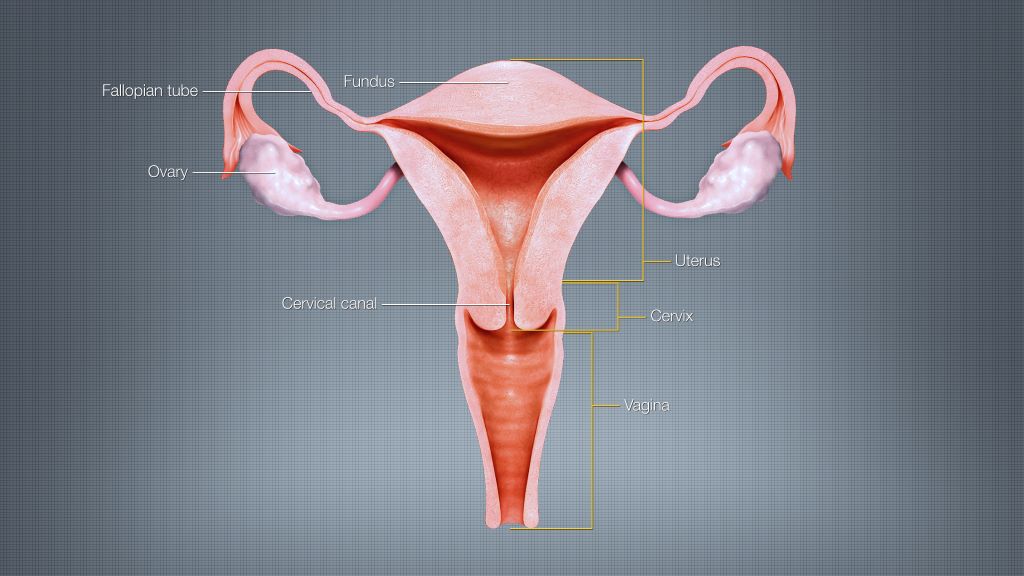
Female reproductive system. Credit: Scientific Animations CC4.0 BY-SA

Contrary to popular belief, the postmenopausal ovary is not defunct.

In a new, paradigm-shifting study of ovaries from 28 women, scientists at Northwestern University and the Buck Institute for Research on Aging found that in the decades after menopause the ovary remains biologically active and continues to remodel with age, showing increased inflammation and changes in tissue structure. The study also identified a molecular link between ovarian ageing and cancer.

The study was published on 14 October in Aging Cell.

“In the ovary, we reach a point of no return with respect to fertility in which it’s no longer producing eggs, and I would venture to say most people assume those ovaries are just sitting there doing nothing, similar to the appendix,” said co-corresponding author Francesca Duncan (associate professor of reproductive science in medicine, Northwestern University Feinberg School of Medicine; associate professor in residence, Buck Institute). “But that’s what makes this study very different.”

The findings challenge the notion that the ovary is an innocent bystander after reproduction ends, raising questions about whether it could play a more active role in ageing and chronic disease in women.

“The ovary is not just a reproductive organ,” said co-corresponding author Birgit Schilling (professor and director of the Mass Spectrometry Center, Buck Institute). “It really influences how somebody ages because it speaks to all these other organs, such as the brain, and also to the bone, and women after menopause experience often a lot of bone problems. So, we think there is a connection between all these organs.”

Women now routinely live for decades after menopause, making it increasingly important to understand what the ovary is doing during this long post-reproductive period, Duncan said.

“Is the ovary the canary in the coal mine in women?” said Duncan. “Could ageing in the ovary set off a cascade of changes that continues throughout the body as women get older?”

Duncan’s group previously found similar evidence of continued remodelling in postmenopausal ovaries of mice. When she embarked on that research and the current study in humans, she expected to see very little activity in the postmenopausal ovary.

“I was predicting that if we took a 50-year-old ovary, a 60-year-old ovary, a 70-year-old, an 80-year-old ovary, they would all look exactly the same, have no eggs, and basically be non-functional,” Duncan said. “The fact that across species we’re seeing this gives us confidence that this isn’t just some random event, but something truly meaningful is happening in the biology.”

What remains unknown is exactly what the ovary is doing after menopause and whether those changes are beneficial or harmful, Duncan said.

“Our data points to progressive ageing features in which the ovary appears to lose its original identity and ends up doing something different,” Duncan said. “We do not yet know if there’s ever a point in a woman’s life when this organ is no longer beneficial and it’s actually harmful. These results really point to the need to ask this question.”

To characterise the changes, Schilling’s team – spearheaded by Buck Institute staff scientist Mark Watson – used advanced proteomic technologies to analyse 5,812 protein groups in normal ovarian tissue from postmenopausal women aged 50 to 75.

Human ovarian tissue is difficult to obtain, making the 28 ovaries an unusually valuable collection. By focusing exclusively on women within a relatively narrow 25-year age range after menopause, the researchers examined molecular changes that continue during the postmenopausal years. The technology allowed examination of thousands of proteins across every sample and identification of patterns using multiple statistical approaches.

Those analyses showed that the ovary continues to change well after its reproductive life ends.

Ovaries from younger postmenopausal women had more proteins involved in maintaining cells and tissue structure, while older ovaries had more proteins associated with inflammation, immune activity and the ongoing breakdown and rebuilding of the extracellular matrix. The differences were most pronounced between ovaries from women in their 50s and those from women 70 and older.

“When we analysed the data in multiple different statistical ways, what became clear across all these methodologies is that the ovary was not stagnant,” Schilling said. “In fact, over the decades, there were quite dramatic differences in terms of some proteins’ levels would go up with age and some proteins’ levels would go down, which tells us this organ is quite dynamic.”

The ageing ovaries also showed some of the same molecular patterns seen in cellular senescence.

The findings build on a discovery Duncan’s lab made a decade ago. In 2016, the lab was the first to report that as ovaries age they become increasingly inflamed, fibrotic and stiff. Because cancer cells can thrive in collagen-rich, stiff environments, the scientists have been investigating whether these age-related changes could make the ovary more hospitable to cancer cells.

The new study provides a molecular view of that remodelling process later in life. As postmenopausal ovaries aged, proteins involved in maintaining tissue structure declined, while proteins involved in inflammation and reshaping the tissue environment became more prominent.

Although all the ovarian tissue in the study appeared normal and cancer-free, some proteins previously associated with ovarian cancer were more abundant in ageing ovaries, with the strongest signal among women in their 60s. The finding does not mean the ovaries were cancerous or precancerous, but suggests healthy ovarian ageing and ovarian cancer share some biological processes.

“It’s not that all postmenopausal women get ovarian cancer, but we know that that fibrotic inflammatory environment is certainly an environment that cancer cells prefer to home in on,” Duncan said. “This is all consistent with the idea that the ageing ovary is a potential third hit for ovarian cancer, with the first hit being genetic and the second hit being environmental. So can we target the ageing ovary therapeutically to lower the risk of ovarian cancer, or can we develop novel biomarkers of ovarian cancer?”

The findings do not answer whether healthy ovaries should ever be removed after menopause. Rather, they raise new questions about how the biological activity of the postmenopausal ovary might influence women’s health systemically and whether the observed changes contribute to disease or are features of normal ageing.

Source: Northwestern University

UP Researchers Call for More Personalised Radiation Dosing in Cancer Treatment

Two cancer patients can receive exactly the same amount of a radioactive medicine, yet their bodies may absorb very different radiation doses.

University of Pretoria (UP) nuclear medicine researchers argue that this is one of the next big challenges precision cancer treatment needs to address.

In a paper published in the inaugural volume of The Lancet Medical Imaging & Theranostics, Prof Mike Sathekge and Dr Kamo Ramonaheng call for greater use of patient-specific dosimetry, which measures how much radiation actually reaches a patient’s tumour and healthy organs.

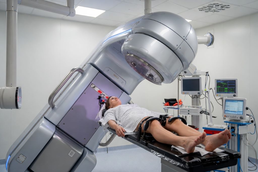
Radiopharmaceutical therapy uses medicines containing small amounts of radioactive material to seek out and attack cancer cells. But treatment is still commonly based on a standard amount of radioactivity, sometimes adjusted for factors such as body weight.

The researchers argue that knowing how much is given is not the same as knowing how much radiation the cancerous cell actually receives.

“Two patients can receive the same treatment, but their bodies may handle it very differently,” explains Prof Sathekge, Head of Nuclear Medicine at UP and Steve Biko Academic Hospital and CEO of the Nuclear Medicine Research Infrastructure (NuMeRI).

“Precision medicine should ultimately mean precision for the individual patient. If we can measure how much radiation reaches the cancer and how much reaches healthy tissue, we have much better information to guide treatment.”

From standard treatment to individual treatment

This measurement is known as dosimetry. The principle is similar to asking not only how much medicine was prescribed, but how much of the treatment reached its intended target.

The researchers propose a practical first step which is to measure the radiation absorbed by the tumour and healthy organs after treatment. This could help doctors understand whether the cancer received enough radiation while limiting unnecessary exposure to healthy tissue.

For patients receiving several treatment cycles, information from the first cycle could eventually help doctors decide whether later treatment should be adjusted or whether continuing the therapy is likely to benefit the patient.

Dr Kamo Ramonaheng elaborates that, “The question should not only be, ‘How much did we give the patient?’ It should also be, ‘How much did this patient actually receive where it mattered?’ Dosimetry brings us much closer to answering that question.”

This could become increasingly important as these therapies are used earlier in cancer care, where patients may live for many years after treatment and reducing long-term side effects becomes particularly important.

New technology is opening the door

Sathekge and Ramonaheng say advances in medical imaging and artificial intelligence are making personalised dosimetry increasingly practical.

More sensitive scanners can provide better information about where radioactive medicines travel in the body, while AI could eventually help analyse images faster and predict how individual patients may respond. The researchers stress that these technologies must be carefully tested before becoming part of routine clinical decision-making.

The ultimate possibility is to use imaging before treatment to predict the radiation dose a patient’s tumour and organs are likely to receive, allowing doctors to plan treatment more precisely before it begins.

Why NuMeRI matters

Much of the infrastructure needed to investigate these approaches already exists at NuMeRI, based at Steve Biko Academic Hospital.

The national research facility brings together advanced PET and SPECT imaging, radiopharmaceutical development, medical physics, radiobiology and clinical research. It is designed to help move discoveries from the laboratory into patient care and to strengthen research and training in personalised nuclear medicine.

Dr Ramonaheng is Head of Medical Physics and Radiobiology at NuMeRI and winner of the prestigious Saul Hertz Award at the Theranostics World Congress 2026. Her research includes patient-specific dosimetry.

Prof Sathekge explains that, “NuMeRI gives us a place where we can develop the science, test it, image patients and build the evidence needed to improve treatment. That is important for dosimetry because personalised treatment depends not only on an idea, but on having the technology, expertise and clinical environment to make it possible.”

The researchers caution that technology alone will not be enough. Wider use of personalised dosimetry will require trained specialists, consistent standards, appropriate funding and stronger clinical evidence.

Their paper describes dosimetry as the “compass” that could guide radiopharmaceutical therapy towards truly personalised cancer treatment.

For Prof Sathekge, the destination is clear, “We want to get closer to giving the right treatment, at the right radiation dose, to the right patient at the right time.”

Skin Sensors Show Link Between Insomnia and Fear Responses

A small study measuring stress-related skin signals found differences in the learned fear and safety responses of adults with and without insomnia.  

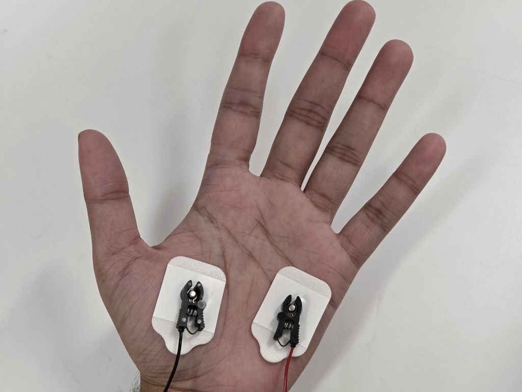
Demonstration of skin conductance sensors attached to the palm, used for estimating physiological arousal. Image credit: Revanth Reddy, CC-BY 4.0

A new computational model using skin conductance measurements to estimate stress-related emotional arousal found a significant difference in learned fear and safety responses over time between adults with and without insomnia. The new study from Dr. Rose T. Faghih of New York University Tandon School of Engineering, US, and collaborators at Harvard Medical School, US, describing the work was published this week in the open-access journal PLOS Mental Health.

Insomnia Disorder, affecting an estimated 10% of the population, has been widely conceptualised as a disorder of chronic hyperarousal linked to emotional and autonomic dysregulation. Sleep is thought to help consolidate fear extinction, the process of learning that a previously threatening stimulus no longer signals danger, forming a new, inhibitory “safety memory” to override an established “fear memory.” 

 Skin conductance, changes in the skin’s electrical properties driven by sweat gland activity, is a well-established marker of stress and emotional arousal. Previous studies looking at skin conductance and fear extinction have focused primarily on sharp, isolated incidences of stress activity.

 In the new study, researchers designed two statistical models estimating stress and arousal from skin conductance: one model using only isolated stress spikes, and one model that also incorporated the signal’s continuous background levels. Both models were applied to skin conductance recordings from an initial cohort of 24 adults with insomnia and 24 good sleepers who completed a task in which they were temporarily conditioned to fear a visual stimulus, then learned it was no longer dangerous.

 Only the model including the tonic background signal detected a significant difference between the two groups. From the conditioning phase, when the visual stimulus was paired with a mild shock, to the extinction phase, when it no longer was, the average ‘high-arousal index’ increased in people with insomnia but slightly decreased in good sleepers. Other differences between the groups — including lower average arousal during conditioning and higher average arousal when the fear and safety memories were recalled on the second day — followed a similar pattern but did not reach statistical significance.

 These findings suggest that because people with insomnia failed to fully acquire the safety memory on the first day or retain it until the second day, the new extinction memory was too weak to oppose the original fear memory during recall. Lead author Tianyu Hwang summarises the experiment as follows: “Our study shows that insomnia isn’t just about poor sleep – it directly relates to the body’s ability to learn and remember that a threat is gone.” 

 “Hyperarousal is thought to be an underlying risk factor for developing insomnia, and we were excited to see that sophisticated mathematical parsing of skin conductance revealed the same effect we had previously observed using fMRI,” co-author Dr Edward Pace-Schott writes. “This hyperarousal could oppose subsequent learning that a stimulus has become safe, which may explain why anxiety treatments like exposure therapy are more successful in people with good sleep.”

 Senior author Dr Rose Faghih adds: “Using our previously developed sympathetic arousal virtual sensors based on microfluctuations in sweat activity, we compared insomnia patients and healthy controls during fear extinction and observed that insomnia is associated with a reduced capacity to extinguish physiological threat responses.” 

 Dr Ryan Bottary, co-author, notes: “In our study, people with insomnia, compared to their good sleeping peers, became more aroused once the danger was taken away. For patients, this may help explain why the fear that ‘a bad night will ruin tomorrow’ survives so many tomorrows that turn out just fine.”

 Co-author Dr Saman Khazaei concludes: “I find it interesting to see that decoded arousal from certain components of skin conductance reflects a signature of a disrupted fear response in people diagnosed with insomnia. This finding may motivate further research on skin conductance and the use of wearable devices that measure it to improve sleep in everyday-life settings.”

Provided by PLOS